. 2022 Mar 4;12(3):jkab453.
doi: 10.1093/g3journal/jkab453.

A conditional glutamatergic synaptic vesicle marker for Drosophila

Affiliations

A conditional glutamatergic synaptic vesicle marker for Drosophila

Sarah J Certel et al. G3 (Bethesda). .

Abstract

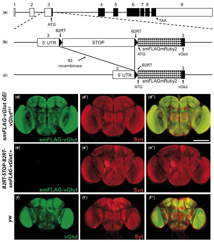
Glutamate is a principal neurotransmitter used extensively by the nervous systems of all vertebrate and invertebrate animals. It is primarily an excitatory neurotransmitter that has been implicated in nervous system development, as well as a myriad of brain functions from the simple transmission of information between neurons to more complex aspects of nervous system function including synaptic plasticity, learning, and memory. Identification of glutamatergic neurons and their sites of glutamate release are thus essential for understanding the mechanisms of neural circuit function and how information is processed to generate behavior. Here, we describe and characterize smFLAG-vGlut, a conditional marker of glutamatergic synaptic vesicles for the Drosophila model system. smFLAG-vGlut is validated for functionality, conditional expression, and specificity for glutamatergic neurons and synaptic vesicles. The utility of smFLAG-vGlut is demonstrated by glutamatergic neurotransmitter phenotyping of 26 different central complex neuron types of which nine were established to be glutamatergic. This illumination of glutamate neurotransmitter usage will enhance the modeling of central complex neural circuitry and thereby our understanding of information processing by this region of the fly brain. The use of smFLAG for glutamatergic neurotransmitter phenotyping and identification of glutamate release sites can be extended to any Drosophila neuron(s) represented by a binary transcription system driver.

Keywords: Drosophila; epitope tag; glutamatergic; synaptic vesicle; vGlut.


Figures

Fig. 1.
Fig. 1.
Strategic design of the conditional glutamatergic synaptic vesicle marker B2RT-STOP-B2RT-smFLAG-vGlut and conditional neuropil-specific expression in adult brain. a) Genomic exon structure of Drosophila vGlut. b) Genome editing at the endogenous vGlut genomic locus included insertion of a transcription STOP cassette flanked by B2 recombinase target sites (B2RTs) in the 5′ UTR upstream of translation start and insertion of the mRuby2_FP FLAG coding sequences in exon 3 at the ATG start codon of vGlut. Prior to excision of the STOP cassette smFLAG-vGlut is not expressed. c) After selective expression of the B2 recombinase in neurons of interest, with a binary transcription system driver and a compatible B2 recombinase responder transgene, the STOP cassette is excised and smFLAG-vGlut is expressed in glutamatergic neurons. d–e) Conditional expression in adult brain. d–d″) smFLAG-vGlut germline excision/vGlutSS1. d) smFLAG-vGlut; d′) Syn; d″) overlay. e–e″) B2RT-STOP-B2RT-smFLAG-vGlut/+. e) smFLAG-vGlut; e′) Syn; e″) overlay. f–f″) yw. f) vGlut; f′) Syt; f″) overlay. smFLAG-vGlut with a germline excision of the STOP cassette exhibits strong neuropil-specific immunostaining in the adult brain only after STOP cassette excision similar to endogenous vGlut. Images in (d) and (e) were collected and processed identically. Syn-Synapsin; Syt-Synaptotagmin. Scale bar: 100 µm.
Fig. 2.
Fig. 2.
Assessment of the conditionality and synaptic vesicle specificity of B2RT-STOP-B2RT-smFLAG-vGlut in larval VNC and third instar larval NMJ. Third instar larval VNC. a–a″) smFLAG-vGlut germline excision/vGlutSS1. a) smFLAG-vGlut; a′) Syn; a″) overlay. b–b″) B2RT-STOP-B2RT-smFLAG-vGlut/+. b) smFLAG-vGlut; b′) Syn; b″) overlay. c–c″) yw. c) vGlut; c′) Syt; c″) overlay. Third instar larval NMJ. d–d″) B2RT-smFLAG-vGlut germline excision/vGlutSS1. d) smFLAG-vGlut; d′) Syn; d″) overlay. e–e″) B2RT-STOP-B2RT-smFLAG-vGlut/+. e) smFLAG-vGlut; e′) Syn; e″) overlay. f–f″) yw. f) vGlut; f′) Syt; f″) overlay. smFLAG-vGlut with a germline excision of the STOP cassette exhibits strong neuropil-specific immunostaining in the larval VNC and at presynaptic terminals of the third larval instar NMJ similar to endogenous vGlut. No detectable anti-FLAG immunostaining is observed in the larval VNC or third instar larval NMJ prior to excision of the STOP cassette. Images in (a) and (b) and images in (d) and (e) were collected and processed identically. Syn, synapsin; Syt, Synaptotagmin. Scale bars: 100 µm.
Fig. 3.
Fig. 3.
Assessment of neurotransmitter and synaptic vesicle specificity of B2RT-STOP-B2RT-smFLAG-vGlut in single neuron types. Neuron anatomy is visualized with the plasma membrane marker CD8-mCherry. a–a″) Glutamatergic neuron MBON-6. a) CD8-mCherry; a′) smFLAG-vGlut; a″) overlay. b–b″) High-resolution 100× images of presynaptic terminals of glutamatergic neuron MBON-6. b) Syt-smHA; b′) smFLAG-vGlut; b″) overlay. High-resolution images of MBON-6 presynaptic terminals reveal near precise overlap of smFLAG-vGlut with the synaptic vesicle marker Syt-smHA. c–c″) Glutamatergic neuron MBON-6. c) CD8-mCherry; c′) GFP-Rab3; c″) overlay. GFP-Rab3 is expressed in glutamatergic neuron MBON-6 and distributes predominantly to presynaptic terminals in a pattern highly similar to that of smFLAG-vGlut. d–d″) GABAergic neuron LH1900. d) CD8-mCherry; d′) smFLAG-vGlut; d″) overlay. e–e″) Cholinergic neuron LH2094. e) CD8-mCherry; e′) smFLAG-vGlut; e″) overlay. No expression of smFLAG-vGlut was detected in the GABAergic neuron LH1900 or the cholinergic neuron LH2094. Large arrows—presynaptic terminals; small arrows—dendrites; arrowheads—cell bodies. Scale bars: a, c, d, e)—50 µm; b)—25µm.
Fig. 4.
Fig. 4.
B2RT-STOP-B2RT-smFLAG-vGlut expression identifies glutamatergic protocerebral bridge neurons. a–a″) PB.b-LAL.s-PS.s/SS52578. b–b″) PBG6–8.sG9.b/SS00117. c–c″) PB18.s-GxΔ7 Gy.b/PB18.s-9i1i8c.b/SS52266. d–d″) PBG1/2-9.b-SPS.s/SS52267. The plasma membrane marker CD8-mCherry (red, left column) allows visualization of the neuroanatomy of each protocerebral neuron. The presence of smFLAG-vGlut expression (arrows, middle column) indicates that each protocerebral neuron is glutamatergic and its subcellular distribution reveals the location of presynaptic terminals where SV fusion and glutamate release occurs. Scale bars: 50 µm.
Fig. 5.
Fig. 5.
B2RT-STOP-B2RT-smFLAG-vGlut expression identifies glutamatergic noduli and asymmetric body neurons. a–a″) LAL.s-GAi.s-NO1i.b/SS46517. b–b″) LAL.s-NO2i.b/SS47398. c–c″) LAL.s-N03Ai.b/SS47432. d–d″) LAL.s-CREc.s-NO3Pc.b/SS46525. e–e″) SLP-AB/SS50464. The plasma membrane marker CD8-mCherry (red, left column) allows visualization of the neuroanatomy of each neuron. The presence of smFLAG-vGlut expression (arrows, middle column) indicates that each neuron is glutamatergic and its subcellular distribution reveals the location of presynaptic terminals where SV fusion and glutamate release occur. Scale bars: 50 µm.

Similar articles

Cited by

References

    1. Adams MD, Celniker SE, Holt RA, Evans CA, Gocayne JD, Amanatides PG, Scherer SE, Li PW, Hoskins RA, Galle RF, et al.The genome sequence of Drosophila melanogaster. Science. 2000;287:2185–2195. - PubMed
    1. Aso Y, Hattori D, Yu Y, Johnston RM, Iyer NA, Ngo T-TB, Dionne H, Abbott LF, Axel R, Tanimoto H, et al.The neuronal architecture of the mushroom body provides a logic for associative learning. Elife. 2014;3:e04577. - PMC - PubMed
    1. Baker DA, Beckingham KM, Armstrong JD.. Functional dissection of the neural substrates for gravitaxic maze behavior in Drosophila melanogaster. J Comp Neurol. 2007;501(5):756–764. - PubMed
    1. Banerjee S, Vernon S, Jiao W, Choi BJ, Ruchti E, Asadzadeh J, Burri O, Stowers RS, McCabe BD.. Miniature neurotransmission is required to maintain Drosophila synaptic structures during ageing. Nat Commun. 2021;12(1):4399. - PMC - PubMed
    1. Buchanan SM, Kain JS, de Bivort BL.. Neuronal control of locomotor handedness in Drosophila. Proc Natl Acad Sci USA. 2015;112(21):6700–6705. - PMC - PubMed

Publication types

LinkOut - more resources