. 2022 Mar 3;220(3):iyac007.
doi: 10.1093/genetics/iyac007.

Assembly-dependent translation of subunits 6 (Atp6) and 9 (Atp9) of ATP synthase in yeast mitochondria

Affiliations

Assembly-dependent translation of subunits 6 (Atp6) and 9 (Atp9) of ATP synthase in yeast mitochondria

Anna M Kabala et al. Genetics. .

Abstract

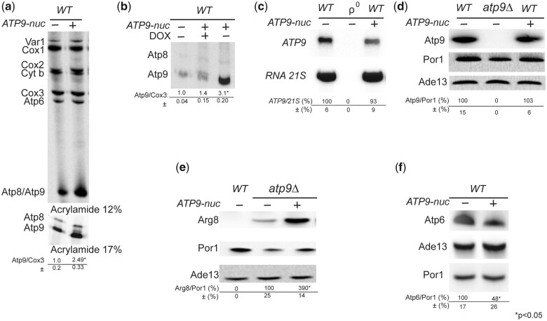
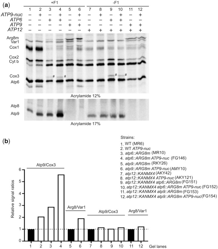
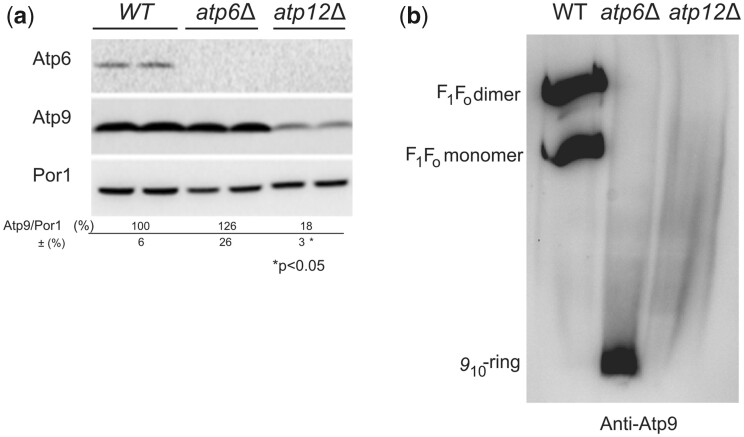
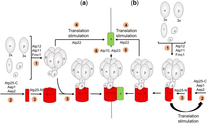
The yeast mitochondrial ATP synthase is an assembly of 28 subunits of 17 types of which 3 (subunits 6, 8, and 9) are encoded by mitochondrial genes, while the 14 others have a nuclear genetic origin. Within the membrane domain (FO) of this enzyme, the subunit 6 and a ring of 10 identical subunits 9 transport protons across the mitochondrial inner membrane coupled to ATP synthesis in the extra-membrane structure (F1) of ATP synthase. As a result of their dual genetic origin, the ATP synthase subunits are synthesized in the cytosol and inside the mitochondrion. How they are produced in the proper stoichiometry from two different cellular compartments is still poorly understood. The experiments herein reported show that the rate of translation of the subunits 9 and 6 is enhanced in strains with mutations leading to specific defects in the assembly of these proteins. These translation modifications involve assembly intermediates interacting with subunits 6 and 9 within the final enzyme and cis-regulatory sequences that control gene expression in the organelle. In addition to enabling a balanced output of the ATP synthase subunits, these assembly-dependent feedback loops are presumably important to limit the accumulation of harmful assembly intermediates that have the potential to dissipate the mitochondrial membrane electrical potential and the main source of chemical energy of the cell.

Keywords: ATP synthase; mitochondria; mitochondria DNA; mitochondrial biogenesis; mitochondrial gene expression; yeast.


Conflict of interest statement

None declared.

Figures

Fig. 1.
Fig. 1.
Mitochondrial translation in various subunit 6 mutants. a) The influence of the subunit 6 (Atp6) L173P variant on mitochondrial translation was investigated in 8 genetically independent recombinant 6-L173P clones issued from crosses between RKY12 (a synthetic ρ with the 6-L173P mutation) and MR10 [a ρ+ strain in which the coding sequence of ATP6 is replaced with ARG8m (atp6::ARG8m)], in comparison to 8 genetically independent WT recombinant clones issued from crosses between SDC30 (a synthetic ρ containing the wild-type ATP6 gene) and MR10 (see Table 1 for strain genotypes). b) Mitochondrial translation in 5 other subunit 6 mutants. Labeling of the mitochondrial translation products was performed in galactose grown cells during 20 min in the presence of [35S]-methionine and [35S]-cysteine and cycloheximide to block cytoplasmic translation. Total cellular protein extracts were then prepared and separated by SDS/PAGE in a 12% polyacrylamide gel containing 4 M urea and 25% glycerol. The gels were dried and the radiolabeled proteins were visualized using a PhosphorImager after 1-week exposure. The subunit 6 (Atp6) and Cox3 signals were quantified and compared to each other within each sample. The Atp6/Cox3 ratio was set to 1 for the WT.
Fig. 2.
Fig. 2.
Upregulation of the subunit 6-L173P variant is F1-dependent. a) ATP synthase in BN-gels. Mitochondria were isolated from the indicated strains grown in rich galactose at the indicated temperature. Proteins were extracted with digitonin (2 gr/gr protein) and separated by BN-PAGE on a 3–10% polyacrylamide gel (50 µg/lane). The proteins were transferred to a PVDF membrane and probed with antibodies against subunit 9. The F1–FO monomers and dimers are identified on the right. b) Cells from WT and fmc1Δ yeasts with or without the subunit 6 mutation L173P were grown in rich galactose at 28°C or 36°C, as indicated, and then incubated for 20 min with [35S]-methionine and [35S]-cysteine in the presence of cycloheximide to block cytoplasmic translation. Total cellular protein extracts were then prepared and separated by SDS/PAGE in a 12% polyacrylamide gel containing 4 M urea and 25% glycerol (with a 30:0.8 ratio of acrylamide and bis-acrylamide). The gels were dried and the radiolabeled proteins were visualized using a PhosphorImager. The subunit 6 (Atp6) and Cox3 signals were quantified and compared to each other within each sample. The Atp6/Cox3 ratio was set to 1.0 for the WT. The identity of the band above Cox3 (designated by a hash sign) in the samples containing the subunit 6 variant L173P is unknown.
Fig. 3.
Fig. 3.
Upregulation of the subunit 6 variant L173P is subunit 9-dependent. a) Growth phenotypes. Fresh glucose cultures of the indicated strains were serially diluted and spotted on rich glucose and glycerol media with or without 5 µg/mL doxycycline (DOX) as indicated. The glucose and glycerol plates were scanned after 3 and 6 days of incubation at 28°C, respectively. b and c) In vivo labeling of mitochondrial translation products. Pulse labeling was performed for 20 min with [35S]-methionine + [35S]-cysteine in the presence of cycloheximide to block cytoplasmic translation, in cells freshly grown in rich galactose medium. Total protein extracts were then prepared and separated by SDS/PAGE in 2 different gels: (1) 12% polyacrylamide containing 4 M urea and 25% glycerol (to resolve Var1, Cox1, Cox2, Cytochrome b, Cox3, and Atp6); (2) 17.5% of polyacrylamide (to resolve Atp8 and Atp9). The 2 gels shown in b) were loaded with equal amounts of radioactivity from the same experiment. After drying of the gels under vacuum, the radiolabeled proteins were visualized using a PhosphorImager after one-week of exposure. The subunit 6 (Atp6) and Cox3 signals were quantified and compared to each other within each sample. The Atp6/Cox3 ratio was set to 1.0 for the WT. The identity of the band above Cox3 (designated by a hash sign) in the samples containing the 6-L173P variant is unknown.
Fig. 4.
Fig. 4.
Pulse labeling of Atp6 and Cox3 and assembly of ATP synthase in strains expressing subunit 6 with and without the L173P mutation from the 5′UTR of COX2. a) Schema of mitochondrial genetic loci. As represented, the ectopic ATP6 gene, with or without the L173P mutation, is located upstream of the COX2 gene under control of the 5′-UTR and 3′-UTR of COX2, in a mitochondrial genome where the coding sequence of the native ATP6 gene is replaced with ARG8m. The corresponding abbreviated genotypes (WT, 5′-UTRCOX2-ATP6WT and 5′-UTRCOX2-ATP6L173P) are indicated on the right. b) Fresh glucose cultures of the strains with the indicated genotypes were serially diluted and spotted on rich glucose and rich glycerol media. The glucose and glycerol plates were scanned after 4 and 6 days of incubation at 28°C, respectively. c) In vivo labeling of Atp6 and Cox3. Pulse labeling of Atp6 and Cox3 was performed for 20 min with [35S]-methionine and [35S]-cysteine in the presence of cycloheximide to block cytoplasmic translation, in cells freshly grown in rich galactose medium. Total protein extracts were then prepared and separated by SDS/PAGE in a 12% polyacrylamide gel containing 4 M urea and 25% glycerol. After drying of the gel under vacuum, the radiolabeled proteins were visualized using a PhosphorImager after 1-week exposure. The subunit 6 (Atp6) and Cox3 signals were quantified and compared to each other within each sample. The Atp6/Cox3 ratio was set to 1.0 for the WT. d) Steady state levels of subunit 6. Total cellular proteins samples (20 µg) extracted from the 4 analyzed strains grown in rich galactose medium were separated by SDS–PAGE in a 12% polyacrylamide gel. The proteins were transferred to a nitrocellulose membrane and probed with antibodies against subunit 6 (Atp6) and porin. The levels of subunit 6 relative to porin are expressed in % of WT. Standard deviation and statistical significance between the 2 strains are indicated (* corresponds to a P-value <0.05) e) Protein complexes were extracted from isolated mitochondria with digitonin (2 gr/gr protein) and separated by BN-PAGE in a 3–10% polyacrylamide gel. After their transfer to a PVDF membrane, they were probed with antibodies against α-F1. The F1FO dimers and monomers, and free F1 are identified in the left-hand margin. f) In vivo labeling of Arg8 in strains RKY112 and RKY116. The rate of Arg8 synthesis in RKY112 and RKY116 from the 5′UTR of ATP6 (atp6::ARG8m) relative to Var1 was probed by pulse labeling in whole galactose grown cells for 20 min with [35S]-methionine and [35S]-cysteine in the presence of cycloheximide to block cytoplasmic translation, with WT and RKY20 strains as controls that do not encode Arg8. Total protein extracts were then prepared and separated by SDS/PAGE in a 12% polyacrylamide gel containing 4 M urea and 25% glycerol. After drying of the gel under vacuum, the radiolabeled proteins were visualized using a PhosphorImager.
Fig. 5.
Fig. 5.
ATP9-nuc stimulates translation at the mitochondrial ATP9 locus. a) Influence of ATP9-nuc on the translation of mitochondrial gene products in WT yeast. Pulse labeling was performed in cells freshly grown in rich galactose medium for 20 min with [35S]-methionine and [35S]-cysteine in the presence of cycloheximide to block cytoplasmic translation. Total cellular proteins were then extracted and separated by SDS/PAGE in 2 different gels: (1) 12% polyacrylamide containing 4 M urea and 25% glycerol (to resolve Var1, Cox1, Cox2, Cytochrome b, Cox3, and Atp6); (2) 17.5% polyacrylamide (to resolve Atp8 and Atp9). The 2 gels were loaded with equal amounts of radioactivity from the same experiment. After drying of the gels under vacuum, the radiolabeled proteins were visualized using a PhosphorImager. The subunit 9 (Atp6) and Cox3 signals were quantified and compared to each other within each sample. The Atp9/Cox3 ratio was set to 1.0 for the WT. Standard deviation and statistical significance of the differences between strains is indicated (* corresponds to a P-value <0.05). b) Evaluation of subunit 9 synthesis in WT yeast after blocking expression of ATP9-nuc with 5 µg/mL doxycycline (DOX), using the same procedure as in a). c) Northern blot analyses. Total RNAs isolated from cells with the indicated genotypes (ρ0 is a derivative of the WT strain totally devoid of mtDNA) freshly grown in rich galactose medium were separated in a 1% agarose gel. After their transfer to a Nytran membrane, the RNAs were hybridized with 32P labeled DNA probes specific to ATP9 and 21S RNA. The ATP9 mRNA signals were normalized to those corresponding to 21S RNA. d–f) Steady state levels of proteins in cells. Total proteins extracted from cells with the indicated genotypes grown in rich galactose medium were separated by SDS–PAGE (20 µg/lane), transferred to a nitrocellulose membrane, and probed with antibodies against the indicated proteins. The levels of subunits 6 (Atp6), 9 (Atp9), and Arg8 (expressed from atp9∷ARG8m) are normalized to porin. Ade13 is used as a cytosolic protein marker. The symbol “*” indicates a statistically significant difference between samples (P-value <0.05).
Fig. 6.
Fig. 6.
Upregulation of the mitochondrial ATP9 locus by ATP9-nuc and/or a lack in Atp6 is F1-dependent. a) Cells with the indicated genotypes were grown in rich galactose medium and their mitochondrially encoded proteins were radioactively labeled for 20 min with [35S]-methionine and [35S]-cysteine in the presence of cycloheximide to inhibit cytosolic translation. Total protein extracts were then prepared and resolved by SDS/PAGE in 2 types of gel loaded with the same amount of radioactivity (as in Fig. 2). After drying of the gels, the proteins were using a PhosphoImager. b) Quantification of subunit 9 and Arg8 expressed from the ATP9 locus (atp9∷ARG8m). The levels of subunit 9 and Cox3 in each sample were quantified and compared to each other. Those in lanes 1–4 are compared to each other after setting to 1.0 the Atp9/Cox3 ratio lane 1. Those in lanes 7–10 are compared to each other after setting to 1.0 the Atp9/Cox3 ratio in lane 7. The levels of Arg8 and Var1 were quantified in lanes 5, 6, 11, 12. The Arg8/Var1 ratio in lane 5 was set to 1.0 and compared to the one in lane 6. The Arg8/Var1 ratio in lane 11 was set to 1.0 and compared to the one in lane 12.
Fig. 7.
Fig. 7.
Influence of a lack in Atp12 or subunit 6 on the steady state levels and assembly of subunit 9. a) Steady states levels of subunit 9 in atp12Δ and atp6Δ yeasts. Total protein extracts from WT, atp6Δ, and atp12Δ strains grown in rich galactose medium were prepared and separated by SDS–PAGE in a 12% polyacrylamide gel. The proteins were transferred to a nitrocellulose membrane, and probed with antibody against subunit 6 (Atp6), subunit 9 (Atp9), and porin (Por1). The reported values (expressed as %WT) were calculated from 3 independent experiments. Statistical significance of the differences between samples is indicated. b) Assembly of subunit 9 in atp12Δ and atp6Δ yeasts. Mitochondria were isolated from WT, atp6Δ, and atp12Δ strains grown in rich galactose medium. Digitonin extracts containing the same quantity of proteins (50 µg) were prepared and separated in 3–10% polyacrylamide BN gel, transferred to a PVDF membrane, and probed with antibodies against subunit 9. The F1–FO monomers and dimers, and subunit 9-ring are identified in the left-hand margin.
Fig. 8.
Fig. 8.
Model of assembly-dependent translation of subunits 6 and 9 of yeast ATP synthase. a) Previously reported model (Rak et al. 2009, 2011). 1: The subunits of F1 (α, β, γ, δ, ε) assemble with the help of Atp11, Atp12, and Fmc1. 2 and 3: The subunit 9 is produced and assembled with the help of Aep1, Aep2, Atp25-C, and Atp25-N independently of any other ATP synthase component. 4: The F1 alone is involved in the activation of subunit 6 translation by Atp22. 5: The F1 and the 910-ring associate to each other. 5: The subunit 6 with the help of Atp10 and Atp23 is incorporated into ATP synthase. b) Model based on the results reported in this study. 1: The subunits of F1 (α, β, γ, δ, ε) assemble with the help of Atp11, Atp12 and Fmc1. 2, 3: The synthesis and assembly of subunit 9 with the help of Aep1, Aep2, Atp25-C, and Atp25-N is F1-dependent. 4: The F1-910 intermediate stimulates translation of subunit 6 by Atp22. 5: The subunit 6 with the help of Atp10 and Atp23 is incorporated into ATP synthase. The peripheral stalk subunits 8, 4, i, j, f, d, and OSCP of the ATP synthase monomer are not represented.

Similar articles

Cited by

References

    1. Ackerman SH. Atp11p and Atp12p are chaperones for F(1)-ATPase biogenesis in mitochondria. Biochim Biophys Acta. 2002;1555(1–3):101–105. - PubMed
    1. Ackerman SH, Gatti DL, Gellefors P, Douglas MG, Tzagoloff A. ATP13, a nuclear gene of Saccharomyces cerevisiae essential for the expression of subunit 9 of the mitochondrial ATPase. FEBS Lett. 1991;278(2):234–238. - PubMed
    1. Ackerman SH, Tzagoloff A. ATP10, a yeast nuclear gene required for the assembly of the mitochondrial F1-F0 complex. J Biol Chem. 1990a;265(17):9952–9959. - PubMed
    1. Ackerman SH, Tzagoloff A. Identification of two nuclear genes (ATP11, ATP12) required for assembly of the yeast F1-ATPase. Proc Natl Acad Sci U S A. 1990b;87(13):4986–4990. - PMC - PubMed
    1. Ackerman SH, Tzagoloff A. Function, structure, and biogenesis of mitochondrial ATP synthase. Prog Nucleic Acid Res Mol Biol. 2005;80:95–133. - PubMed

Publication types

MeSH terms

Substances