

Repression of lncRNA PART1 attenuates ovarian cancer cell viability, migration and invasion through the miR-503-5p/FOXK1 axis
- PMID: 35100978
- PMCID: PMC8802513
- DOI: 10.1186/s12885-021-09005-x
Repression of lncRNA PART1 attenuates ovarian cancer cell viability, migration and invasion through the miR-503-5p/FOXK1 axis
Abstract
Background: Ovarian cancer (OC) is a female malignant tumor with a high fatality rate. Long non-coding RNAs (lncRNAs) are deeply involved in OC progression. The aim of this study is to explore the specific mechanism of lncRNA prostate androgen-regulated transcript 1 (PART1) in OC.
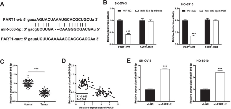
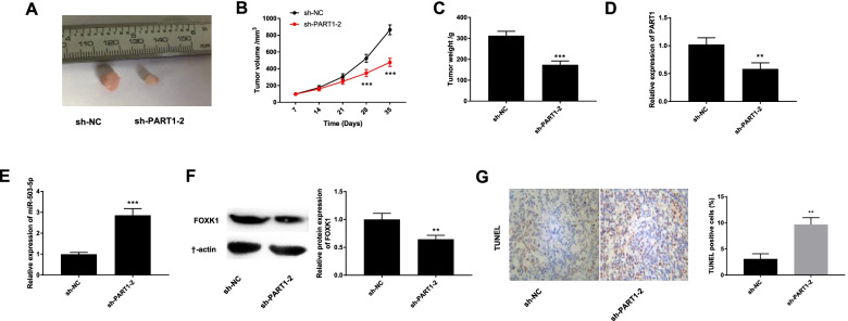
Methods: Quantitative real time PCR was utilized to determine the expression levels of PART1, microRNA (miR)-503-5p and forkhead-box k1 (FOXK1) in OC tissues and/or cells. The cell viability, migration, and invasion in OC were evaluated by 3-(4, 5-dimethyl-2-thiazolyl)-2, 5-diphenyl-2-h-tetrazolium bromide assay, wound healing assay and transwell invasion assay, respectively. Flow cytometry was used to analyze the cell apoptosis. The xenograft tumor was conducted in nude mice to verify the effect of PART1 knockdown on OC in vivo. The target relationships among PART1, miR-503-5p and FOXK1 were predicted by StarBase, and verified by luciferase reporter assay. The level of FOXK1 was assessed by western blot.
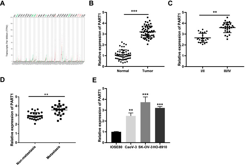
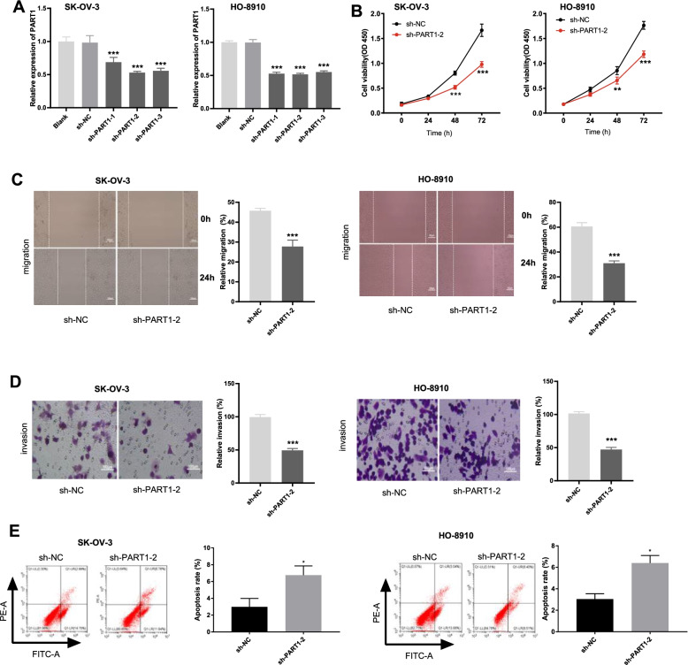
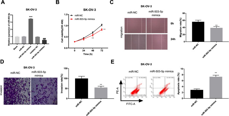
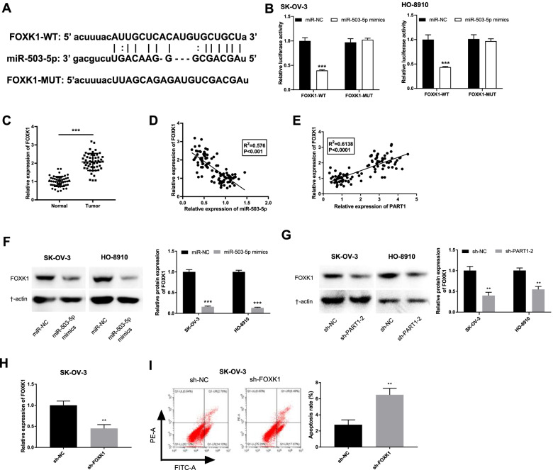
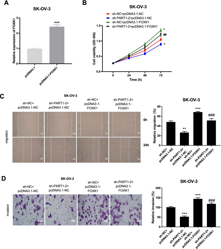
Results: Increased expression of PART1 and FOXK1 was observed in OC tissues or cells, whereas miR-503-5p was downregulated. PART1 silencing or miR-503-5p overexpression repressed the cell viability, migration and invasion, and protomed apoptosis. Meanwhile, miR-503-5p was a target of PART1, and FOXK1 was a direct target gene of miR-503-5p. Both downregulation of miR-503-5p and upregulation of FOXK1 partly relieved the suppressive effects of PART1 knockdown on the oncogenicity of OC in vitro.
Conclusion: Decreased PART1 represses the cell viability, migration and invasion of OC via regulating the miR-503-5p/FOXK1 axis, which provided an underlying target for treating OC.
Keywords: FOXK1; Ovarian cancer; lncRNA PART1; miR-503-5p.
© 2022. The Author(s).
Conflict of interest statement
The authors declare that they have no competing interests
Figures
Similar articles
-
Knockdown of circAPLP2 Inhibits Progression of Colorectal Cancer by Regulating miR-485-5p/FOXK1 Axis.Cancer Biother Radiopharm. 2021 Nov;36(9):737-752. doi: 10.1089/cbr.2019.3310. Epub 2020 Apr 24. Cancer Biother Radiopharm. 2021. PMID: 32343603
-
Long non-coding RNA MCM3AP-AS1 promotes growth and migration through modulating FOXK1 by sponging miR-138-5p in pancreatic cancer.Mol Med. 2019 Dec 12;25(1):55. doi: 10.1186/s10020-019-0121-2. Mol Med. 2019. PMID: 31830901 Free PMC article.
-
LINC-PINT suppresses tumour cell proliferation, migration and invasion through targeting miR-374a-5p in ovarian cancer.Cell Biochem Funct. 2020 Dec;38(8):1089-1099. doi: 10.1002/cbf.3565. Epub 2020 Jul 7. Cell Biochem Funct. 2020. PMID: 32638404
-
Getting to know ovarian cancer: Focusing on the effect of LncRNAs in this cancer and the effective signaling pathways.Pathol Res Pract. 2024 Feb;254:155084. doi: 10.1016/j.prp.2023.155084. Epub 2024 Jan 4. Pathol Res Pract. 2024. PMID: 38244434 Review.
-
MiR-4492, a New Potential MicroRNA for Cancer Diagnosis and Treatment: A Mini Review.Chonnam Med J. 2024 Jan;60(1):21-26. doi: 10.4068/cmj.2024.60.1.21. Epub 2024 Jan 25. Chonnam Med J. 2024. PMID: 38304137 Free PMC article. Review.
Cited by
-
PART1 facilitates tumorigenesis and inhibits ferroptosis by regulating the miR-490-3p/SLC7A11 axis in hepatocellular carcinoma.Aging (Albany NY). 2024 Jul 5;16(14):11339-11358. doi: 10.18632/aging.206009. Epub 2024 Jul 5. Aging (Albany NY). 2024. PMID: 39029955 Free PMC article.
-
Diverse activity of miR-150 in Tumor development: shedding light on the potential mechanisms.Cancer Cell Int. 2023 Nov 3;23(1):261. doi: 10.1186/s12935-023-03105-3. Cancer Cell Int. 2023. PMID: 37924077 Free PMC article. Review.
-
Long non‑coding RNA PART1: dual role in cancer.Hum Cell. 2022 Sep;35(5):1364-1374. doi: 10.1007/s13577-022-00752-y. Epub 2022 Jul 21. Hum Cell. 2022. PMID: 35864416 Review.
-
A review on the role of long non-coding RNA prostate androgen-regulated transcript 1 (PART1) in the etiology of different disorders.Front Cell Dev Biol. 2023 Feb 15;11:1124615. doi: 10.3389/fcell.2023.1124615. eCollection 2023. Front Cell Dev Biol. 2023. PMID: 36875771 Free PMC article. Review.
-
Hsa_circ_0102899 promotes epithelial-mesenchymal transition in non-small cell lung cancer.Clin Transl Oncol. 2023 Nov;25(11):3252-3262. doi: 10.1007/s12094-023-03220-7. Epub 2023 Jul 1. Clin Transl Oncol. 2023. PMID: 37393417
References
-
- Rustin GJ, van der Burg ME, Griffin CL, Guthrie D, Lamont A, Jayson GC, Kristensen G, Mediola C, Coens C, Qian W, et al. Early versus delayed treatment of relapsed ovarian cancer (MRC OV05/EORTC 55955): a randomised trial. Lancet. 2010;376(9747):1155–1163. doi: 10.1016/S0140-6736(10)61268-8. - DOI - PubMed
MeSH terms
Substances
Grants and funding
LinkOut - more resources
Full Text Sources
Medical
