Case Reports
. 2022 Jan 31;17(1):29.
doi: 10.1186/s13023-021-02068-w.

Novel insights into PORCN mutations, associated phenotypes and pathophysiological aspects

Affiliations
Case Reports

Novel insights into PORCN mutations, associated phenotypes and pathophysiological aspects

Annabelle Arlt et al. Orphanet J Rare Dis. .

Abstract

Background: Goltz syndrome (GS) is a X-linked disorder defined by defects of mesodermal- and ectodermal-derived structures and caused by PORCN mutations. Features include striated skin-pigmentation, ocular and skeletal malformations and supernumerary or hypoplastic nipples. Generally, GS is associated with in utero lethality in males and most of the reported male patients show mosaicism (only three non-mosaic surviving males have been described so far). Also, precise descriptions of neurological deficits in GS are rare and less severe phenotypes might not only be caused by mosaicism but also by less pathogenic mutations suggesting the need of a molecular genetics and functional work-up of these rare variants.

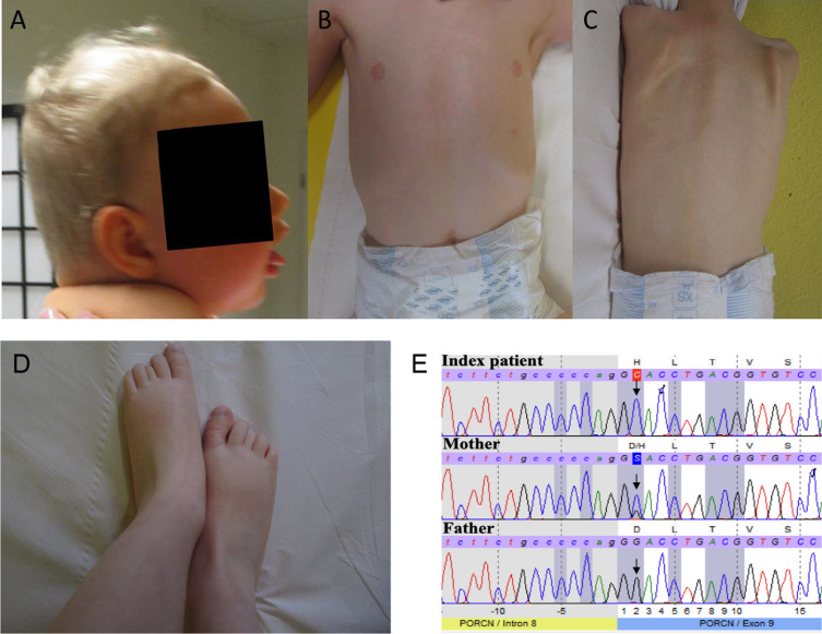
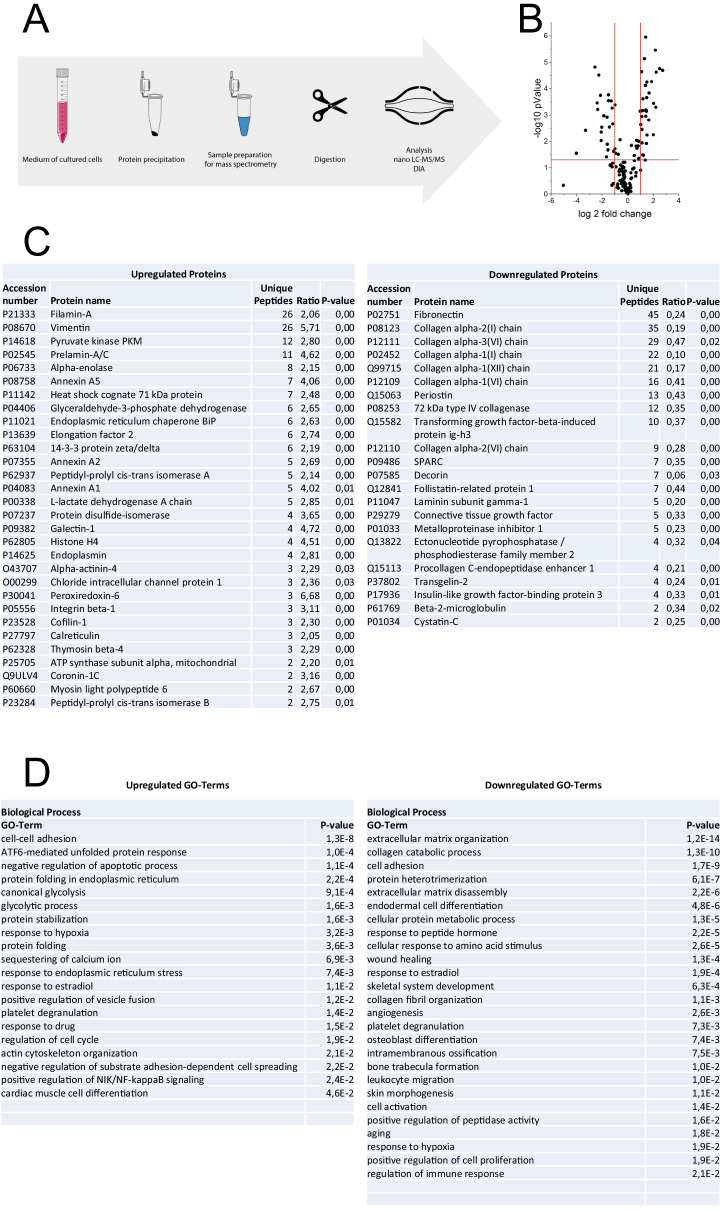
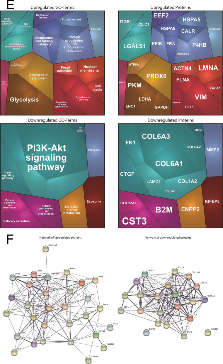
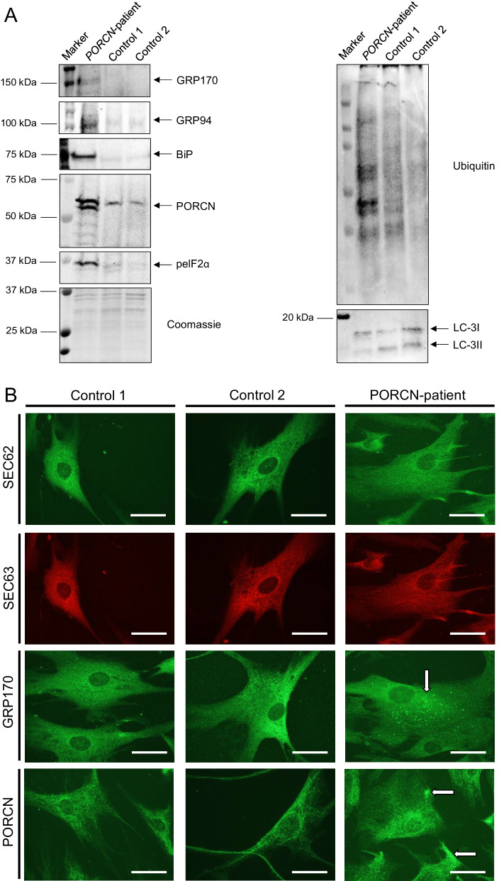
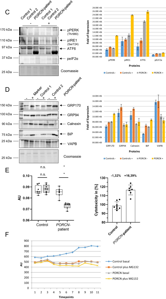
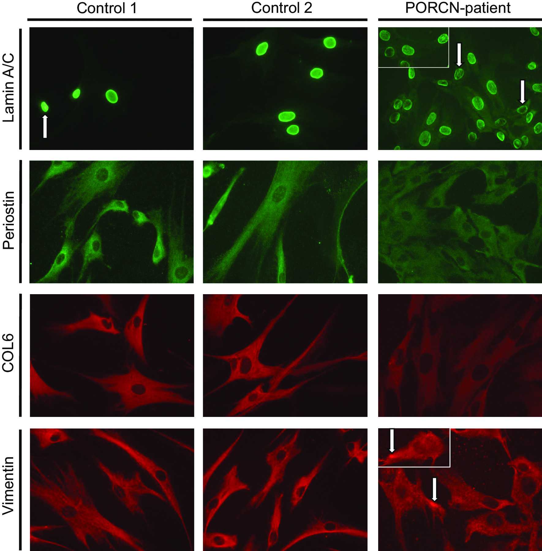
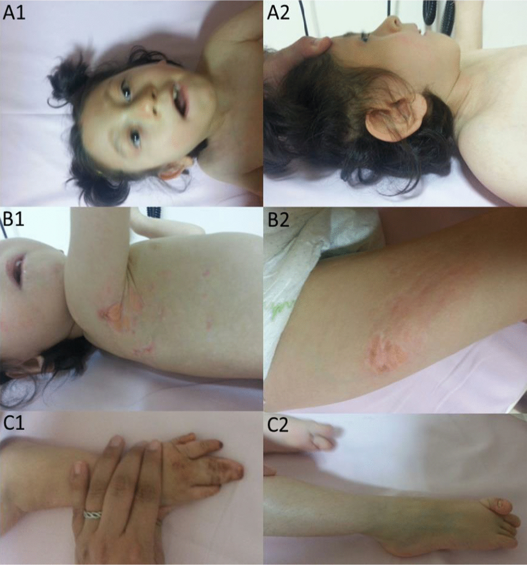
Results: We report two cases: one girl suffering from typical skin and skeletal abnormalities, developmental delay, microcephaly, thin corpus callosum, periventricular gliosis and drug-resistant epilepsy caused by a PORCN nonsense-mutation (c.283C > T, p.Arg95Ter). Presence of these combined neurological features indicates that CNS-vulnerability might be a guiding symptom in the diagnosis of GS patients. The other patient is a boy with a supernumerary nipple and skeletal anomalies but also, developmental delay, microcephaly, cerebral atrophy with delayed myelination and drug-resistant epilepsy as predominant features. Skin abnormalities were not observed. Genotyping revealed a novel PORCN missense-mutation (c.847G > C, p.Asp283His) absent in the Genome Aggregation Database (gnomAD) but also identified in his asymptomatic mother. Given that non-random X-chromosome inactivation was excluded in the mother, fibroblasts of the index had been analyzed for PORCN protein-abundance and -distribution, vulnerability against additional ER-stress burden as well as for protein secretion revealing changes.

Conclusions: Our combined findings may suggest incomplete penetrance for the p.Asp283His variant and provide novel insights into the molecular etiology of GS by adding impaired ER-function and altered protein secretion to the list of pathophysiological processes resulting in the clinical manifestation of GS.

Keywords: Connective tissue disorder; ER-stress; Fibroblast proteomics; Focal dermal hypoplasia; Goltz syndrome; Lamin a/c; Protein-serine O-palmitoleoyltransferase porcupine.


Conflict of interest statement

The authors declare not to have any competing conflict of interest.

Figures

Fig. 1
Fig. 1
Clinical findings in patient 1 and molecular genetic findings in the family of patient 1: A triangular face, broad forehead, protruding eyebrow arches, narrow chin, full cheeks and lips, large ears and plagiocephaly. B Chest with supernumerary nipple. C Dorsal skin without pigmental or structural anomalies. D Feet with partial syndactyly D II/III and brachydaktyly DIII. E Electropherogram of Sanger sequencing reveals for patient 1 point mutations at position c.847G > C (p.Asp283His) in exon 9 of the PORCN gene. The mother shows a wildtype and a mutant signal. The father indicates only a wildtype signal (indicated by the arrows)
Fig. 2
Fig. 2
Proteomic studies: A Scheme of the workflow. B Volcano plot displaying fold of changes of dysregulated (up- and down-regulations) proteins. C List of up- and downregulate proteins. D List of biological processes affected by up- and down-regulation of the different protein classes. E Proteomaps-based in silico analysis of cellular functions affected by the respective dysregulated proteins. F STRING-based analysis of functional interaction of dysregulated proteins
Fig. 2
Fig. 2
Proteomic studies: A Scheme of the workflow. B Volcano plot displaying fold of changes of dysregulated (up- and down-regulations) proteins. C List of up- and downregulate proteins. D List of biological processes affected by up- and down-regulation of the different protein classes. E Proteomaps-based in silico analysis of cellular functions affected by the respective dysregulated proteins. F STRING-based analysis of functional interaction of dysregulated proteins
Fig. 3
Fig. 3
Immunological studies of ER-homeostasis and protein clearance on fibroblasts: A Immunoblot studies revealed increase level of GRP170, GRP94, BiP and peIF2a in protein extracts derived from the PORCN-patient compared to controls. Immunoblot studies of PORCN revealed an increase along with the presence of an additional band of lower molecular weight compared to the analysis of the PORCN protein in control fibroblasts. Additionally, immunoblot studies of protein-ubiquitination revealed an increase in patient-derived fibroblasts along with a decrease of LC3-I to LC3-II conversion. Coomassie blue staining was performed to demonstrate equal protein loading. B Immunofluorescence studies of SEC62, SEC63, two ER-membrane resident proteins, revealed a blurred immunoreactivity in fibroblasts derived from the patient compared to the respective protein distributions observed on control cells. Immunofluorescence of GRP170 revealed a cytoplasmic punctuate accumulation in patient-derived fibroblasts (white arrow) compared to the reticular protein distribution detected in control cells. Immunofluorescence studies of PORCN showed focal cytoplasmic accumulations in patient-derived cells (white arrows) compared to the observed immunoreactivity in control cells. Scale bars = 30 µm. C Immunoblot-based investigation of ER-stress response of Tunicamycin treatment (− = without treatment, +  = with treatment) focussing on the three major transducers of the UPR revealed increased phosphorylation of PERK and IRE1 in patient-derived fibroblasts whereby on PERK-phosphorylation was enhanced after Tunicamycin-treatment. Along this line, increased proteolytic cleavage of ATF6 toward its activation is visible in patient-derived cells. This effect is not significantly further pronounced in patient-derived cells after Tunicamycin-treatment. Controls were merged in the diagrams. D Immunoblot-based investigation of ER-stress response of Tunicamycin-treatment (− = without treatment, +  = with treatment) focussing on down-stream factors revealed a more pronounced increase of BiP and GRP170 in patient-derived cells in treated cells. Whereas GRP94 showed an increased in stressed control fibroblasts, patient-derived cells did not present with elevated level after stressing. Along this line, increase of Calnexin in stressed patient fibroblasts was less pronounced compared to control cells. In stressed patient-derived fibroblasts VAPB-level were decreased whereas in control cells level remained unchanged after Tunicamycin-application. Controls were merged in the diagrams. E Results of MTT-assay based studies focussing on cellular metabolic activity. Left panel visualizes the metabolic activity of control and patient-derived cells under basal conditions (white box plots) and after Tunicamycin-treatment (grey box plots). AU: arbitury unit. Right panel: Tunicamycin-treatment results in a slight decrease of cytotoxicity in control fibroblasts (most likely based on the activation of compensatory mechanisms) whereas a profound increase of cytotoxicity is detectable in PORCN-patient derived cells. F Studies of proteasomal activity in control (blue and orange) and patient-derived (grey and yellow) cells reveal an activity-decrease under basal conditions. In comparison to control cells, patient-derived cells do not show a fundamental decrease of this activity after MG132-treatment. Y-axis: 350/440 nm; X-axis: time points in 5 min intervals between measurements
Fig. 3
Fig. 3
Immunological studies of ER-homeostasis and protein clearance on fibroblasts: A Immunoblot studies revealed increase level of GRP170, GRP94, BiP and peIF2a in protein extracts derived from the PORCN-patient compared to controls. Immunoblot studies of PORCN revealed an increase along with the presence of an additional band of lower molecular weight compared to the analysis of the PORCN protein in control fibroblasts. Additionally, immunoblot studies of protein-ubiquitination revealed an increase in patient-derived fibroblasts along with a decrease of LC3-I to LC3-II conversion. Coomassie blue staining was performed to demonstrate equal protein loading. B Immunofluorescence studies of SEC62, SEC63, two ER-membrane resident proteins, revealed a blurred immunoreactivity in fibroblasts derived from the patient compared to the respective protein distributions observed on control cells. Immunofluorescence of GRP170 revealed a cytoplasmic punctuate accumulation in patient-derived fibroblasts (white arrow) compared to the reticular protein distribution detected in control cells. Immunofluorescence studies of PORCN showed focal cytoplasmic accumulations in patient-derived cells (white arrows) compared to the observed immunoreactivity in control cells. Scale bars = 30 µm. C Immunoblot-based investigation of ER-stress response of Tunicamycin treatment (− = without treatment, +  = with treatment) focussing on the three major transducers of the UPR revealed increased phosphorylation of PERK and IRE1 in patient-derived fibroblasts whereby on PERK-phosphorylation was enhanced after Tunicamycin-treatment. Along this line, increased proteolytic cleavage of ATF6 toward its activation is visible in patient-derived cells. This effect is not significantly further pronounced in patient-derived cells after Tunicamycin-treatment. Controls were merged in the diagrams. D Immunoblot-based investigation of ER-stress response of Tunicamycin-treatment (− = without treatment, +  = with treatment) focussing on down-stream factors revealed a more pronounced increase of BiP and GRP170 in patient-derived cells in treated cells. Whereas GRP94 showed an increased in stressed control fibroblasts, patient-derived cells did not present with elevated level after stressing. Along this line, increase of Calnexin in stressed patient fibroblasts was less pronounced compared to control cells. In stressed patient-derived fibroblasts VAPB-level were decreased whereas in control cells level remained unchanged after Tunicamycin-application. Controls were merged in the diagrams. E Results of MTT-assay based studies focussing on cellular metabolic activity. Left panel visualizes the metabolic activity of control and patient-derived cells under basal conditions (white box plots) and after Tunicamycin-treatment (grey box plots). AU: arbitury unit. Right panel: Tunicamycin-treatment results in a slight decrease of cytotoxicity in control fibroblasts (most likely based on the activation of compensatory mechanisms) whereas a profound increase of cytotoxicity is detectable in PORCN-patient derived cells. F Studies of proteasomal activity in control (blue and orange) and patient-derived (grey and yellow) cells reveal an activity-decrease under basal conditions. In comparison to control cells, patient-derived cells do not show a fundamental decrease of this activity after MG132-treatment. Y-axis: 350/440 nm; X-axis: time points in 5 min intervals between measurements
Fig. 4
Fig. 4
Immunological studies to confirm proteomic data: Immunofluorescence studies revealed the presence of irregular nucleoplasmic Lamin A/C-depositions (often appearing as rods) in cells derived from the patient in addition to nuclei with remarkable decreased immunoreactivity (white arrows). Periostin and Collagen alpha-3 (VI) decreased cellular level in patient-derived cells. Vimentin staining revealed a uniform distribution throughout the cytoplasm of control fibroblasts, in patient-derived cells, the fluorescence intensity of reticular Vimentin staining is less intense. Moreover, focal increase of Vimentin within the cytoplasm and adjacent to the plasma membrane was frequently identified in patient-derived fibroblasts. Scale bars = 30 µm
Fig. 5
Fig. 5
Clinical features of patient 2: A1, A2 Facial features including microphthalmia, short neck, low hairline, highly arched eyebrows, hypoplastic helix, retrognathia, high arched palate, thin lips, dental abnormalities, and large ears. B1, B2 Typical skin abnormalities. Atrophic erythematous linear lesions and focal herniations of subcutaneous tissue. C1, C2 Skeletal malformations: include syndactyly of third and fourth fingers of the right hand, sacral dimple, pes planovalgus and overriding of toes

References

    1. Goltz RW, Peterson WC, Gorlin RJ, Ravits HG. Focal dermal hypoplasia. Arch Dermatol. 1962;86:708–717. doi: 10.1001/archderm.1962.01590120006002. - DOI - PubMed
    1. Wang X, Reid Sutton V, Omar Peraza-Llanes J, Yu Z, Rosetta R, Kou YC, Eble TN, Patel A, Thaller C, Fang P, et al. Mutations in X-linked PORCN, a putative regulator of Wnt signaling, cause focal dermal hypoplasia. Nat Genet. 2007;39(7):836–838. doi: 10.1038/ng2057. - DOI - PubMed
    1. Grzeschik KH, Bornholdt D, Oeffner F, Konig A, del Carmen BM, Enders H, Fritz B, Hertl M, Grasshoff U, Hofling K, et al. Deficiency of PORCN, a regulator of Wnt signaling, is associated with focal dermal hypoplasia. Nat Genet. 2007;39(7):833–835. doi: 10.1038/ng2052. - DOI - PubMed
    1. Paller AS. Wnt signaling in focal dermal hypoplasia. Nat Genet. 2007;39(7):820–821. doi: 10.1038/ng0707-82. - DOI - PubMed
    1. Clements SE, Mellerio JE, Holden ST, McCauley J, McGrath JA. PORCN gene mutations and the protean nature of focal dermal hypoplasia. Br J Dermatol. 2009;160(5):1103–1109. doi: 10.1111/j.1365-2133.2009.09048.x. - DOI - PubMed

Publication types

LinkOut - more resources