. 2022 Mar:282:121391.
doi: 10.1016/j.biomaterials.2022.121391. Epub 2022 Jan 28.

Rapid 3D bioprinting of a multicellular model recapitulating pterygium microenvironment

Affiliations

Rapid 3D bioprinting of a multicellular model recapitulating pterygium microenvironment

Zheng Zhong et al. Biomaterials. 2022 Mar.

Abstract

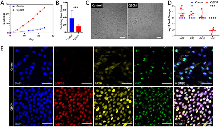
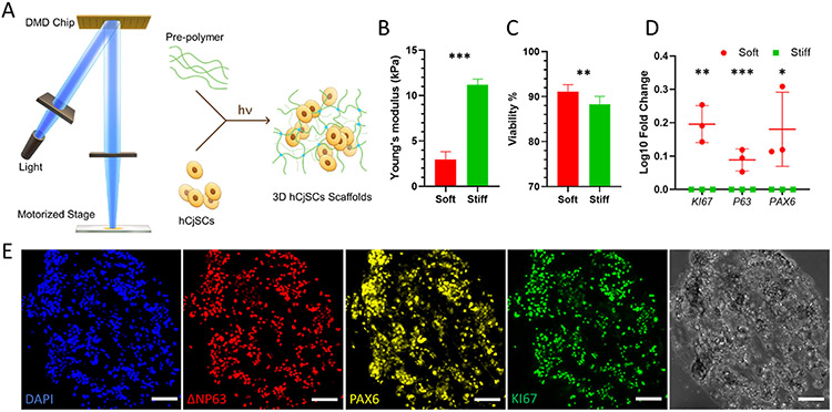
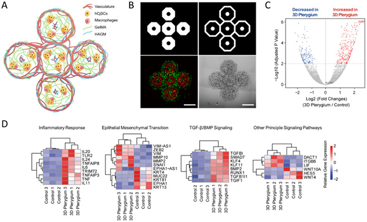
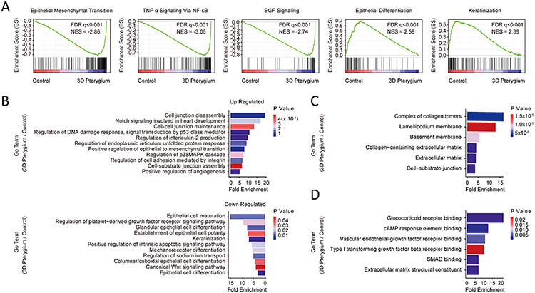
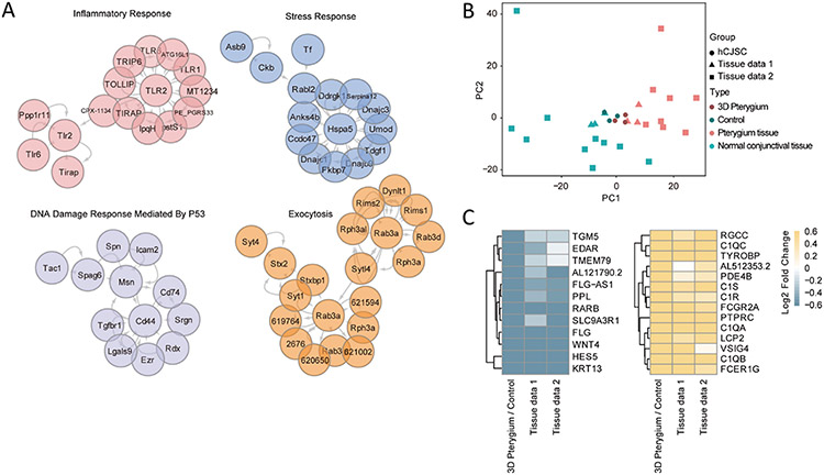
Pterygium is an ocular surface disorder with high prevalence that can lead to vision impairment. As a pathological outgrowth of conjunctiva, pterygium involves neovascularization and chronic inflammation. Here, we developed a 3D multicellular in vitro pterygium model using a digital light processing (DLP)-based 3D bioprinting platform with human conjunctival stem cells (hCjSCs). A novel feeder-free culture system was adopted and efficiently expanded the primary hCjSCs with homogeneity, stemness and differentiation potency. The DLP-based 3D bioprinting method was able to fabricate hydrogel scaffolds that support the viability and biological integrity of the encapsulated hCjSCs. The bioprinted 3D pterygium model consisted of hCjSCs, immune cells, and vascular cells to recapitulate the disease microenvironment. Transcriptomic analysis using RNA sequencing (RNA-seq) identified a distinct profile correlated to inflammation response, angiogenesis, and epithelial mesenchymal transition in the bioprinted 3D pterygium model. In addition, the pterygium signatures and disease relevance of the bioprinted model were validated with the public RNA-seq data from patient-derived pterygium tissues. By integrating the stem cell technology with 3D bioprinting, this is the first reported 3D in vitro disease model for pterygium that can be utilized for future studies towards personalized medicine and drug screening.

Keywords: 3D bioprinting; Disease model; Epithelial mesenchymal transition; Hydrogels; Pterygium; Stem cells; Tissue engineering.


Conflict of interest statement

Declaration of competing interest

The authors declare the following financial interests/personal relationships which may be considered as potential competing interests: SC is a co-founder of and has an equity interest in Allegro 3D, Inc., and he serves on the scientific advisory board. Some of his research grants, including those acknowledged here, have been identified for conflict-of-interest management based on the overall scope of the project and its potential benefit to Allegro 3D, Inc. The author is required to disclose this relationship in publications acknowledging the grant support, however the research subject and findings reported here did not involve the company in any way and have no relationship with the business activities or scientific interests of the company. The terms of this arrangement have been reviewed and approved by the University of California San Diego in accordance with its conflict-of-interest policies. The other authors have no competing interests to declare.

Figures

Fig. 1.
Fig. 1.
In vitro expansion of primary hCjSCs using CjSCM. (A) Representative cumulative quantification plot showed the cell doublings versus the culture time of the human primary conjunctival epithelial cells in culture with CjSCM or control medium. (B) Average cell doubling time of human conjunctival epithelial cells in culture with control medium and CjSCM from passage 1 to 8 (mean ± sd, n = 3; ***: P < 0.001). (C) Cell morphologies of nonconfluent primary human conjunctival epithelial cells cultured with CjSCM or control medium at passage 3. Scale bars: 100 μm. (D) Real time qPCR showing the relative mRNA expression of KI67 (proliferation), P63 (stemness), PAX6 (ocular lineage), VIM (mesenchymal lineage) in the cells expanded in CjSCM or control medium (mean ± sd, n = 4, *: P < 0.05, ***: P < 0.001.). (E) Immunofluorescence staining of ΔNP63, PAX6 and KI67 on hCjSCs expanded in CjSCM or control medium at passage 3. Scale bars: 50 μm.
Fig. 2.
Fig. 2.
DLP-based 3D bioprinting of hydrogel scaffolds supporting the stemness and functionality of the encapsulated hCjSCs. (A) Schematics of the DLP bioprinter setup and the photopolymerization process to fabricate hydrogel scaffolds encapsulating hCjSCs. (B) Compressive modulus of the hCjSCs encapsulated in soft and stiff bioprinted scaffolds (mean ± sd, n = 3). (C) The ratio of PI-negative population measured with flow cytometry representing the percentage of viable cells in soft and stiff bioprinted scaffolds cultured for 5 days (mean ± sd, n = 3). (D) Real time qPCR showing the relative mRNA expression of KI67, P63 and PAX6 of hCjSCs in 2D culture condition (2D control) or 3D hydrogel scaffolds with different stiffness (mean ± sd, n = 3, *: P < 0.05, **: P < 0.01, ***: P < 0.001.). (E) Representative immunofluorescence staining and corresponding bright field images of bioprinted hydrogel scaffolds encapsulating hCjSCs after 2 days of culture expressing ΔNP63, PAX6 and KI67. Scale bars: 100 μm.
Fig. 3.
Fig. 3.
DLP-based 3D bioprinting of multicellular pterygium model with distinct transcriptomic profiles. (A) Illustration of the bioprinted multicellular 3D pterygium model. (B) Representative images of the 3D pterygium model. Red: hCjSCs and macrophages; green: HUVECs and fibroblasts. Scale bars: 1 mm. (C) Volcano plot of global transcriptomic landscape comparing the bioprinted 3D pterygium model with the 2D control. The x-axis represents log2 transformed fold changes, and the y-axis shows the −log10 transformed p-value adjusted for multiple test correction (n = 3 per condition). (D) Heatmap of representative DEGs correlated to inflammatory response, epithelial mesenchymal transition, TGF-β/BMP signaling, and other principal signaling pathways in the 3D pterygium model versus the 2D control. Scale bars represent relative gene expression (log2 fold changes).
Fig. 4.
Fig. 4.
GSEA and GO analysis revealed the pterygium-related pathological features in the 3D pterygium model. (A) Representative GSEA results comparing the 3D pterygium model with the control. FDR: false discovery rate, NES: normalized enrichment score. (B) GO terms enriched in hCjSCs cultured in the 3D pterygium model versus 2D control. (C) Selected upregulated GO terms from the cellular component domain in the 3D pterygium model. (D) Selected upregulated GO terms from the molecular function domain in the 3D pterygium model.
Fig. 5.
Fig. 5.
Transcriptome profiles of 3D pterygium model resemble a patient-derived pterygium tissue. (A) PPI enrichment analysis based on the DEGs between the 3D pterygium model and the control. (B) PCA of the global transcriptomic profiles of the hCjSCs from the bioprinted model (3D pterygium) and 2D culture (Control), and human tissues from healthy individuals (normal conjunctival tissue) and pterygium patients (Pterygium tissue). (C) Heatmap of consistent DEGs correlated to activation of immune response and epithelial cell differentiation. Tissue data 1 (X. Liu et al.) and tissue data 2 (Y. Chen et al.) represent human tissue data from two independent studies. Scale bar represents normalized fold change.

References

    1. Harvey TM, Fernandez AGA, Patel R, Goldman D, Ciralsky J, Conjunctival anatomy and physiology, in: Ocul. Surf. Dis. Cornea, Conjunctiva Tear Film, Elsevier Inc., 2013, pp. 23–27, 10.1016/B978-1-4557-2876-3.00004-3. - DOI
    1. Akpek EK, Gottsch JD, Immune of defense at the ocular surface, Eye 17 (2003) 949–956, 10.1038/sj.eye.6700617. - DOI - PubMed
    1. Foster JB, Lee WB, The tear film: anatomy, structure and function, in: Ocul. Surf. Dis. Cornea, Conjunctiva Tear Film, Elsevier Inc., 2013, pp. 17–21, 10.1016/B978-1-4557-2876-3.00003-1. - DOI
    1. Gipson IK, Goblet cells of the conjunctiva: a review of recent findings, Prog. Retin. Eye Res 54 (2016) 49–63, 10.1016/j.preteyeres.2016.04.005. - DOI - PMC - PubMed
    1. Thorel D, Ingen-Housz-Oro S, Royer G, Delcampe A, Bellon N, Bodemer C, Welfringer-Morin A, Bremond-Gignac D, Robert MP, Tauber M, Malecaze F, Dereure O, Daien V, Colin A, Bernier C, Couret C, Vabres B, Tetart F, Milpied B, Cornut T, Ben Said B, Burillon C, Cordel N, Beral L, De Prost N, Wolkenstein P, Muraine M, Gueudry J, Management of ocular involvement in the acute phase of Stevens-Johnson syndrome and toxic epidermal necrolysis: French national audit of practices, literature review, and consensus agreement, Orphanet J. Rare Dis 15 (2020) 259, 10.1186/s13023-020-01538-x. - DOI - PMC - PubMed

Publication types

Supplementary concepts