. 2022 Feb 8;119(6):e2114092119.
doi: 10.1073/pnas.2114092119.

Sequence-specific RNA recognition by an RGG motif connects U1 and U2 snRNP for spliceosome assembly

Affiliations

Sequence-specific RNA recognition by an RGG motif connects U1 and U2 snRNP for spliceosome assembly

Tebbe de Vries et al. Proc Natl Acad Sci U S A. .

Abstract

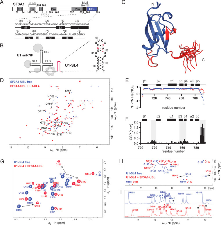
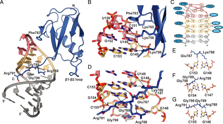
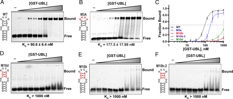
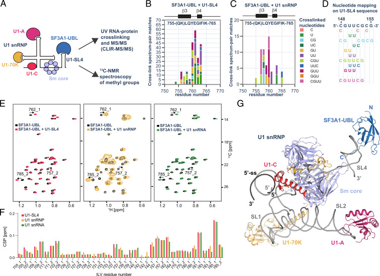
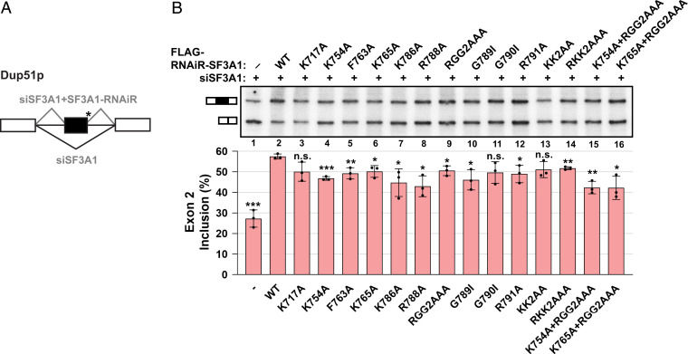
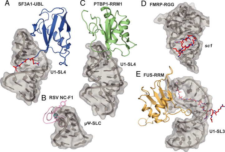
In mammals, the structural basis for the interaction between U1 and U2 small nuclear ribonucleoproteins (snRNPs) during the early steps of splicing is still elusive. The binding of the ubiquitin-like (UBL) domain of SF3A1 to the stem-loop 4 of U1 snRNP (U1-SL4) contributes to this interaction. Here, we determined the 3D structure of the complex between the UBL of SF3A1 and U1-SL4 RNA. Our crystallography, NMR spectroscopy, and cross-linking mass spectrometry data show that SF3A1-UBL recognizes, sequence specifically, the GCG/CGC RNA stem and the apical UUCG tetraloop of U1-SL4. In vitro and in vivo mutational analyses support the observed intermolecular contacts and demonstrate that the carboxyl-terminal arginine-glycine-glycine-arginine (RGGR) motif of SF3A1-UBL binds sequence specifically by inserting into the RNA major groove. Thus, the characterization of the SF3A1-UBL/U1-SL4 complex expands the repertoire of RNA binding domains and reveals the capacity of RGG/RG motifs to bind RNA in a sequence-specific manner.

Keywords: RGG motif; spliceosome assembly; splicing; ubiquitin-like domain.


Conflict of interest statement

The authors declare no competing interest.

Figures

Fig. 1.
Fig. 1.
Interaction of SF3A1-UBL with U1-SL4. (A) Domain organization of SF3A1 and primary sequences of the SF3A1-UBL construct used in this study, including the secondary structure elements shown below. S1 and S2, SURP1 and SURP2; Pro, proline-rich sequence; +/−, charged residues; NLS, nuclear localization signal. Interaction sites for SF3A2 and SF3A3 are highlighted in light gray. (B) Schematic representation of U1 snRNP with U1-SL4 shown in red and the predicted secondary structure of U1-SL4 on the right. Nucleotides with the strongest CSP in 2D 1H-1H TOCSY are shown in red. (C) Solution structure of the free SF3A1-UBL. Overlay of the 20 lowest energy structures is shown. Amide CSPs of D and F shown in red. (D) Overlay of 2D 1H-15N heteronuclear single quantum coherence (HSQC) spectra of 15N-labeled SF3A1-UBL in the free (blue) and U1-SL4 RNA-bound (red) 1:1 complex form. CSPs of C-terminal residues are indicated with black arrows. (E) Backbone dynamics data of SF3A1-UBL in the free (blue) and U1-SL4 bound states (red). (F) Plot of the combined chemical shift difference between amide group resonances of the free and bound forms of SF3A1-UBL. (G) Overlay of 2D 1H-1H TOCSY spectra of U1-SL4 free (blue) and bound to SF3A1-UBL (red). Asterisk indicates U145, which was not identified in the bound state in 2D 1H-1H TOCSY. (H and I) Overlay of 2D 1H-15N HSQC and 1D 1H spectra, respectively, of imino signals of U1-SL4 free (blue) and bound to SF3A1-UBL (red). Black asterisk in I indicates an imino signal deriving from a duplex conformation. Black circles in I indicate protein amide signals.
Fig. 2.
Fig. 2.
Molecular basis of the interaction between SF3A1-UBL and U1-SL4. (A) Overall view of the crystal structure of SF3A1-UBL (residues 704 to 791) (blue) and U1-SL4 (UUCG tetraloop in red, GCG base pairs in yellow, and the rest of the SL in gray). (B) Close-up views of the contacts to the UUCG tetraloop of U1-SL4. Putative hydrogen bonds are shown as dashed lines. Solid lines indicate electrostatic interactions. (C) Schematic representation of the intermolecular interactions; side chain–mediated contacts are written in black, while amino acids using the main chain are written in white. (D) Specific recognition of RNA by carboxyl-terminal residues Lys786, Glu787, Arg788, Gly789, Gly790, and Arg791. (EG) Base-specific recognition of the RNA base pairs in the upper part of the RNA duplex by carboxyl-terminal residues of SF3A1-UBL.
Fig. 3.
Fig. 3.
Mutational analysis of the U1-SL4 RNA. (A) EMSA experiments performed with SF3A1-UBL and U1-SL4. (B) Loop mutant of U1-SL4 probed for binding to SF3A1-UBL. Bases different from WT U1-SL4 are shown in red. (C) Binding curves of the indicated U1-SL4 variants. (DF) Mutants of the upper helical part of U1-SL4. Respective Kd values were derived from curve fitting to the relative bound fraction per lane. GST, glutathione-S-transferase.
Fig. 4.
Fig. 4.
Analysis of SF3A1-UBL binding to U1 snRNP. (A) Schematic representation of the SF3A1-UBL/U1 snRNP complex analysis. (B) Protein–RNA cross-links identified for 1:1 complex of SF3A1-UBL and U1-SL4 by CLIR-MS/MS plotted on the sequence of SF3A1-UBL. The bar colors represent the nucleotide composition of the RNA adducts. Protein–RNA cross-links are shown as counts of cross-link spectrum matches. (C) Cross-links identified for SF3A1-UBL bound to in vitro–reconstituted U1 snRNP. (D) Mapping of nucleotides cross-linked to SF3A1-UBL on the sequence of U1-SL4. (E) Overlay of the 2D 1H-13C HMQC spectra of the free SF3A1-UBL protein (black) and in complex with U1-SL4 (red), U1 snRNP (yellow), and U1 snRNA (green). (F) Plot showing the CSPs of the methyl groups of isoleucine, leucine, and valine (ILV) of SF3A1-UBL observed upon addition of U1-SL4 (red), U1 snRNP (yellow), or U1 snRNA (green). Methyl groups are labeled according to the residue number; 1 or 2 stands for HD1/CD1 and HD2/CD2 in the case of leucine or HG1/CG1 and HG2/CG2 in the case of valine. (G) Structural model of SF3A1-UBL bound to U1 snRNP.
Fig. 5.
Fig. 5.
Mutations in SF3A1-UBL interfere with splicing rescue of the Dup51p minigene reporter under SF3A1 knockdown conditions. (A) Schematic representation of three-exon/two-intron Dup51p reporters depicting the splicing pattern upon siRNA-mediated knockdown and rescue with an siRNA-resistant construct. The asterisk indicates a mutant 5′-ss. (B) Primer extension analysis monitors the inclusion of exon 2 in RNA isoforms of the Dup51p minigene reporter. The mRNA products are shown schematically to the left of the gel image. All cells were transfected with siSF3A1 and plasmid harboring WT or mutant FLAG-RNAiR (RNA interference-resistant)-SF3A1. In the absence of the RNAi-resistant clone, exon 2 inclusion is inhibited (lane 1). Cotransfection with the WT RNAi-resistant SF3A1 clone rescues exon 2 inclusion under siSF3A1 treatment (lane 2), which is reduced if splicing rescue is performed using mutant RNAi-resistant SF3A1 (lanes 3 to 16). Percent exon 2 inclusion (n =3; *P < 0.05, **P < 0.01, ***P < 0.001) is plotted below the gel.
Fig. 6.
Fig. 6.
Structural comparison of UNCG tetraloop recognition and RNA binding RGG/RG motifs. (A) SF3A1-UBL bound to U1-SL4. (B) Solution structure of RSV nucleocapsid protein (NC) zinc knuckle motif (F1) bound to μΨ RNA packaging signal containing a UNCG-type tetraloop. Zinc atom shown in cyan (PDB ID: 2IHX). (C) Structural model of PTBP1-RRM1 bound to U1-SL4 based on Ref. . (D) Crystal structure of RGG motif of FMRP bound to sc1 RNA (PDB ID: 5DEA). (E) Solution structure of FUS-RRM bound to U1-SL3 (PDB ID: 6SNJ). RGG/RG motifs are highlighted in red.

Similar articles

Cited by

References

    1. Nesic D., Krämer A., Domains in human splicing factors SF3a60 and SF3a66 required for binding to SF3a120, assembly of the 17S U2 snRNP, and prespliceosome formation. Mol. Cell. Biol. 21, 6406–6417 (2001). - PMC - PubMed
    1. Zhang Z., et al. , Molecular architecture of the human 17S U2 snRNP. Nature 583, 310–313 (2020). - PubMed
    1. Mount S. M., Pettersson I., Hinterberger M., Karmas A., Steitz J. A., The U1 small nuclear RNA-protein complex selectively binds a 5′ splice site in vitro. Cell 33, 509–518 (1983). - PubMed
    1. Sharma S., Wongpalee S. P., Vashisht A., Wohlschlegel J. A., Black D. L., Stem-loop 4 of U1 snRNA is essential for splicing and interacts with the U2 snRNP-specific SF3A1 protein during spliceosome assembly. Genes Dev. 28, 2518–2531 (2014). - PMC - PubMed
    1. Zhan X., Yan C., Zhang X., Lei J., Shi Y., Structures of the human pre-catalytic spliceosome and its precursor spliceosome. Cell Res. 28, 1129–1140 (2018). - PMC - PubMed

Publication types

MeSH terms

Substances