. 2022 Feb 1;8(1):10.
doi: 10.1038/s41421-022-00373-7.

Robust induction of B cell and T cell responses by a third dose of inactivated SARS-CoV-2 vaccine

Affiliations

Robust induction of B cell and T cell responses by a third dose of inactivated SARS-CoV-2 vaccine

Yihao Liu et al. Cell Discov. .

Abstract

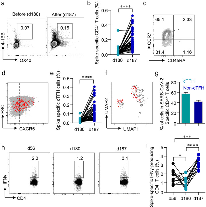
SARS-CoV-2 inactivated vaccines have shown remarkable efficacy in clinical trials, especially in reducing severe illness and casualty. However, the waning of humoral immunity over time has raised concern over the durability of immune memory following vaccination. Thus, we conducted a nonrandomized trial among the healthcare workers (HCWs) to investigate the long-term sustainability of SARS-CoV-2-specific B cells and T cells stimulated by inactivated vaccines and the potential need for a third booster dose. Although neutralizing antibodies elicited by the standard two-dose vaccination schedule dropped from a peak of 29.3 arbitrary units (AU)/mL to 8.8 AU/mL 5 months after the second vaccination, spike-specific memory B and T cells were still detectable, forming the basis for a quick recall response. As expected, the faded humoral immune response was vigorously elevated to 63.6 AU/mL by 7.2 folds 1 week after the third dose along with abundant spike-specific circulating follicular helper T cells in parallel. Meanwhile, spike-specific CD4+ and CD8+ T cells were also robustly elevated by 5.9 and 2.7 folds respectively. Robust expansion of memory pools by the third dose potentiated greater durability of protective immune responses. Another key finding in this trial was that HCWs with low serological response to two doses were not truly "non-responders" but fully equipped with immune memory that could be quickly recalled by a third dose even 5 months after the second vaccination. Collectively, these data provide insights into the generation of long-term immunological memory by the inactivated vaccine, which could be rapidly recalled and further boosted by a third dose.


Conflict of interest statement

The authors declare no competing interests.

Figures

Fig. 1
Fig. 1. Humoral responses induced by a third dose of inactivated SARS-CoV-2 vaccine BBIBP-CorV.
a Protocol of the prospective cohort and the nonrandomized trial. Healthcare workers (HCWs) were recruited from a perspective cohort who received two doses of an inactivated vaccine either in the morning (AM, n = 25) or afternoon (PM, n = 25) on day (d) 0 and d28. They were assigned to morning or afternoon vaccination to receive a third dose of the inactivated vaccine on day 180 according to their previous vaccinating time. Blood samples were collected on different time points d0 (T1), d14 (T2), d21 (T3), d28 (T4), d56 (T5), d180 (T6), d187 (T7), d194 (T8), and d208 (T9). be Neutralizing antibodies (NAbs) against SARS-CoV-2 in the serum of each volunteer was measured by Chemifluorescence Assay longitudinally. b The concentration of NAbs in the sera at different time points was summarized and shown in the box plot. c The longitudinal changes of NAbs in the sera before and after the third dose of vaccination. d NAbs in the sera from the morning or afternoon group at different time points. e NAb concentrations in the sera of female or male. Medians of the data were shown. f, g Neutralizing activities of sera against SARS-CoV-2 were measured by pseudovirus neutralizing assay. Half-maximal inhibitory dilutions (ID50) of sera from T6 and T8 against SARS-CoV-2 WT and Delta variant (B.1.167.2) were summarized. h, i Comparison of neutralizing activities between the morning and afternoon group on T8. Comparisons were done by Wilcoxon rank-sum test in be or Student’s t-test in fh. **P < 0.01, ****P < 0.0001; ns, not significant.
Fig. 2
Fig. 2. SARS-CoV-2-specific memory B cell after a third dose of inactivated vaccine.
Recombinant spike or receptor-binding domain (RBD) proteins were used to label SARS-CoV-2-specific BCR. Data were acquired by flow cytometry. a, c Representative FACS plots for the spike- or RBD-specific BCR were shown. b, d Longitudinal changes of spike- or RBD-specific memory B cells were summarized. e IgG+, IgA+, and IgM+ B cells were measured by flow cytometry. The UMAP plot demonstrated the distribution of spike- and RBD-specific memory B cells in IgG+, IgA+, and IgM+ B cells. f Mean percentages of each BCR subtype in spike- and RBD-specific B cells were summarized. g Spike- or RBD-specific IgG-secreting B cells were measured by ELISpot after incubating PBMCs in spike- or RBD-coated plates for 18 h. Representative ELISpot images were shown. h, i Numbers of Spike- or RBD-specific IgG-secreting B cells per 106 PBMCs were expressed as spot-forming units (s.f.u.) and summarized. Comparisons were done by one-way ANOVA corrected for multiple comparisons in b, d and Student’s paired t-test in h, i. ***P < 0.001, ****P < 0.0001; ns, not significant.
Fig. 3
Fig. 3. Cellular responses induced by a third dose of inactivated SARS-CoV-2 vaccine.
a PBMCs were stimulated with or without the spike peptide pool (2 μg/mL). Anti-CD3/CD28 dynabeads served as a positive control. IFNγ-producing T cells were detected by ELISpot after 24 h incubation. Representative images of ELISpot wells were shown. b The number of IFNγ-producing T cells was quantified as the spot-forming units (s.f.u.) per 106 PBMCs. cg PBMCs were stimulated with a spike peptide pool (2 μg/mL) for 12 h. Activation-induced markers (AIM) CD69 and 4-1BB were measured by flow cytometry. c Representative FACS plots showing the SARS-CoV-2-specific AIM+CD8+ T cells. d Percentages of AIM+CD8+ T cells were summarized. e TN (CD45RA+CCR7+), TEM (CD45RACCR7), TCM (CD45RACCR7+), TERMA (CD45RA+CCR7) populations in AIM+CD8+ T cells. f, g SARS-CoV-2-specific CD3+CD4CD8 (DN) T cells were identified by CD69 and 4-1BB after spike peptide pool stimulation. h, i Intracellular cytokine staining assays were performed to confirm the presence of SARS-CoV-2-specific CD8+ T cells in PBMCs by measuring intracellular IFNγ expression after peptide pool stimulation. Comparisons were done by Student’s paired t-test in b, d, and g, and one-way ANOVA corrected for multiple comparisons in i. ****P < 0.0001; ns, not significant.
Fig. 4
Fig. 4. Rapid induction of CD4+ T cells by the third dose.
a, b Activation-induced markers (AIM) OX40 and 4-1BB in CD4+ T cells were detected after incubating PBMCs with spike peptide pool (2 μg/mL) for 12 h. Percentages of AIM+CD4+ T cells in the PBMCs were summarized. c TN (CD45RA+CCR7+), TEM (CD45RACCR7), TCM (CD45RACCR7+), TERMA (CD45RA+CCR7) populations in AIM+CD4+ T cells were gated. d Expression of CXCR5 in AIM+CD4+ T cells was measured by flow cytometry. e Percentages of circulating follicular helper T cells (cTFH) in total CD4+ T cells were summarized. f UMAP plot showed the distribution of cTFH cells among AIM+CD4+ T cells. g Percentage of cTFH cells in AIM+CD4+ T cells. h, i Intracellular cytokine staining assays were performed to confirm the presence of SARS-CoV-2-specific CD4+ T cells in PBMCs by measuring intracellular IFNγ expression after peptide pool stimulation. Comparisons were done by Student’s paired t-test in b, e and one-way ANOVA corrected for multiple comparison in i. *P < 0.05, ***P < 0.001, ****P < 0.0001.
Fig. 5
Fig. 5. Immune responses in HCWs with minimal serologic response to a standard two-dose vaccination schedule.
a Serum NAbs in HCWs with minimal serologic response to a standard two-dose vaccination schedule were analyzed longitudinally at different time points before and after the third dose (red arrow). b Comparison of NAbs between vaccinees with negative or positive serological responses to a standard two-dose vaccination schedule. c Percentages of spike-specific memory B cells in HCWs with a negative serological response to a standard two-dose vaccination schedule. df Numbers of spike- or RBD-specific IgG-producing B cells in HCWs with a negative serological response to a standard two-dose vaccination schedule. g, h Numbers of spike-specific IFNγ-producing T cells as detected by ELISpot in HCWs with a negative serological response to a standard two-dose vaccination schedule. ik Spike-specific T cells, including CD69+4-1BB+CD8+ T cells, OX40+4-1BB+CD4+ T cells, and CXCR5+OX40+4-1BB+ cTFH in HCWs with a negative serological response to a standard two-dose vaccination schedule. Comparisons were done by Wilcoxon rank-sum test in b and one-way ANOVA corrected for multiple comparisons in the rest panels. *P < 0.05, **P < 0.01, ****P < 0.0001; ns, not significant.

References

    1. Crook H, Raza S, Nowell J, Young M, Edison P. Long covid-mechanisms, risk factors, and management. BMJ. 2021;374:n1648. - PubMed
    1. COVID-19 Map – Johns Hopkins Coronavirus Resource Center. https://coronavirus.jhu.edu/map.html (2021).
    1. Xia S, et al. Safety and immunogenicity of an inactivated SARS-CoV-2 vaccine, BBIBP-CorV: a randomised, double-blind, placebo-controlled, phase 1/2 trial. Lancet Infect. Dis. 2021;21:39–51. - PMC - PubMed
    1. Jackson, L. A. et al. An mRNA vaccine against SARS-CoV-2 – Preliminary report. N. Engl. J. Med.383, 1920–1931 (2020). - PMC - PubMed
    1. Krause, P. R. & Gruber, M. F. Emergency use authorization of covid vaccines – Safety and efficacy follow-up considerations. N. Engl. J. Med.383, e107 (2020). - PubMed