. 2022 Apr;9(10):e2103939.
doi: 10.1002/advs.202103939. Epub 2022 Feb 1.

Quantifying Cell-Derived Changes in Collagen Synthesis, Alignment, and Mechanics in a 3D Connective Tissue Model

Affiliations

Quantifying Cell-Derived Changes in Collagen Synthesis, Alignment, and Mechanics in a 3D Connective Tissue Model

Benjamin T Wilks et al. Adv Sci (Weinh). 2022 Apr.

Abstract

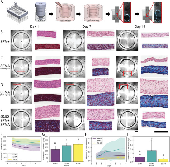
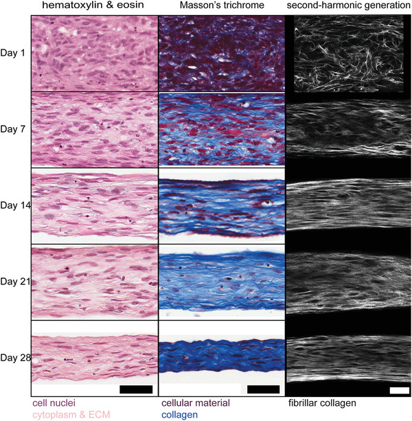
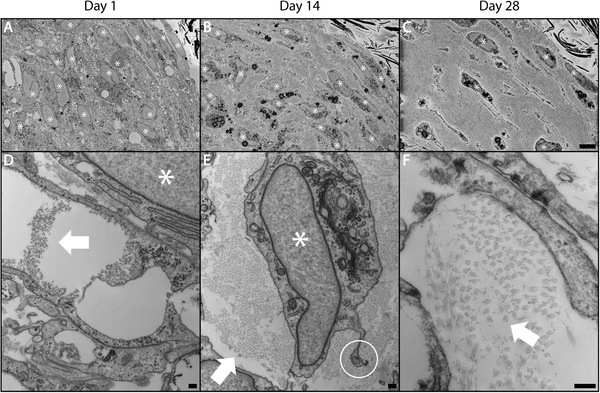
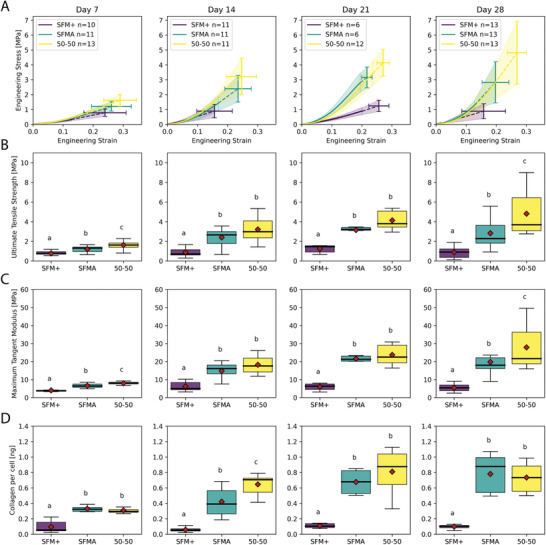
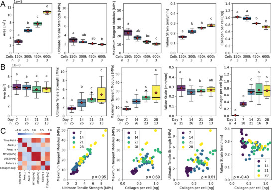
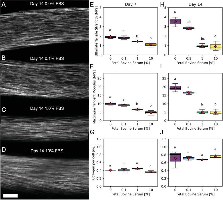
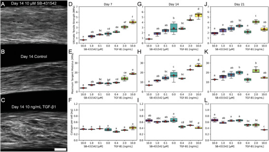
Dysregulation of extracellular matrix (ECM) synthesis, organization, and mechanics are hallmark features of diseases like fibrosis and cancer. However, most in vitro models fail to recapitulate the three-dimensional (3D) multi-scale hierarchical architecture of collagen-rich tissues and as a result, are unable to mirror native or disease phenotypes. Herein, using primary human fibroblasts seeded into custom fabricated 3D non-adhesive agarose molds, a novel strategy is proposed to direct the morphogenesis of engineered 3D ring-shaped tissue constructs with tensile and histological properties that recapitulate key features of fibrous connective tissue. To characterize the shift from monodispersed cells to a highly-aligned, collagen-rich matrix, a multi-modal approach integrating histology, multiphoton second-harmonic generation, and electron microscopy is employed. Structural changes in collagen synthesis and alignment are then mapped to functional differences in tissue mechanics and total collagen content. Due to the absence of an exogenously added scaffolding material, this model enables the direct quantification of cell-derived changes in 3D matrix synthesis, alignment, and mechanics in response to the addition or removal of relevant biomolecular perturbations. To illustrate this, the effects of nutrient composition, fetal bovine serum, rho-kinase inhibitor, and pro- and anti-fibrotic compounds on ECM synthesis, 3D collagen architecture, and mechanophenotype are quantified.

Keywords: 3D tissue engineering; TGF-β1; collagen; connective tissue; extracellular matrix; fibroblast; fibrosis; mechanics; mechanophenotype.


Conflict of interest statement

J.R.M. has an equity interest in Microtissues, Inc. This relationship has been reviewed and is managed by Brown University in accordance with its conflict of interest policies.

Figures

Figure 1
Figure 1
Human fibroblasts form stable 3D ring‐shaped tissues constructs whose thickness and uniformity are dependent on the composition of serum‐free media. To fabricate ring‐shaped tissues, molten agarose was added to all wells of a 24‐well plate followed by custom stainless steel inserts. After 10–15 min, the stainless steel inserts were removed and wells were left with 3D non‐adhesive agarose gels with a 5 mm diameter central inner peg surrounded by a ring‐shaped trough in which cells could be seeded, settle, self‐assemble into ring‐shaped tissue constructs, and be used for downstream assays like mechanical testing to failure at 7 to 28 day timepoints (A) After equilibrating gels for at least 24 h in serum‐free DMEM, human fibroblasts were seeded in either serum‐free media supplemented with collagen promoting supplements (SFM+), advanced DMEM (SFMA), or a 50:50 combination of the two and imaged daily (B–E) and the x‐y thickness of the tissues was measured over time (F,G). Tissues in SFM+ (B) and 50:50 (E) maintained a uniform thickness, but tissues in 50:50 were significantly thicker. Conversely, tissues in SFMA developed non‐uniform thick and thin regions as a function of time (C,D). This variation in tissue thick uniformity was quantified over 7 days (H). The average x‐y thickness (G) and the coefficient of variation of tissue thickness (I) were quantified as a function of media composition and time. Tissues in SFMA and 50:50 were significantly thicker than tissues in SFM+ at day 7 (G, One‐way ANOVA with post‐hoc Tukey HSD, p<0.05). The coefficient of variation of the thickness was significantly higher for SFMA compared to SFM+ and 50:50 media groups (I, One‐way ANOVA with post‐hoc Tukey HSD, p<0.05). Data presented as mean ± S.D., n=24. Scale bars = 1000, 200 µm.
Figure 2
Figure 2
Tissue construct histology and SHG suggest an inverse relationship between cellularity and collagen content over time. Tissues cultured in 50:50 media were examined by histology (H&E and Mason's trichrome) and multiphoton second‐harmonic generation (SHG) microscopy at days 1, 7, 14, 21 and 28. In the first 14 days, there was a decrease in cellularity over time paired with an increase in cell alignment and collagen density. This shift in cellularity and cellular alignment continued through 28 days with cells increasingly elongated in the same direction as synthesized collagen. Scale bars = 100 µm.
Figure 3
Figure 3
Human tissue rings secrete de novo collagen fibrils over time. Tissues cultured for 1, 14 and 28 days in 50:50 media were fixed, embedded and tissue cross‐sections were examined by serial block‐face (SBF) scanning electron microscopy (SEM) (A–C) and transmission electron microscopy (TEM) (D‐F). These images confirm a shift from purely cellular aggregates to highly‐aligned, collagen‐rich ring‐shaped tissue constructs over a period of 28 days in culture. At day 1, constructs were mostly cellular with the appearance of collagen fibrils beginning in the extracellular space (D). Day 14 revealed a shift in cellularity with individual cells clearly enmeshed in cell‐synthesized collagen and intercellular contacts maintained via long projections (E). Present are some autophagic and apoptotic cell features such as lysosomal vacuoles and nuclear degradation (A–C). By day 28, collagen fibrils appeared more separated from one another and increasing numbers of fibrils were out of plane suggesting less organization along with a continued decrease in cellularity (F). White stars indicate cell nuclei. White arrows indicate collagen fibrils. White circle highlights fibripositor. Scale bars = 500 µm (A–C), 200 nm (D–F).
Figure 4
Figure 4
Mechanical properties and total collagen of tissue rings increase over time and is dependent on the composition of the serum‐free media. Tissue rings cultured for 7, 14, 21, and 28 days in SFM+, SFMA or 50:50 media were removed from the molds and subjected to tensile testing and analyzed for total collagen. Tissue constructs exhibited media and time dependent mechanical properties (A) that scaled with total collagen content (D). At all time points, tissue strength (B), stiffness (C), and collagen content (D) were significantly greater in 50:50 and SFMA tissues compared to SFM+ corroborating the differences visualized in tissue architecture via histology, SHG, and electron microscopy (Kruskal‐Wallis with post‐hoc Conover, p<0.05). While the stiffness and collagen content of 50:50 and SFMA tissue constructs were relatively consistent, by day 28, the ultimate tensile strength and maximum tangent modulus of 50:50 tissue constructs (4.81 ± 2.10, 27.8 ± 10.8 MPa) were significantly greater than SFMA (2.82 ± 1.38, 19.8 ± 6.84 MPa) and SFM+ (0.88 ± 0.50, 5.50 ± 1.91 MPa) (B‐C, Kruskal‐Wallis with post‐hoc Conover, p<0.05).
Figure 5
Figure 5
Mechanical properties of tissue rings are dependent on the number of cells seeded and maturation time. Tissue rings seeded with 1.5, 3.0, 4.5 or 6.0 x 10⁵ cells were cultured for 7, 14, 21, and 28 days in 50:50 media were measured for cross sectional area, collagen content per cell and three mechanical properties: ultimate tensile strength, maximum tangent modulus, and failure strain. Increasing the number of cells significantly increased the cross‐sectional area of the tissues (Kruskal‐Wallis with post‐hoc Conover, p<0.05). However, as cell number was increased, ultimate tensile strength, maximum tangent modulus, and collagen synthesis per cell all decreased, whereas failure strain increased by day 28 (A, Kruskal‐Wallis with post‐hoc Conover, p<0.05). A close examination of the time course of tissues formed with 3.0 x 10⁵ cells showed no significant change in cross‐sectional area or failure strain over 7 to 28 days (B). However, there was a significant increase in ultimate tensile strength and stiffness between 7, 14, and 21 day tissues corresponding to a significant increase in collagen content from 1 to 14 days (Kruskal‐Wallis with post‐hoc Conover, p<0.05). Spearman correlation matrix revealed strong correlations between tissue mechanics and collagen content as a function of maturation time (C). Stiffness and strength were highly correlated with ρ = 0.95. Similarly, stiffness, strength, and failure strain were significantly correlated with collagen content with ρ = 0.69, ρ = 0.61, and ρ = ‐0.40, and corresponding P‐values of 6.79 × 10⁻⁹, 2.49 × 10⁻⁶, and 0.019, respectively (Spearman Correlation with post‐hoc Bonferonni, p<0.05).
Figure 6
Figure 6
Fetal bovine serum (FBS) decreases strength and stiffness of tissues, but not total collagen content. Tissue rings seeded with 3 x 10⁵ cells that were cultured in 50:50 media with varying levels of FBS (0.0 (A), 0.1 (B), 1.0 (C) or 10% (D)) were imaged by SHG at day 14 (A–D) and measured for ultimate tensile strength, maximum tangent modulus and collagen per cell at days 7 and 14 (E–F). The SHG images of fibrillar collagen revealed qualitative differences in collagen architecture characterized by increased interfibrillar spacing in the no serum control compared to a more diffuse signal without visible dark space between fibrillar collagen in samples with serum. This shift in tissue architecture was reflected in the tissue mechanics. Ultimate tensile strength and maximum tangent modulus were highest in the no serum control at both time points. While day 14 untreated tissues had a stiffness of 19.3 ± 1.99 MPa, tissues cultured with 10% serum had a stiffness of 4.91 ± 1.37 MPa and as little as 1.0% FBS significantly decreased tissue stiffness and strength (Kruskal‐Wallis with post‐hoc Conover, p = 0.0045). However, there were no significant changes in collagen content at any FBS dose suggesting alternate mechanisms to changes in tissue mechanics such as crosslinking. Scale bar = 50 µm.
Figure 7
Figure 7
TGF‐β1 increases, whereas SB‐431542 decreases tissue mechanics. Tissue rings seeded with 3 x 10⁵ cells that were cultured in 50:50 media with varying levels of SB‐431542 (0.1, 1.0, 10.0 (A) µm), control (B), or TGF‐β1 (0.4, 2.0 or 10 ng mL−1 (C)) were measured for ultimate tensile strength , maximum tangent modulus, and collagen per ring at days 7 (D), 14 (E), and 21 (F). TGF‐β1 increased tissue strength and stiffness as a function of concentration after 7 and 14 days, whereas the TGF‐β1 inhibitor SB‐431542 resulted in a decrease in stiffness and strength compared to control at all time points. By day 21, the dose‐response curve abruptly shifted with a large decrease in strength and stiffness for rings treated with the highest concentration, 10 ng mL−1, of TGF‐β1. The highest concentration of SB‐431542, 10 µm, had the largest negative effect on tissue mechanics. However, drug treatment did not completely abrogate the development of construct strength and stiffness suggesting that there was either incomplete inhibition of TGF‐β1 signaling or TGF‐β1 independent pathways contributed to the development of mechanical properties. Interestingly, SB‐431542 had no effect on the total pepsin‐acid soluble collagen content of ring constructs at any dose or time point. There was a slight decrease in pepsin‐acid soluble collagen content of TGF‐β1 treated tissues at later time points. This may be explained by increased covalently crosslinked insoluble collagen. Scale bar = 50 µm.

Similar articles

Cited by

References

    1. Connizzo B. K., Yannascoli S. M., Soslowsky L. J., Matrix Biol. 2013, 32, 106. - PMC - PubMed
    1. Goh K. L., Holmes D. F., Lu H.‐Y., Richardson S., Kadler K. E., Purslow P. P., Wess T. J., J. Biomech. Eng. 2008, 130, 2. - PubMed
    1. Vanakker O., Callewaert B., Malfait F., Coucke P., Annual Review of Genomics and Human Genetics 2015, 16, 229. - PubMed
    1. Tilstra D. J., Byers P. H., Annu. Rev. Med. 1994, 45, 149. - PubMed
    1. Roca‐Cusachs P., Conte V., Trepat X., Nat. Cell Biol. 2017, 19, 742. - PubMed

Publication types

LinkOut - more resources