. 2022 Jan;27(1):016003.
doi: 10.1117/1.JBO.27.1.016003.

In vivo safety study using radiation at wavelengths and dosages relevant to intravascular imaging

Affiliations

In vivo safety study using radiation at wavelengths and dosages relevant to intravascular imaging

Timothy Sowers et al. J Biomed Opt. 2022 Jan.

Abstract

Significance: Intravascular photoacoustic (IVPA) imaging can identify native lipid in atherosclerotic plaques in vivo. However, the large number of laser pulses required to produce 3D images is a safety concern that has not been fully addressed.

Aim: We aim to evaluate if irradiation at wavelengths and dosages relevant to IVPA imaging causes target vessel damage.

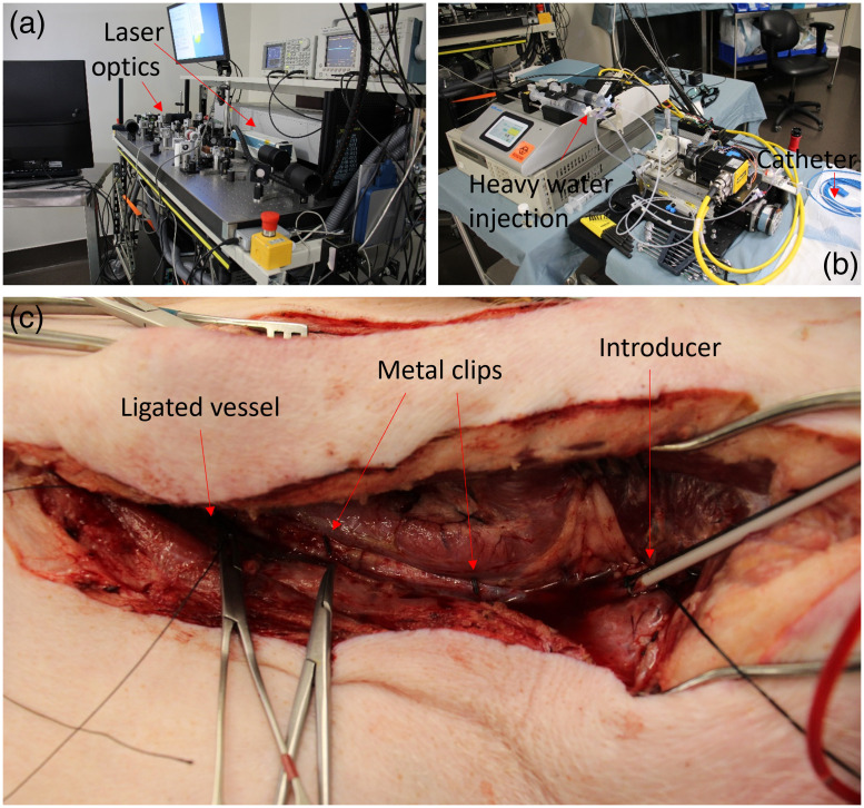
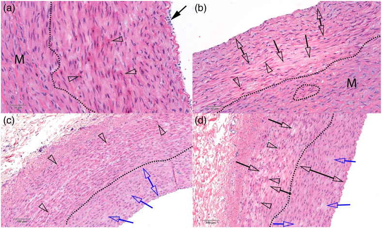
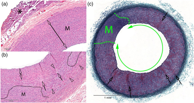
Approach: We irradiate the carotid artery of swine at one of several energy dosages using radiation at 1064 or 1720 nm and use histological evaluation by a pathologist to identify dose-dependent damage.

Results: Media necrosis was the only dose-dependent form of injury. Damage was present at a cumulative fluence of 50 J / cm2 when using 1720 nm light. Damage was more equivocally identified at 700 J / cm2 using 1064 nm.

Conclusions: In prior work, IVPA imaging of native lipid in swine has been successfully conducted below the damage thresholds identified. This indicates that it will be possible to use IVPA imaging in a clinical setting without damaging vessel tissue. Future work should determine if irradiation causes an increase in blood thrombogenicity and confirm whether damaged tissue will heal over longer time points.

Keywords: imaging; in vivo; intravascular; laser; photoacoustics; safety.


Figures

Fig. 1
Fig. 1
(a) An image of the laser and imaging system used for IVPA imaging, including the laser, optics, and computational hardware. (b) An image of components of the imaging system kept at the side of the operating table. The components include linear and rotational motors, a pumping system for injection of heavy water, and the catheter, which is extending to the bed at the right of the image. (c) Image of one of the carotid arteries after tissue dissection. The vessel has been ligated on the inferior (left) side to isolate the artery after catheter introduction (right). Two metal clips are attached to the artery for registration of fluoroscopy images.
Fig. 2
Fig. 2
Examples of carotid artery vessels subjected to various dosages of light radiation after staining with H&E. The dosages by panel are (a) negative control, no irradiation; (b) 1064 nm, 8.3  J/cm2; (c) 1064 nm, 100  J/cm2; and (d) 1064 nm, 700J/cm2. The dotted lines separate intact regions (M) of vessel tissue from damaged regions. Clear arrowheads indicate hypereosinophilic and contracted SMCs. The blue and black single arrows show regions of SMC effacement/loss. The double blue area in (c) shows pyknosis of SMCs from compressive necrosis, whereas the double black arrow indicates intact region of vessel in (d). The solid black arrow in (a) shows some intact media. The trend shows a nonzero baseline of damage in the control condition, with the incidence of damage increasing at higher light dosages. Additional notated images of samples subjected to these dosages are shown in the pathology report in the Supplementary Material.
Fig. 3
Fig. 3
Examples of carotid artery vessels subjected to various dosages of light radiation after staining with (a), (b) H&E and (c) GET. The dosages by panel are (a) 1720 nm, 8.3  J/cm2; (b) 1720 nm, 50  J/cm2; and (c) 1720 nm, 200  J/cm2. The dotted lines separate intact regions (M) of vessel tissue from damaged regions. The asterisk in (a) marks collagen denaturation, likely from electrocauterization used to remove tissue during the surgery. Otherwise, no damage was detected in (a). In (b), clear arrowheads indicate hypereosinophilic and contracted SMCs, whereas clear arrows show regions with cell effacement. The double arrow indicates a region with pyknotic nuclei and hypereosinophilic SMCs, consistent with compressive injury. Damage is present throughout the vessel in (c), in the form of widespread media necrosis (double green arrow). In addition, clear arrows show radial clusters of hyperchromatic and shrunken SMCs evoking contraction bands. Scale bar shows physical size of carotid artery. Additional notated images of samples subjected to these dosages are shown in the pathology report in the Supplementary Material.

References

    1. Iskander-Rizk S., et al. , “In vivo intravascular photoacoustic imaging of plaque lipid in coronary atherosclerosis,” EuroIntervention 15(5), 452–456 (2019).10.4244/EIJ-D-19-00318 - DOI - PubMed
    1. Jansen K., et al. , “Intravascular photoacoustic imaging of human coronary atherosclerosis,” Opt. Lett. 36(5), 597–599 (2011).OPLEDP10.1364/OL.36.000597 - DOI - PubMed
    1. Karpiouk A. B., Wang B., Emelianov S. Y., “Development of a catheter for combined intravascular ultrasound and photoacoustic imaging,” Rev. Sci. Instrum. 81(1), 014901 (2010).RSINAK10.1063/1.3274197 - DOI - PMC - PubMed
    1. Kole A., et al. , “Comparative quantification of arterial lipid by intravascular photoacoustic-ultrasound imaging and near-infrared spectroscopy-intravascular ultrasound,” J. Cardiovasc. Transl. Res. 12(3), 211–220 (2019).10.1007/s12265-018-9849-2 - DOI - PMC - PubMed
    1. Sethuraman S., et al. , “Intravascular photoacoustic imaging using an IVUS imaging catheter,” IEEE Trans. Ultrason. Ferr. 54(5), 978–986 (2007).ITUCER10.1109/TUFFC.2007.343 - DOI - PubMed

Publication types