. 2021 Dec 21;13(12):e20566.
doi: 10.7759/cureus.20566. eCollection 2021 Dec.

Can the Dynamic External Pelvimetry Test in Late Pregnancy Reveal Obstructed and Prolonged Labor? Results From a Pilot Study

Affiliations

Can the Dynamic External Pelvimetry Test in Late Pregnancy Reveal Obstructed and Prolonged Labor? Results From a Pilot Study

Marco Siccardi et al. Cureus. .

Abstract

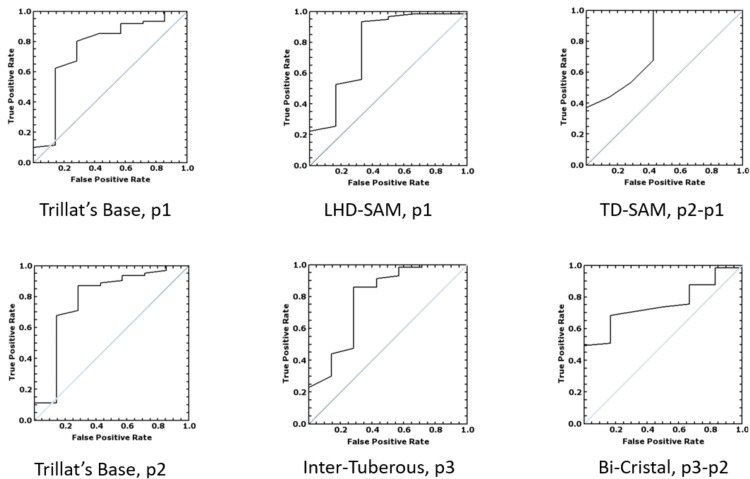
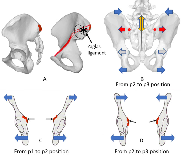
Background The size and mobility of the maternal pelvic space are fundamental factors in successful childbirth and can allow operators to screen for dystocia. This pilot study including a group of 70 pregnant women aimed to test whether the external dynamic pelvimetry test can be used to predict the likelihood of obstructed labor. Methodology The study cohort consisted of 70 pregnant women in their third trimester. The cohort was divided retrospectively into an obstructed labor group and a control group. Obstructed labor was defined using the following obstetric outcomes: augmentation with oxytocin from the first phase of the dilating period, Kristeller's maneuvers, vacuum extractor (kiwi), forceps, and the cesarean section following the onset of labor. Results The measurements obtained for the longitudinal hemi-diameter of Michaelis, the inter-tuberous diameter, and the base of the Trillat's triangle were statistically significant in every position. The difference in the measurements of the transverse diameter of Michaelis between standing and hands-and-knees position and the difference in the sizes of the bi-cristal diameter between hands-and-knees and squatting position were statistically significant. Conclusions Dimension and biomechanical properties of the pelvic tissue and spaces influence the evolutionary childbirth process. After clinical confirmation on a large population, hypomobility of specified external pelvic diameters measured in shifting positions can become a screening tool to detect the contracted pelvis and prevent damage caused by dystocia and prolonged labor in women and newborns.

Keywords: biomechanics; childbirth; dystocia; fetal-pelvic disproportion; operative delivery; pelvimetry; pelvis; pregnancy.


Conflict of interest statement

Marco Siccardi has intellectual property on the CE patented “DDI” instrument. Cristina Valle denies any relevant issues.

Figures

Figure 1
Figure 1. Instruments of dynamic external pelvimetry test and measurements of external pelvic diameters.
A: The Digital Distance Indicator (DDI) (Metrica, Milan, Italy, EU, https://www.metrica.it/) and the prototype accessory Dynamic Pelvimeter. B-E: Small pelvic diameters. B: The transverse diameter of Michaelis’ sacral area. C: The longitudinal hemi-diameter of Michaelis’ sacral area. D: The inter-tuberous diameter. E: The base of the Trillat’s triangle. F-O: Large pelvic diameters. F: The bi-trochanteric diameter. G: The bi-cristal diameter. H: The external conjugate. I: The bi-spinous diameter.
Figure 2
Figure 2. The different positions used for the dynamic external pelvimetry test.
The maternal positions chosen for the dynamic external pelvimetry test were defined by different degrees of hip flexion. A-C: Kneeling positions. A: p1, straight-leg erect position. B: p2, hands-and-knees position. C: p3, kneeling squat position. D-E: Supine position. D: p1, straight-leg supine position. E: p2, flexed-leg position.
Figure 3
Figure 3. ROC curve plot of the most relevant diameters.
Positions: straight leg (p1), flexed leg (p2), and kneeling squat (p3); the degree of change when the measurements taken in p2 and p1 are compared (p2-p1), as well as the measurements taken in p3 and p2 positions (p3-p2). TD-SAM: transverse diameter of the sacral area of Michaelis; LHD-SAM: longitudinal hemi-diameter of the sacral area of Michaelis; Trillat’s base: the base of the Trillat’s triangle
Figure 4
Figure 4. Zaglas ligament, pelvic mobility, and the inclination (angle) of the pelvic brim.
The inclination of the pelvic inlet bone edge changes with maternal postures. Moving into the squat position enlarges inferior diameters and narrows the superior diameter. The change of the inclination of the pelvic brim appears to favor fetal descent. Moreover, the region of the transverse diameter of Michaelis is anatomically related to the pelvic inlet. The inter-tuberous diameter of the outlet is functionally associated with the transverse diameter between the ischial spines in the midpelvis [17]. Zaglas ligament is one of the bands of the posterior sacroiliac ligament [23]. A: Lateral and medial view of the ilia. B: Posterior view of the pelvis. C, D: A simplified figure of the left and right ilioischial complexes of the pelvis (pubis and sacrum are removed) passing perpendicularly through the innominate line of the pelvic brim. p1: kneeling erect position; p2: all-fours position; p3: kneeling squat position. Asterisk: reported attachment site of the Zaglas ligament on the medial ileum; red line: pelvic brim; red dots: posterior superior iliac spines; red arrows: mobility of the transverse diameter of the Michaelis area; yellow double-headed arrow: mobility of the longitudinal hemi-diameter of the Michaelis area; gray line: the longitudinal diameter of the Michaelis area; blue arrows: mobility of the bi-cristal and inter-tuberous diameter; light gray arrows: inter-spinous diameter of the midpelvis; small black arrows pointing to red marks in C and D panels: together, they indicate the pelvic brim slope in each position.

Similar articles

Cited by

References

    1. Abnormal labor: diagnosis and management. Drennan Drennan, K K, Blackwell Blackwell, S S, Sokol RJ. Glob Libr Women's Med. 2008:0.
    1. World Health Organization. Geneva, Switzerland: WHO; 2005. Make every mother and child count. World Health Report 2005.
    1. MR obstetric pelvimetry: effect of birthing position on pelvic bony dimensions. Michel SC, Rake A, Treiber K, et al. AJR Am J Roentgenol. 2002;179:1063–1067. - PubMed
    1. Does pregnancy and/or shifting positions create more room in a woman's pelvis? Reitter A, Daviss BA, Bisits A, et al. Am J Obstet Gynecol. 2014;211:662–669. - PubMed
    1. The effects of squatting while pregnant on pelvic dimensions: a computational simulation to understand childbirth. Hemmerich A, Bandrowska T, Dumas GA. J Biomech. 2019;87:64–74. - PubMed

LinkOut - more resources