. 2022 Feb;11(2):61-72.
doi: 10.1302/2046-3758.112.BJR-2020-0482.R2.

CircSCAPER contributes to IL-1β-induced osteoarthritis in vitro via miR-140-3p/EZH2 axis

Affiliations

CircSCAPER contributes to IL-1β-induced osteoarthritis in vitro via miR-140-3p/EZH2 axis

Zhaxi Luobu et al. Bone Joint Res. 2022 Feb.

Abstract

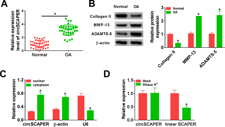
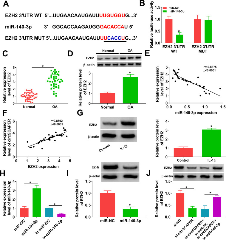
Aims: Circular RNA (circRNA) S-phase cyclin A-associated protein in the endoplasmic reticulum (ER) (circSCAPER, ID: hsa_circ_0104595) has been found to be highly expressed in osteoarthritis (OA) patients and has been associated with the severity of OA. Hence, the role and mechanisms underlying circSCAPER in OA were investigated in this study.

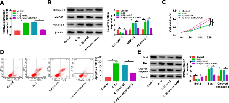
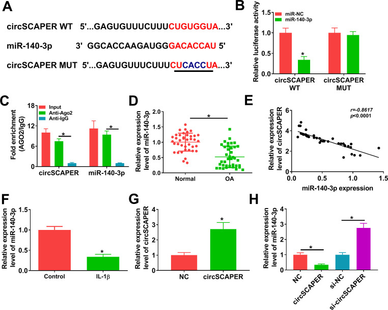
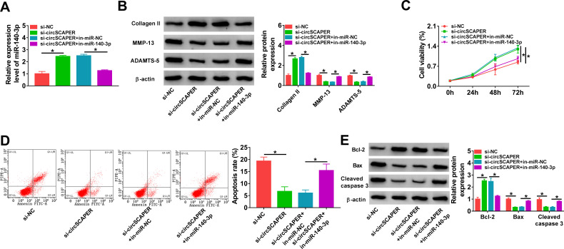
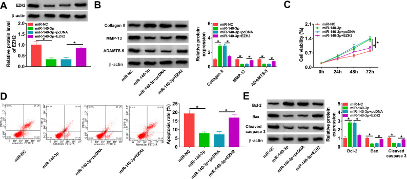
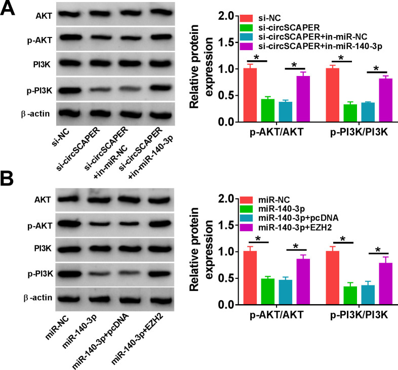
Methods: In vitro cultured human normal chondrocyte C28/I2 was exposed to interleukin (IL)-1β to mimic the microenvironment of OA. The expression of circSCAPER, microRNA (miR)-140-3p, and enhancer of zeste homolog 2 (EZH2) was detected using quantitative real-time polymerase chain reaction and Western blot assays. The extracellular matrix (ECM) degradation, proliferation, and apoptosis of chondrocytes were determined using Western blot, cell counting kit-8, and flow cytometry assays. Targeted relationships were predicted by bioinformatic analysis and verified using dual-luciferase reporter and RNA immunoprecipitation (RIP) assays. The levels of phosphoinositide 3-kinase (PI3K)/protein kinase B (AKT) pathway-related protein were detected using Western blot assays.

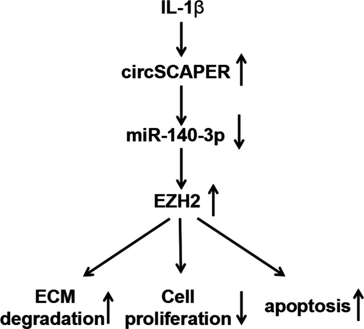
Results: CircSCAPER was highly expressed in OA cartilage tissues and IL-1β-induced chondrocytes. Knockdown of circSCAPER reduced IL-1β-evoked ECM degradation, proliferation arrest, and apoptosis enhancement in chondrocytes. Mechanistically, circSCAPER directly bound to miR-140-3p, and miR-140-3p inhibition reversed the effects of circSCAPER knockdown on IL-1β-induced chondrocytes. miR-140-3p was verified to target EZH2, and overexpression of miR-140-3p protected chondrocytes against IL-1β-induced dysfunction via targeting EZH2. Additionally, we confirmed that circSCAPER could regulate EZH2 through sponging miR-140-3p, and the circSCAPER/miR-140-3p/EZH2 axis could activate the PI3K/AKT pathway.

Conclusion: CircSCAPER promoted IL-1β-evoked ECM degradation, proliferation arrest, and apoptosis enhancement in chondrocytes via regulating miR-140-3p/EZH2 axis, which gained a new insight into the pathogenesis of OA. Cite this article: Bone Joint Res 2022;11(2):61-72.

Keywords: EZH2; Flow cytometry; IL-1β; MicroRNA; Osteoarthritis (OA); Quantitative real-time polymerase chain reaction; RNAs; Western blot; apoptosis; cartilage tissues; chondrocyte; chondrocytes; circSCAPER; interleukin; miR-140-3p; osteoarthritis.


Figures

Fig. 1
Fig. 1
CircSCAPER expression is elevated in osteoarthritis (OA) cartilage tissues. a) Quantitative real-time polymerase chain reaction (qRT-PCR) analysis of circSCAPER expression level in OA cartilage tissues and normal cartilage tissues. b) Detection of matrix metallopeptidase 13 (MMP-13), a disintegrin and metalloproteinase with thrombospondin motifs (ADAMTS)-5, and collagen II protein levels in OA cartilage tissues using Western blot. c) qRT-PCR analysis of circSCAPER, β-actin, and U6 in purified C28/I2 nuclear and cytoplasmic fractions. d) qRT-PCR analysis of circSCAPER and SCAPER messenger RNA (mRNA) levels in C28/I2 cells treated with or without RNase R. Independent-samples t-test, *p < 0.05. All of the experiments were performed in triplicate.
Fig. 2
Fig. 2
CircSCAPER knockdown attenuates interleukin (IL)-1β-induced chondrocyte dysfunction. a) to e) C28/I2 cells were transfected with circSCAPER-specific small interfering RNA (siRNA) (si-circSCAPER) and negative control (si-NC), followed by treatment with IL-1β (10 ng/l) for 24 hours. a) Quantitative real-time polymerase chain reaction (qRT-PCR) analysis of circSCAPER expression in cells. b) Western blot analysis of matrix metallopeptidase 13 (MMP-13), a disintegrin and metalloproteinase with thrombospondin motifs (ADAMTS-5), and collagen II protein levels in cells. c) Cell counting kit-8 (CCK-8) assay for cell proliferation. d) Flow cytometry for cell apoptosis. e) Western blot analysis of B-cell lymphoma-2 (Bcl-2), Bcl-2-associated X (Bax), and cleaved caspase 3 protein levels in cells. One-way analysis of variance (ANOVA), *p < 0.05. All of the experiments were performed in triplicate.
Fig. 3
Fig. 3
MicroRNA (miR)-140-3p is a target of circSCAPER. a) The potential binding sites between circSCAPER and miR-140-3p. b) The relative luciferase activity analysis in C28/I2 cells after transfection with circSCAPER wild-type (WT) or circSCAPER mutant (MUT) and miR-140-3p mimics or miR-negative control (NC) using dual-luciferase reporter assay. c) Quantitative real-time polymerase chain reaction (qRT-PCR) analysis of circSCAPER and miR-140-3p levels in C28/I2 cells after RNA immunoprecipitation (RIP) assay. d) qRT-PCR analysis of miR-140-3p expression level in osteoarthritis (OA) cartilage tissues and normal cartilage tissues. e) Correlation analysis between miR-140-3p and circSCAPER expression in OA cartilage tissues. f) Detection of miR-140-3p level in C28/I2 cells treated with interleukin (IL)-1β (10 ng/l). g) Detection of circSCAPER level in C28/I2 cells transfected with circSCAPER or NC. h) qRT-PCR analysis of miR-140-3p expression level in C28/I2 cells transfected with circSCAPER, NC, circSCAPER-specific small interfering RNA (siRNA) (si-circSCAPER), or si-NC. Independent-samples t-test, *p < 0.05. All of the experiments were performed in triplicate. IgG, immunoglobulin G.
Fig. 4
Fig. 4
CircSCAPER knockdown attenuates interleukin (IL)-1β-induced chondrocyte dysfunction via microRNA (miR)-140-3p. a) to e) C28/I2 cells were transfected with circSCAPER-specific small interfering RNA (siRNA) (si-circSCAPER), si-negative control (NC), si-circSCAPER+in-miR-NC, or si-circSCAPER+in-miR-140-3p, followed by treatment with IL-1β (10 ng/l) for 24 hours. a) Quantitative real-time polymerase chain reaction (qRT-PCR) analysis of miR-140-3p level in cells. b) Western blot analysis of matrix metallopeptidase 13 (MMP-13), a disintegrin and metalloproteinase with thrombospondin motifs (ADAMTS)-5, and collagen II protein levels in cells. c) Cell counting kit-8 (CCK-8) assay for cell proliferation. d) Flow cytometry for cell apoptosis. e) Western blot analysis of B-cell lymphoma-2 (Bcl-2), Bcl-2-associated X (Bax), and cleaved caspase 3 protein levels in cells. One-way analysis of variance (ANOVA), *p < 0.05. All of the experiments were performed in triplicate. NC, negative control.
Fig. 5
Fig. 5
CircSCAPER functions as a sponge for microRNA (miR)-140-3p to regulate the expression of its target enhancer of zeste homolog 2 (EZH2). a) The potential binding sites between miR-140-3p and EZH2. b) The analysis of relative luciferase activity in C28/I2 cells after transfection with EZH2 3' untranslated region (3'UTR) wild-type (WT) or EZH2 3’UTR mutant (MUT) and miR-140-3p mimics or miR-negative control (NC) using dual-luciferase reporter assay. c) and d) Quantitative real-time polymerase chain reaction (qRT-PCR) and Western blot analysis of EZH2 level in osteoarthritis (OA) cartilage tissues. e) and f) Correlation analysis between miR-140-3p and EZH2 messenger RNA (mRNA) or circSCAPER expression in OA cartilage tissues. g) Measurement of EZH2 protein level in C28/I2 cells treated with interleukin (IL)-1β (10 ng/l). h) qRT-PCR analysis of miR-140-3p expression level in C28/I2 cells transfected with in-miR-NC, in-miR-140-3p, miR-NC, or miR-140-3p. i) Western blot analysis of EZH2 level in C28/I2 cells transfected with miR-NC or miR-140-3p. j) Western blot analysis of EZH2 level in C28/I2 cells transfected with circSCAPER-specific small interfering RNA (siRNA) (si-circSCAPER), si-NC, si-circSCAPER+ in-miR-NC, or si-circSCAPER+ in-miR-140-3p. Independent-samples t-test, *p < 0.05. All of the experiments were performed in triplicate. IgG, immunoglobulin G.
Fig. 6
Fig. 6
MicroRNA (miR)-140-3p protects chondrocytes against interleukin (IL)-1β-induced dysfunction through enhancer of zeste homolog 2 (EZH2). a) to e) C28/I2 cells were transfected with miR-NC, miR-140-3p, miR-140-3p+pcDNA, or miR-140-3p+EZH2, followed by treatment with IL-1β (10 ng/l) for 24 hours. a) Western blot analysis of EZH2 protein level in cells. b) Western blot analysis of matrix metallopeptidase 13 (MMP-13), a disintegrin and metalloproteinase with thrombospondin motifs (ADAMTS)-5, and collagen II protein levels in cells. c) Cell counting kit-8 (CCK-8) assay for cell proliferation. d) Flow cytometry for cell apoptosis. e) Detection of B-cell lymphoma-2 (Bcl-2), Bcl-2-associated X (Bax), and cleaved caspase 3 protein levels in cells using Western blot. One-way analysis of variance (ANOVA), *p < 0.05. All of the experiments were performed in triplicate.
Fig. 7
Fig. 7
CircSCAPER can activate the phosphoinositide 3-kinase (PI3K)/protein kinase B (AKT) pathway through the microRNA (miR)-140-3p/enhancer of zeste homolog 2 (EZH2) axis. a) Western blot analysis of p-AKT and p-PI3K levels in C28/I2 cells transfected with circSCAPER-specific small interfering RNA (siRNA) (si-circSCAPER), si-negative control (NC), si-circSCAPER+in-miR-NC, or si-circSCAPER+in-miR-140-3p in the presence of interleukin (IL)-1β. b) Western blot analysis of p-AKT and p-PI3K levels in C28/I2 cells transfected with miR-NC, miR-140-3p, miR-140-3p+pcDNA, or miR-140-3p+EZH2 in the presence of IL-1β. One-way analysis of variance, *p < 0.05. All of the experiments were performed in triplicate.
Fig. 8
Fig. 8
Graphical abstract of how circSCAPER promotes interleukin (IL)-1β-induced chondrocyte dysfunction. CircSCAPER promotes IL-1β-stimulated extracellular matrix (ECM) degradation, proliferation arrest, and apoptosis enhancement in chondrocytes via regulating the microRNA (miR)-140-3p/enhancer of zeste homolog 2 (EZH2) axis.

Similar articles

Cited by

References

    1. Glyn-Jones S, Palmer AJR, Agricola R, et al. . Osteoarthritis. Lancet. 2015;386(9991):376–387. - PubMed
    1. He Z, Nie P, Lu J, et al. . Less mechanical loading attenuates osteoarthritis by reducing cartilage degeneration, subchondral bone remodelling, secondary inflammation, and activation of NLRP3 inflammasome. Bone Joint Res. 2020;9(10):731–741. - PMC - PubMed
    1. Wang Q, Rozelle AL, Lepus CM, et al. . Identification of a central role for complement in osteoarthritis. Nat Med. 2011;17(12):1674–1679. - PMC - PubMed
    1. Chen D, Shen J, Zhao W, et al. . Osteoarthritis: toward a comprehensive understanding of pathological mechanism. Bone Res. 2017;5:16044. - PMC - PubMed
    1. Akhbari P, Karamchandani U, Jaggard MKJ, et al. . Can joint fluid metabolic profiling (or “metabonomics”) reveal biomarkers for osteoarthritis and inflammatory joint disease? Bone Joint Res. 2020;9(3):108–119. - PMC - PubMed