

Tau positron emission tomography, cerebrospinal fluid and plasma biomarkers of neurodegeneration, and neurocognitive testing: an exploratory study of participants with myotonic dystrophy type 1
- PMID: 35103843
- PMCID: PMC9217820
- DOI: 10.1007/s00415-022-10970-x
Tau positron emission tomography, cerebrospinal fluid and plasma biomarkers of neurodegeneration, and neurocognitive testing: an exploratory study of participants with myotonic dystrophy type 1
Abstract
Objective: To investigate Tau pathology using multimodal biomarkers of neurodegeneration and neurocognition in participants with myotonic dystrophy type 1 (DM1).
Methods: We recruited twelve participants with DM1 and, for comparison, two participants with Alzheimer's Disease (AD). Participants underwent cognitive screening and social cognition testing using the Dépistage Cognitif de Québec (DCQ), among other tests. Biomarkers included Tau PET with [18F]-AV-1451, CSF (Aβ, Tau, phospho-Tau), and plasma (Aβ, Tau, Nf-L, GFAP) studies.
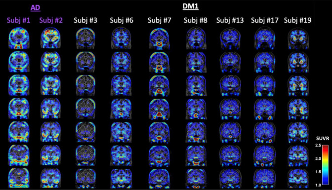
Results: Of the twelve DM1 participants, seven completed the full protocol (Neurocognition 11/12; PET 7/12, CSF 9/12, plasma 12/12). Three DM1 participants were cognitively impaired (CI). On average, CI DM1 participants had lower scores on the DCQ compared to cognitively unimpaired (CU) DM1 participants (75.5/100 vs. 91.4/100) and were older (54 vs. 44 years old) but did not differ in years of education (11.3 vs. 11.1). The majority (6/7) of DM1 participants had no appreciable PET signal. Only one of the CI participants presented with elevated Tau PET SUVR in bilateral medial temporal lobes. This participant was the eldest and most cognitively impaired, and had the lowest CSF Aβ 1-42 and the highest CSF Tau levels, all suggestive of co-existing AD. CSF Tau and phospho-Tau levels were higher in the 3 CI compared to CU DM1 participants, but with a mean value lower than that typically observed in AD. Nf-L and GFAP were elevated in most DM1 participants (9/11 and 8/11, respectively). Finally, CSF phospho-Tau was significantly correlated with plasma Nf-L concentrations.
Conclusions and relevance: We observed heterogenous cognitive and biomarker profiles in individuals with DM1. While some participants presented with abnormal PET and/or CSF Tau, these patterns were highly variable and only present in a small subset. Although DM1 may indeed represent a non-AD Tauopathy, the Tau-PET tracer used in this study was unable to detect an in vivo Tau DM1 signature in this small cohort. Interestingly, most DM1 participants presented with elevated plasma Nf-L and GFAP levels, suggestive of other, possibly related, central brain alterations which motivate further research. This pioneering study provides novel insights towards the potential relationship between biomarkers and neurocognitive deficits commonly seen in DM1.
Keywords: CSF; Cognition; Imaging; Myotonic dystrophy type 1; Social cognition; Tau PET.
© 2022. The Author(s).
Conflict of interest statement
The first and last authors have no disclosure.
Figures

References
-
- Clark C, Lewczuk P, Kornhuber J, Richiardi J, Marechal B, Karikari TK, Blennow K, Zetterberg H, Popp J. Plasma neurofilament light and phosphorylated tau 181 as biomarkers of Alzheimer's disease pathology and clinical disease progression. Alzheimers Res Ther. 2021;13:65. doi: 10.1186/s13195-021-00805-8. - DOI - PMC - PubMed
-
- Dakterzada F, Lopez-Ortega R, Arias A, Riba-Llena I, Ruiz-Julian M, Huerto R, Tahan N, Pinol-Ripoll G. Assessment of the concordance and diagnostic accuracy between elecsys and lumipulse fully automated platforms and innotest. Front Aging Neurosci. 2021;13:604119. doi: 10.3389/fnagi.2021.604119. - DOI - PMC - PubMed
MeSH terms
Substances
LinkOut - more resources
Full Text Sources
Medical
Miscellaneous
