Observational Study
. 2022 Apr;54(4):763-772.
doi: 10.1007/s11255-022-03130-7. Epub 2022 Feb 1.

The role of occlusion and micro-incontinence in the pathogenesis of penile lichen sclerosus: an observational study of pro-inflammatory cytokines' gene expression

Affiliations
Observational Study

The role of occlusion and micro-incontinence in the pathogenesis of penile lichen sclerosus: an observational study of pro-inflammatory cytokines' gene expression

M Czajkowski et al. Int Urol Nephrol. 2022 Apr.

Abstract

Purpose: To assess the expression of selected cytokines in penile lichen sclerosus (PLS) and associate them with the occurrence of micro-incontinence (MI) in different stages of PLS.

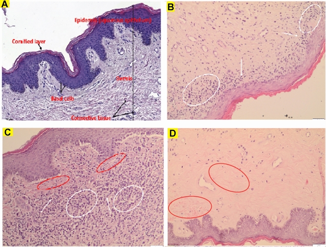
Methods: The skin biopsies from 49 PLS affected, and 13 from nonlesional foreskins (healthy control adult males undergoing circumcision due to phimosis caused by short frenulum) were obtained. All specimens were used for RNA extraction and RT-qPCR. Quantitative assessment of the gene expression of interleukin 1-A (IL-1A), interleukin 1-B (IL-1B), interleukin 1 receptor antagonist (IL-1RN), interleukin 6 (IL-6), transforming growth factor β1 (TGF-β1), and interferon-gamma (INF-γ) was performed. To determinate the presence of MI, the patients were asked about voiding patterns, especially leaking tiny drops of urine from the urethral meatus after urination.

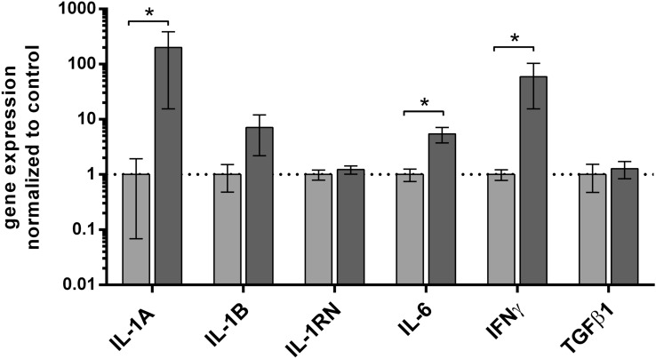
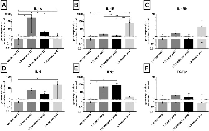
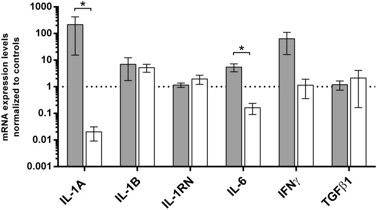
Results: IL-1A, IL-6, and INF-γ mRNA levels were approximately 150, 16, and 59 times higher in PLS than in control samples, respectively. The highest IL-1A mRNA levels were observed in early PLS (n = 13), INF-γ in moderate PLS (n = 32), while IL-6 in severe PLS (n = 4). MI was noted in 45 PLS patients vs. 0 in control (p < 0.0001). IL-1A and IL-6 vs control ratios were concentration (ca.) 400 and 30 times higher, respectively, in MI PLS samples than in PLS without MI.

Conclusion: Occlusion and irritating urine effect are associated with the clinical progression of penile LS with increased mRNA expression of IL-1A, INF-γ, and IL-6 pro-inflammatory cytokines in the foreskin.

Keywords: IFN-γ; IL-1; IL-6; Micro-incontinence; Penile lichen sclerosus; TGF-β1.


Conflict of interest statement

The authors have nothing to disclose.

Figures

Fig. 1
Fig. 1
Cytokines’ gene expression at the mRNA level in foreskin samples of penile lichen sclerosus and control patients. Gene expression was assessed as described in methods. The ordinate axis is shown on a logarithmic scale. Bars and whiskers represent the mean ± standard deviation of the mean (SEM) of genes’ expression levels in PLS (dark grey bars) normalized to control foreskin (light grey bars) samples (presented as 1; dotted horizontal line at 1). *P < 0.05 (Mann–Whitney U test between each group, solid line). PLS penile lichen sclerosus
Fig. 2
Fig. 2
Cytokines’ mRNA levels in penile lichen sclerosus stages in relation to control samples. Gene expression was assessed as described in “Materials and methods”. The ordinate axis is shown on a logarithmic scale. Bars and whiskers represent the mean ± SEM normalized to control foreskin samples (presented as 1). *P < 0.05, **P < 0.01, ***P < 0.001 (Mann–Whitney U test between each group, solid lines above bars; Kruskal–Wallis ANOVA test between all groups, dotted line above bars). LS lichen sclerosus
Fig. 3
Fig. 3
Representative microphotographs of normal foreskin and LS histological sections. A Normal control foreskin. BD Early, moderate and severe LS stages, respectively. Symbols for BC: white arrows—lymphocytes, white ellipses—dense inflammatory infiltrate, red ellipses—dermal hyalinization. Hematoxylin and eosin staining, scale bars represent 100 μm for (A) and 50 μm for (BE)
Fig. 4
Fig. 4
Summary of cytokines’ gene expression levels in penile lichen sclerosus to the occurrence of micro-incontinence. Gene expression was assessed as described in “Materials and methods”. The ordinate axis is shown on a logarithmic scale. Bars and whiskers represent the mean ± standard deviation of the mean (SEM) normalized to control foreskin samples (dotted horizontal line at 1), grouped by each analyzed gene. Bar legends: light grey bars represent PLS patients with micro-incontinence (MI) while white ones represent PLS patients without MI, respectively. *P < 0.05, Mann–Whitney U test between MI and lack of MI subgroups

References

    1. Clouston D, Hall A, Lawrentschuk N. Penile lichen sclerosus (balanitis xerotica obliterans) BJU Int. 2011;108(Suppl 2):14–19. doi: 10.1111/j.1464-410X.2011.10699.x. - DOI - PubMed
    1. Bunker CB, Patel N, Shim TN. Urinary voiding symptomatology (micro-incontinence) in male genital lichen sclerosus. Acta Derm Venereol. 2013;93(2):246–248. doi: 10.2340/00015555-1481. - DOI - PubMed
    1. Shah VS, Jung NL, Lee DK, Nepple KG. Does routine pathology analysis of adult circumcision tissue identify penile cancer. Urology. 2015;85(6):1431–1434. doi: 10.1016/j.urology.2014.12.065. - DOI - PMC - PubMed
    1. Czajkowski M, Żawrocki A, Czajkowska K, Kłącz J, Sokołowska-Wojdyło M, Biernat W, et al. Lichen sclerosus and phimosis - discrepancies between clinical and pathological diagnosis and its consequences. Urology. 2021 doi: 10.1016/j.urology.2020.11.027. - DOI - PubMed
    1. Fergus KB, Lee AW, Baradaran N, Cohen AJ, Stohr BA, Erickson BA, et al. Pathophysiology, clinical manifestations, and treatment of lichen sclerosus: a systematic review. Urology. 2020;135:11–19. doi: 10.1016/j.urology.2019.09.034. - DOI - PubMed

Publication types

LinkOut - more resources