. 2022 Mar 8;7(5):e151683.
doi: 10.1172/jci.insight.151683.

Accumulation of meningeal lymphocytes correlates with white matter lesion activity in progressive multiple sclerosis

Affiliations

Accumulation of meningeal lymphocytes correlates with white matter lesion activity in progressive multiple sclerosis

Shanzeh M Ahmed et al. JCI Insight. .

Abstract

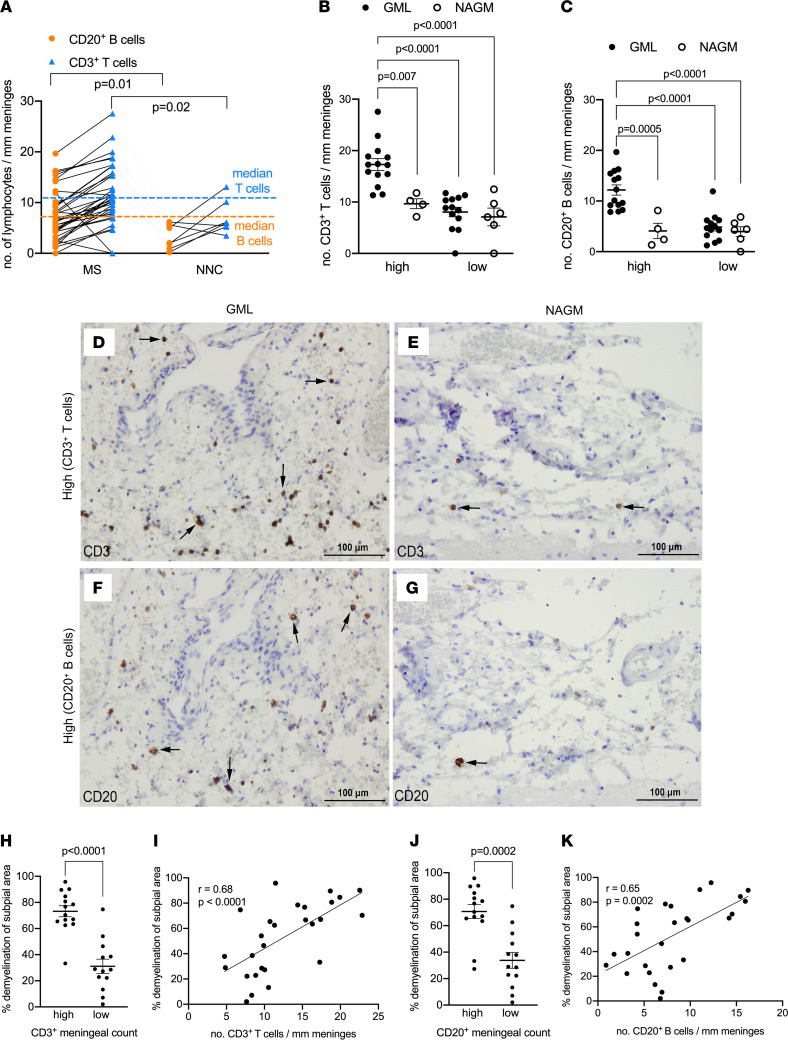
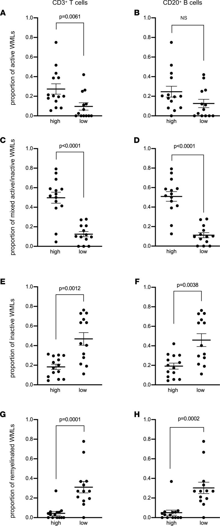
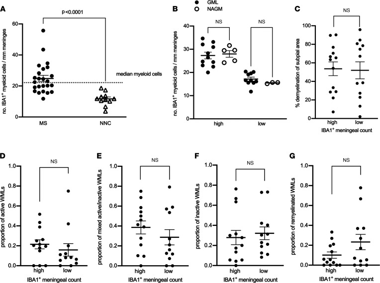
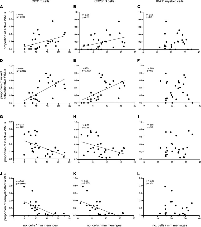
Subpial cortical demyelination is an important component of multiple sclerosis (MS) pathology contributing to disease progression, yet mechanism(s) underlying its development remain unclear. Compartmentalized inflammation involving the meninges may drive this type of injury. Given recent findings identifying substantial white matter (WM) lesion activity in patients with progressive MS, elucidating whether and how WM lesional activity relates to meningeal inflammation and subpial cortical injury is of interest. Using postmortem FFPE tissue blocks (range, 5-72 blocks; median, 30 blocks) for each of 27 patients with progressive MS, we assessed the relationship between meningeal inflammation, the extent of subpial cortical demyelination, and the state of subcortical WM lesional activity. Meningeal accumulations of T cells and B cells, but not myeloid cells, were spatially adjacent to subpial cortical lesions, and greater immune cell accumulation was associated with larger subpial lesion areas. Patients with a higher extent of meningeal inflammation harbored a greater proportion of active and mixed active/inactive WM lesions and an overall lower proportion of inactive and remyelinated WM lesions. Our findings support the involvement of meningeal lymphocytes in subpial cortical injury and point to a potential link between inflammatory subpial cortical demyelination and pathological mechanisms occurring in the subcortical WM.

Keywords: Adaptive immunity; Demyelinating disorders; Immunology; Multiple sclerosis.


Conflict of interest statement

Conflict of interest: JLG is a consultant for Roche (Canada) and held grants from Novartis, EMD Serono, and Roche. ABO has participated as a speaker in meetings sponsored by and received consulting fees from Accure Therapeutics, Atara Biotherapeutics, Biogen, Bristol Myers Squibb/Celgene/Receptos, GlaxoSmithKline, Gossamer, Janssen/Actelion, Medimmune, Merck/EMD Serono, Novartis, Roche/Genentech, and Sanofi and has received grant support to the University of Pennsylvania from Biogen Idec, Roche/Genentech, Merck/EMD Serono, and Novartis. VR is coinventor of a patent (WO2008044928) that describes the use of inhibitors of the terminal complement pathway for therapeutic purposes; she received consulting honoraria from Merck/EMD Serono and Fluidigm.

Figures

Figure 1
Figure 1. Meningeal T cells and B cells are enriched in MS and topographically linked to cortical subpial demyelination.
(A) Quantification of meningeal CD20+ B cell count and CD3+ T cell count in 27 MS donors and 9 nonneurological controls (NNCs) showing enrichment in MS donors versus NNCs. The median for meningeal CD20+ B cell count (orange dotted line) and meningeal CD3+ T cell count (blue dotted line) was used to stratify MS donors in high versus low meningeal B or T cell count. (B) Quantification of meningeal CD3+ T cell count showing significant enrichment of T cells in meninges adjacent to GML versus NAGM in MS donors with high (n = 14) but not low (n = 13) meningeal T cell count. (C) Quantification of meningeal CD20+ B cell count showing significant enrichment of B cells in meninges adjacent to GML versus NAGM in MS donors with high (n = 14) but not low (n = 13) meningeal T cell count. (DG) Representative immunohistochemical staining for CD3 (D and E, arrows) and CD20 (F and G, arrows) in meninges adjacent to subpial GML or NAGM in MS donors with high CD3+ or CD20+ meningeal cell count. (H) Quantification of the proportion of subpial gray matter that did not stain positive for myelin in MS donors with high versus low CD3+ T cell meningeal count. (I) Spearman’s correlation coefficient between meningeal CD3+ T cell count and the proportion of subpial gray matter that did not stain positive for myelin. (J) Quantification of the proportion of subpial gray matter that did not stain positive for myelin in MS donors with high versus low CD20+ B cell meningeal count. (K) Spearman’s correlation coefficient between meningeal CD20+ B cell count and the proportion of subpial gray matter that did not stain positive for myelin. In AC each data point represents mean cell count (mean ± SD) in all fields analyzed per case. Statistically significant differences determined by nonparametric Mann-Whitney test (A, H, and J) or nonparametric Kruskal-Wallis test followed by Dunn’s correction for multiple comparisons (B and C). Scale bars: 100 μm (DG).
Figure 2
Figure 2. Enrichment of meningeal T cells and B cells is linked to subcortical white matter lesion activity.
Quantification of (A and B) the proportion of active white matter lesions (WMLs), (C and D) mixed active/inactive WMLs, (E and F) inactive WMLs, and (G and H) remyelinated WMLs in MS donors with high (n = 14) versus low (n = 13) meningeal CD3+ T cells (A, C, E, and G) or CD20+ B cell (B, D, F, and H) count. Each data point represents the proportion of WMLs in all tissue blocks analyzed per case (range 5–72 blocks, median: 30 blocks per donor). Statistically significant differences were determined by the nonparametric Mann-Whitney test.
Figure 3
Figure 3. Meningeal myeloid cells are enriched in MS but not topographically linked to subpial demyelination or to the extent of cortical subpial demyelination or to subcortical white matter lesion activity.
(A) Quantification of meningeal IBA1+ myeloid cell count in MS donors and nonneurological controls (NNCs) showing enrichment in 27 MS donors compared with 9 NNCs. The median for meningeal IBA1+ myeloid cell count (shown by the dotted line) was used to stratify MS donors in high versus low meningeal myeloid cell count. (B) Quantification of meningeal IBA1+ myeloid cell count showing no significant changes in the number of meningeal myeloid cells adjacent to GML versus NAGM in MS donors with high (n = 14) or low (n = 13) meningeal myeloid cell count. (C) Quantification of the proportion of subpial gray matter that did not stain positive for myelin in MS donors with high versus low meningeal IBA1+ myeloid cell count. (DG) Quantification of (D) proportion of active white matter lesions (WMLs), (E) mixed active/inactive WMLs, (F) number of inactive WMLs, and (G) number of remyelinated WMLs in MS donors with high versus low meningeal IBA1+ myeloid cell count. Each data point represents the proportion of WMLs in all tissue blocks analyzed per case (range 5–72 blocks, median: 30 blocks per donor). In A and CG, statistically significant differences were determined by nonparametric Mann-Whitney test. In B, statistically significant differences were determined by nonparametric Kruskal-Wallis test followed by Dunn’s correction for multiple comparisons.
Figure 4
Figure 4. The density of meningeal CD3+ T cells and CD20+ B cells but not IBA1+ myeloid cells correlates with subcortical white matter lesion activity.
Spearman’s correlation coefficient between meningeal CD3+ T cell, CD20+ B cell, and IBA1+ myeloid cell count with proportion of active white matter lesions (WMLs) (AC), proportion of mixed active/inactive WMLs (DF), proportion of inactive WMLs (GI), and proportion of remyelinated WMLs (JL) in each of 27 MS donors. Each data point represents the proportion of WMLs obtained from all tissue blocks analyzed per case (range 5–72 blocks, median: 30 blocks per donor) and the mean meningeal cell count in all fields analyzed per case.

References

    1. Peterson JW, et al. Transected neurites, apoptotic neurons, and reduced inflammation in cortical multiple sclerosis lesions. Ann Neurol. 2001;50(3):389–400. doi: 10.1002/ana.1123. - DOI - PubMed
    1. Bo L, et al. Subpial demyelination in the cerebral cortex of multiple sclerosis patients. J Neuropathol Exp Neurol. 2003;62(7):723–732. doi: 10.1093/jnen/62.7.723. - DOI - PubMed
    1. Geurts JJ, et al. Extensive hippocampal demyelination in multiple sclerosis. J Neuropathol Exp Neurol. 2007;66(9):819–827. doi: 10.1097/nen.0b013e3181461f54. - DOI - PubMed
    1. Lucchinetti CF, et al. Inflammatory cortical demyelination in early multiple sclerosis. N Engl J Med. 2011;365(23):2188–2197. doi: 10.1056/NEJMoa1100648. - DOI - PMC - PubMed
    1. Popescu BF, et al. A case of multiple sclerosis presenting with inflammatory cortical demyelination. Neurology. 2011;76(20):1705–1710. doi: 10.1212/WNL.0b013e31821a44f1. - DOI - PMC - PubMed

Publication types