. 2022 Mar 8;7(5):e155569.
doi: 10.1172/jci.insight.155569.

Increased Ca2+ influx through CaV1.2 drives aortic valve calcification

Affiliations

Increased Ca2+ influx through CaV1.2 drives aortic valve calcification

Maiko Matsui et al. JCI Insight. .

Abstract

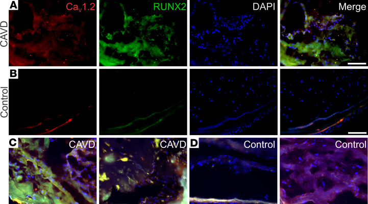
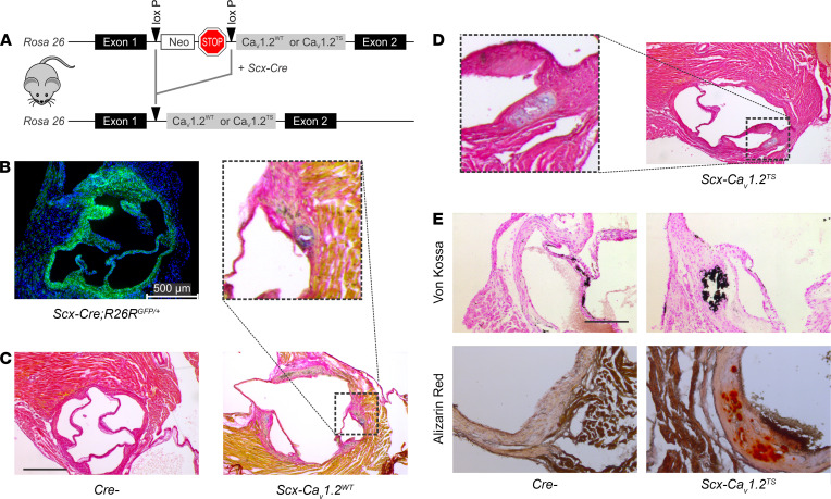
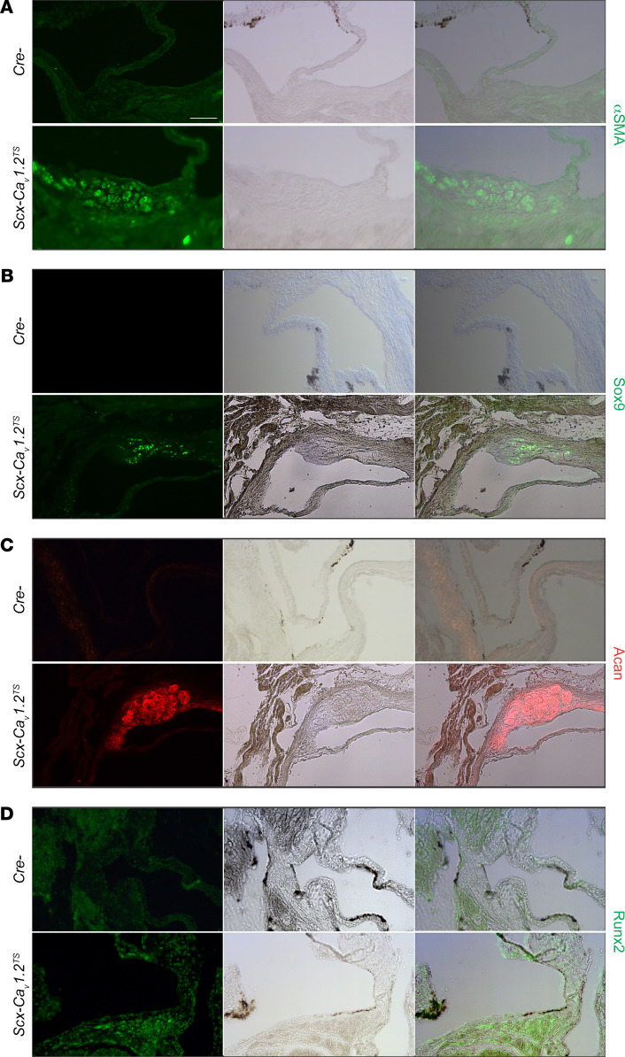
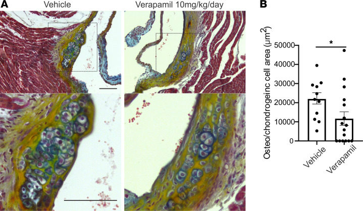
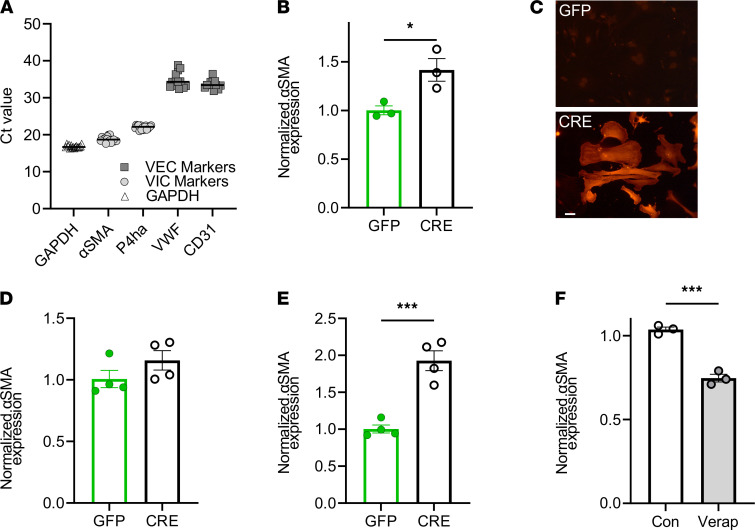
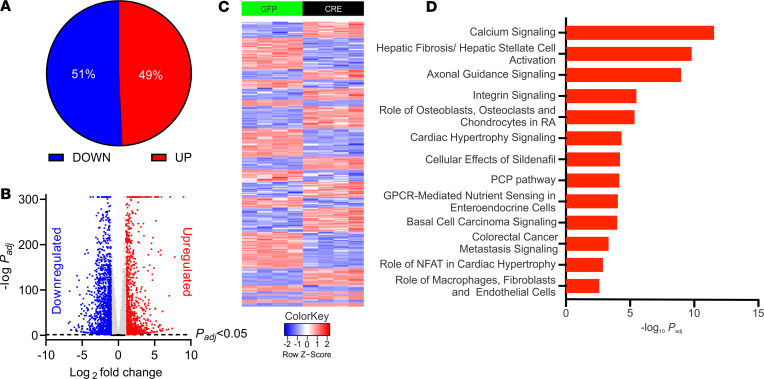
Calcific aortic valve disease (CAVD) is heritable, as revealed by recent GWAS. While polymorphisms linked to increased expression of CACNA1C - encoding the CaV1.2 L-type voltage-gated Ca2+ channel - and increased Ca2+ signaling are associated with CAVD, whether increased Ca2+ influx through the druggable CaV1.2 causes CAVD is unknown. We confirmed the association between increased CaV1.2 expression and CAVD in surgically removed aortic valves from patients. We extended our studies with a transgenic mouse model that mimics increased CaV1.2 expression within aortic valve interstitial cells (VICs). In young mice maintained on normal chow, we observed dystrophic valve lesions that mimic changes found in presymptomatic CAVD and showed activation of chondrogenic and osteogenic transcriptional regulators within these valve lesions. Chronic administration of verapamil, a CaV1.2 antagonist used clinically, slowed the progression of lesion development in vivo. Exploiting VIC cultures, we demonstrated that increased Ca2+ influx through CaV1.2 drives signaling programs that lead to myofibroblast activation of VICs and upregulation of genes associated with aortic valve calcification. Our data support a causal role for Ca2+ influx through CaV1.2 in CAVD and suggest that early treatment with Ca2+ channel blockers is an effective therapeutic strategy.

Keywords: Cardiology; Ion channels.


Conflict of interest statement

Conflict of interest: The authors have declared that no conflict of interest exists.

Figures

Figure 1
Figure 1. Increased CaV1.2 and RUNX2 expression in calcified segments of aortic valves from individuals with CAVD.
(A and B) IHC analysis of CaV1.2 (red) and RUNX2 (green) of valve tissue excised from a patient with CAVD (A) or a valve excised from a heart harvested for transplant and without CAVD (B). The blue signal is DAPI, and merged images are shown in the right-most panels for each. (C and D) Merged images for 2 additional valves from patients with CAVD (C) and from valves excised from hearts harvested for transplant and without CAVD (D). Scale bars: 100 μm.
Figure 2
Figure 2. Increased CaV1. 2 expression in the aortic valve is causal for CAVD.
(A) Transgenic strategy for inducible CaV1.2 expression. (B) GFP immunofluorescence (DAPI counterstain) showing aortic valve targeting with Scx-Cre. (C and D) Pentachrome staining shows lesions (inset) in CaV1.2WT (C) and CaV1.2TS (D) but not in Cre control mice (C). Scale bar: 200 μm. (E) Von Kossa and alizarin red stains demonstrate calcification of valve lesions in mice overexpressing CaV1.2TS in the valve. Scale bar: 100 μm.
Figure 3
Figure 3. Lesion-specific upregulation of chondrogenic and osteogenic makers in vivo.
Immunofluorescence for α-SMA (A), Sox9 (B), Aggrecan (C), and Runx2 (D) with accompanying light transmission and merged images of valves from Cre control and Scx-CaV1.2TS mice shows expression of the specific marker within the valve lesion but not in the unaffected valve. For each marker, examples from ≥3 samples. Scale bar: 100 μm.
Figure 4
Figure 4. Blockade of Ca2+ influx through CaV1.2 reduces aortic valve lesions in Scx-CaV1.2TS mice.
(A) Pentachrome stain of aortic valves from 3-month-old Scx-CaV1.2TS mice. Valves were isolated after 4 weeks of treatment by mini-osmotic pumps with verapamil or vehicle control. Inset shows magnification of lesion. (B) Quantification of lesion area. *P = 0.04 (2-sided t test). Scale bars: 100 μm. Osteo, osteogenic.
Figure 5
Figure 5. Blockade of Ca2+ influx through CaV1.2 reduces aortic valve lesions in Scx-CaV1.2TS mice.
(A) qPCR showing expression of VIC markers (α-SMA and P4ha) compared with endothelial cell markers (VWF and CD31) in cultured VICs. (B) Normalized (to GAPDH) α-SMA expression after 48 hours in CaV1.2WT-expressing VICs (after infection with virus expression GFP or Cre recombinase). *P < 0.05. (C) Anti–α-SMA immunofluorescence in CaV1.2TS VIC cultures expressing Cre or GFP. Scale bar: 50 μm. (D) Normalized (to GAPDH) α-SMA expression after 48 hours in WT VICs (after infection with virus expression GFP or Cre recombinase). P = NS. (E) Normalized (to GAPDH) α-SMA expression after 48 hours in CaV1.2TS expressing VICs (after infection with virus expression GFP or Cre recombinase). P < 0.001. (F) qPCR shows verapamil decreases relative expression of α-SMA in CaV1.2TS expressing VICs. ***P < 0.001. (BF) Statistical comparisons were performed with 2-sided t tests. Con, control; VEC, valve endothelial cell.
Figure 6
Figure 6. RNA-Seq of VICs infected with adenovirus expressing GFP or Cre.
(A) Distribution of upregulated (Up) and downregulated (Down) differentially expressed genes 48 hours after viral infection. (B) Volcano plot of differentially expressed genes 48 hours after infection. Blue and red symbols indicate genes differentially expressed (Padj < 0.05) and log2-fold greater than 1.0 or less than –1.0, respectively. Gray indicates genes differentially expressed (Padj < 0.05) and log2-fold less than 1.0 and greater than –1.0. Black indicates genes not differentially expressed (Padj > 0.05). (C) Heat map of the 2724 transcripts for which expression in the Cre infected VICs was upregulated or downregulated by more than 2-fold. (D) Gene ontology analysis by IPA of upregulated genes. PCP, planar cell polarity; RA, rheumatoid arthritis

References

    1. Nishimura RA, et al. 2014 AHA/ACC guideline for the management of patients with valvular heart disease: a report of the American College of Cardiology/American Heart Association task force on practice guidelines. Circulation. 2014;129(23):e521–e643. - PubMed
    1. Lindman BR, Merryman WD. Unloading the stenotic path to identifying medical therapy for calcific aortic valve disease: barriers and opportunities. Circulation. 2021;143(15):1455–1457. doi: 10.1161/CIRCULATIONAHA.120.052531. - DOI - PMC - PubMed
    1. Akat K, et al. Aortic valve calcification: basic science to clinical practice. Heart. 2009;95(8):616–623. - PubMed
    1. Rutkovskiy A, et al. Valve interstitial cells: the key to understanding the pathophysiology of heart valve calcification. J Am Heart Assoc. 2017;6(9):e006339. - PMC - PubMed
    1. Rajamannan NM, et al. Human aortic valve calcification is associated with an osteoblast phenotype. Circulation. 2003;107(17):2181–2184. doi: 10.1161/01.CIR.0000070591.21548.69. - DOI - PMC - PubMed

Supplementary concepts