. 2022 Jan;151(1):17.
doi: 10.1121/10.0009169.

Triangular body-cover model of the vocal folds with coordinated activation of the five intrinsic laryngeal muscles

Affiliations

Triangular body-cover model of the vocal folds with coordinated activation of the five intrinsic laryngeal muscles

Gabriel A Alzamendi et al. J Acoust Soc Am. 2022 Jan.

Abstract

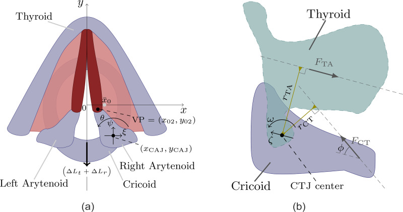
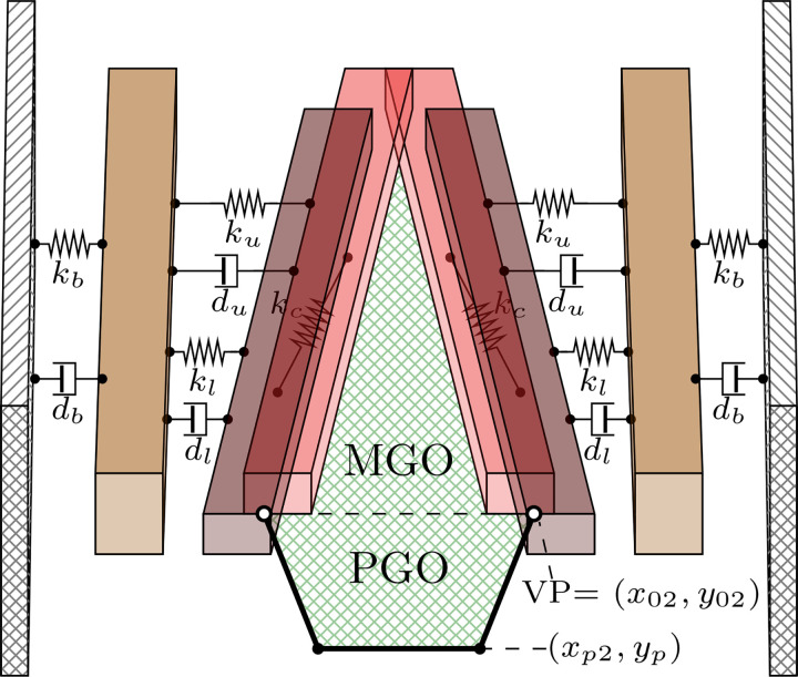
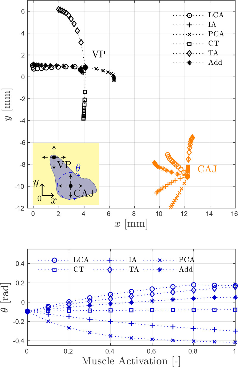
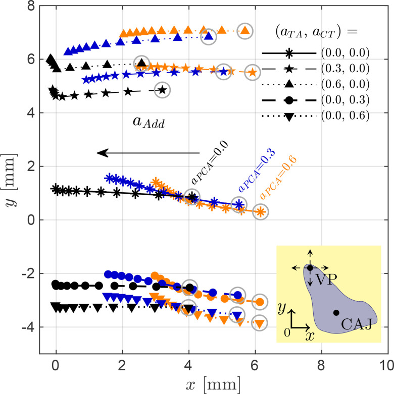
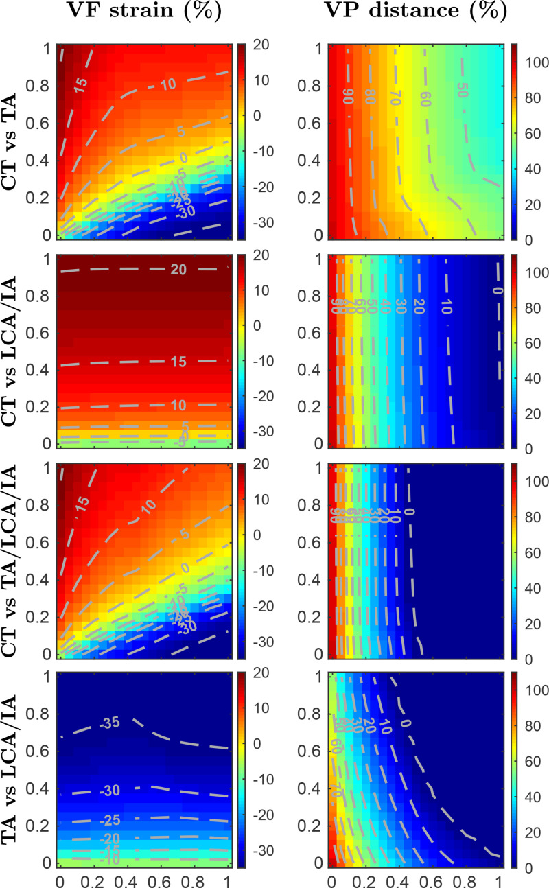
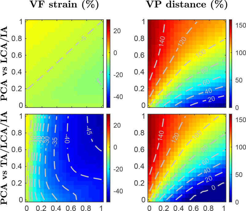
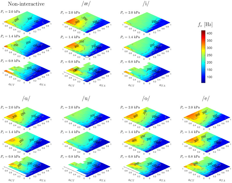
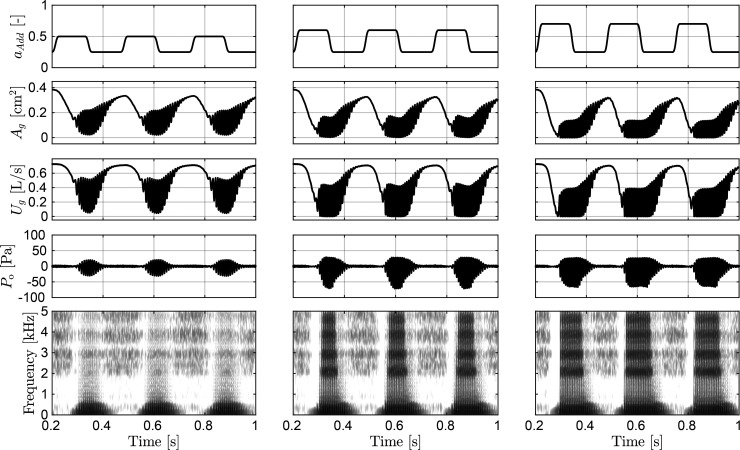
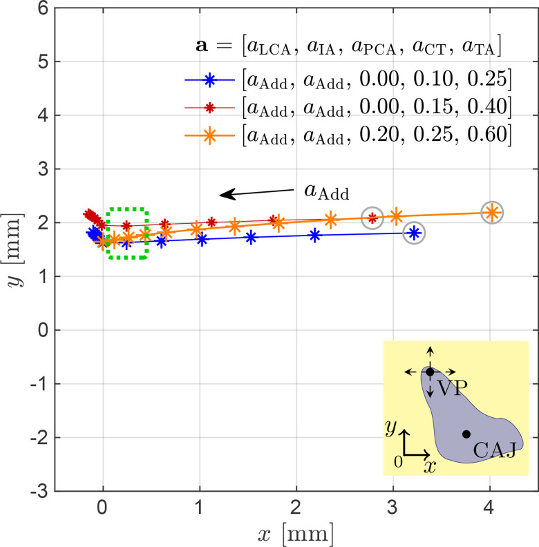
Poor laryngeal muscle coordination that results in abnormal glottal posturing is believed to be a primary etiologic factor in common voice disorders such as non-phonotraumatic vocal hyperfunction. Abnormal activity of antagonistic laryngeal muscles is hypothesized to play a key role in the alteration of normal vocal fold biomechanics that results in the dysphonia associated with such disorders. Current low-order models of the vocal folds are unsatisfactory to test this hypothesis since they do not capture the co-contraction of antagonist laryngeal muscle pairs. To address this limitation, a self-sustained triangular body-cover model with full intrinsic muscle control is introduced. The proposed scheme shows good agreement with prior studies using finite element models, excised larynges, and clinical studies in sustained and time-varying vocal gestures. Simulations of vocal fold posturing obtained with distinct antagonistic muscle activation yield clear differences in kinematic, aerodynamic, and acoustic measures. The proposed tool is deemed sufficiently accurate and flexible for future comprehensive investigations of non-phonotraumatic vocal hyperfunction and other laryngeal motor control disorders.


Figures

FIG. 1.
FIG. 1.
(Color online) Main laryngeal structures involved in the prephonatory posturing. (a) Cricoarytenoid accommodation at the glottal plane; and (b) Projection of effective cricothyroid accommodation on the glottal plane. Glottal geometry and vocal fold adjustment are controlled via the relative accommodation of major laryngeal cartilages. Figures adapted from Titze (2006). CAJ: cricoarytenoid joint, CTJ: cricothyroid joint.
FIG. 2.
FIG. 2.
(Color online) Schematic of the triangular body-cover model of the vocal folds.
FIG. 3.
FIG. 3.
(Color online) Simulated accommodation of the (right) arytenoid cartilage obtained by the independent activation of the five intrinsic muscles and the adductory complex. Top: Cartesian displacement of the cricoarytenoid joint (CAJ) center, (xCAJ+ξ,yCAJ+ψ), and the vocal process (VP), (x02,y02). The inset schematic illustrates the VP movements resulting from the displacement and rotation of the CAJ. Bottom: Rotation angle θ for the CAJ. Concurrent beginning of the paths indicates zero muscle activation.
FIG. 4.
FIG. 4.
(Color online) Effects of laryngeal muscle coactivation on glottal adduction. Vocal process (VP) coordinates, (x02,y02), produced through the activation of the adductory complex for the non-coactivation case (solid line with medium markers), and for two activation levels for both TA muscle (dashed and dotted lines with small markers) and CT muscle (dashed and dotted lines with large markers). Three PCA activations are drawn in black ( aPCA=0.0), dark color ( aPCA=0.3), and light color ( aPCA=0.6) lines. Markers enclosed in circles indicate the results for null adductory complex activation ( aAdd=0.0). The inset schematic illustrates the VP Cartesian movements.
FIG. 5.
FIG. 5.
(Color online) Muscle activation plots for parametric coactivation of main phonatory intrinsic muscles. Vocal fold strain (left column) and vocal process distance (right column) are depicted as functions of the paired coactivation of muscle groups. The rows show four activation scenarios with contour lines included for clarity. For each row, A vs B refers to the abscissa and ordinate axes indicating activation levels for muscle groups A and B, respectively; null activations are set for the remaining intrinsic muscles.
FIG. 6.
FIG. 6.
(Color online) Muscle activation plots for parametric coactivation of adductory/abductory intrinsic muscles. Vocal fold strain (left column) and vocal process distance (right column) are depicted as functions of the paired coactivation of muscle groups. The rows show two activation scenarios with contour lines included for clarity. For each row, A vs B refers to the abscissa and ordinate axes indicating activation levels for muscle groups A and B, respectively; null activations are set for the remaining intrinsic muscles.
FIG. 7.
FIG. 7.
(Color online) Muscle activation plots with fundamental frequency, f0, for CT versus TA activation, considering different vocal tract shapes and subglottal pressures Ps=[0.8,1.4,2.0] kPa. In all the cases aLCA=aIA=0.5, and aPCA=0.0. The non-interactive case has no vocal tract and represents an excised larynx scenario. Isofrequency contours are drawn for clarity.
FIG. 8.
FIG. 8.
Simulated muscle control of voicing-devoicing for a /hi-hi-hi-hi/ gesture. From left to right, columns correspond to simulations for weak, moderate, and strong VF adduction with maximum activation levels 0.5, 0.6, and 0.7, respectively. Adductory signals aAdd are shown in the top row. The following three rows show the output signals for the glottal area Ag, glottal volume velocity Ug, and the radiated acoustic pressure Po. The bottom row depicts the wideband spectrogram of Po.
FIG. 9.
FIG. 9.
(Color online) Displacement paths for the right vocal process (VP), (x02,y02), for three simulated tension states in the larynx. Trajectories correspond to laryngeal postures produced by parametric activation of the intrinsic muscles following the activation set a=[aLCA,aIA,aPCA,aCT,aTA]. The paths describe variations for the normalized activations aAdd in the range of 0 to 1 in steps of 0.1. The inset schematic illustrates the VP Cartesian movements. Configurations enclosed in the dotted rectangle are considered for voiced sound simulations.

References

    1. Alzamendi, G. A. , Manríquez, R. , Hadwin, P. J. , Deng, J. J. , Peterson, S. D. , Erath, B. D. , Mehta, D. D. , Hillman, R. E. , and Zañartu, M. (2020). “ Bayesian estimation of vocal function measures using laryngeal high-speed videoendoscopy and glottal airflow estimates: An in vivo case study,” J. Acoust. Soc. Am. 147(5), EL434–EL439.10.1121/10.0001276 - DOI - PMC - PubMed
    1. Birkholz, P. , Kröger, B. J. , and Neuschaefer-Rube, C. (2011a). “ Articulatory synthesis of words in six voice qualities using a modified two-mass model of the vocal folds,” in First International Workshop on Performative Speech and Singing Synthesis, p3s-2011, March 14–15, Vancouver, Canada.
    1. Birkholz, P. , Kröger, B. J. , and Neuschaefer-Rube, C. (2011b). “ Synthesis of breathy, normal, and pressed phonation using a two-mass model with a triangular glottis,” in Proceedings of Interspeech 2011: 12th Annual Conference of the International Speech Communication Association, August 27–31, Florence, Italy, pp. 2681–2684.
    1. Chhetri, D. K. , Neubauer, J. , and Berry, D. A. (2012). “ Neuromuscular control of fundamental frequency and glottal posture at phonation onset,” J. Acoust. Soc. Am. 131(2), 1401–1412.10.1121/1.3672686 - DOI - PMC - PubMed
    1. Chhetri, D. K. , Neubauer, J. , Sofer, E. , and Berry, D. A. (2014). “ Influence and interactions of laryngeal adductors and cricothyroid muscles on fundamental frequency and glottal posture control,” J. Acoust. Soc. Am. 135(4), 2052–2064.10.1121/1.4865918 - DOI - PMC - PubMed

Publication types