. 2022 Feb 8;119(6):e2024508119.
doi: 10.1073/pnas.2024508119.

Executioner caspases 3 and 7 are dispensable for intestinal epithelium turnover and homeostasis at steady state

Affiliations

Executioner caspases 3 and 7 are dispensable for intestinal epithelium turnover and homeostasis at steady state

Farzaneh Ghazavi et al. Proc Natl Acad Sci U S A. .

Abstract

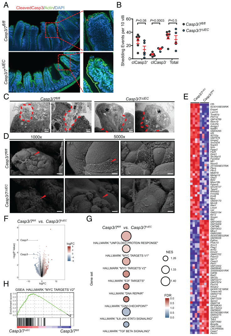
Apoptosis is widely believed to be crucial for epithelial cell death and shedding in the intestine, thereby shaping the overall architecture of the gastrointestinal tract, but also regulating tolerance induction, pinpointing a role of apoptosis intestinal epithelial cell (IEC) turnover and maintenance of barrier function, and in maintaining immune homeostasis. To experimentally address this concept, we generated IEC-specific knockout mice that lack both executioner caspase-3 and caspase-7 (Casp3/7ΔIEC), which are the converging point of the extrinsic and intrinsic apoptotic pathway. Surprisingly, the overall architecture, cellular landscape, and proliferation rate remained unchanged in these mice. However, nonapoptotic cell extrusion was increased in Casp3/7ΔIEC mice, compensating apoptosis deficiency, maintaining the same physiological level of IEC shedding. Microbiome richness and composition stayed unaffected, bearing no sign of dysbiosis. Transcriptome and single-cell RNA sequencing analyses of IECs and immune cells revealed no differences in signaling pathways of differentiation and inflammation. These findings demonstrate that during homeostasis, apoptosis per se is dispensable for IEC turnover at the top of intestinal villi intestinal tissue dynamics, microbiome, and immune cell composition.

Keywords: apoptosis; caspases; cell death; mucosal immunology; regeneration.


Conflict of interest statement

The authors declare no competing interest.

Figures

Fig. 1.
Fig. 1.
Epithelial deletion of Caspase-3 and -7 does not affect intestinal homeostasis at steady state. (A) The length of small intestine and its normalized value to the body weight in Casp3/7ΔIEC (n = 7) were compared to Casp3/7fl/fl mice (n = 7). (B) Representative micrographs of H&E staining of small intestinal sections from Casp3/7ΔIEC and Casp3/7fl/fl mice. Scale bars are indicated. (C) Quantification of the average length of around 200 villi measured in Casp3/7ΔIEC (n = 4) and Casp3/7fl/fl (n = 4) mice. (D and E) Alcian Blue staining (as a proxy for goblet cells) and automated quantification thereof in small intestinal sections from Casp3/7ΔIEC (n = 6) and Casp3/7fl/fl (n = 7) mice. (F and G) Lysozyme staining (as a proxy for Paneth cells) and automated quantification thereof in small intestinal sections from Casp3/7ΔIEC (n = 6) and Casp3/7fl/fl (n = 7). (H) Ultramicroscopic analysis of Paneth cell compartment via TEM. (I and J) Ki67 staining (as a proxy for proliferating cells) and automated quantification thereof in small intestinal sections from Casp3/7ΔIEC (n = 6) and Casp3/7fl/fl (n = 7) mice. (K and L) The organoid cultures derived from LGR5+ intestinal stem cells of Casp3/7ΔIEC and Casp3/7fl/fl mice and organoid forming capacity calculated after the first five passages (P1 to P5). (Scale bars, 50 µm.) A two-tailed unpaired t test was conducted to compare Casp3/7ΔIEC with Casp3/7fl/fl mice. Each point represents an individual mouse, and the line represents mean ± SEM. For bar plots in L, the error bars are the SEM and three mice per group per passage were analyzed. (M) UMAP of intraepithelial (IEL) cells extracted from three Casp3/7fl/fl and Casp3/7ΔIEC (n = 3 per genotype) mice. Identified populations are indicated. In total, 679 Casp3/7fl/fl vs. 2,227 Casp3/7ΔIEC enterocytes; when looking at total cells per sample: 7,584 Casp3/7fl/fl cells and 11,956 Casp3/7ΔIEC cells, equaling a relative share of 9.0% in WT and 18.6% in Casp3/7ΔIEC. The relative higher share of enterocytes in the case of Casp3/7ΔIEC cells is probably due to the stressful conditions during the isolation procedure resulting in higher levels of apoptosis in WT cells, while this occurs less in enterocytes derived from Casp3/7ΔIEC mice. (N) Overlay of Casp3/7fl/fl and Casp3/7ΔIEC cells from IEL cells. (O) UMAP with normalized expression of Mki67. (ns: non significant P-value.)
Fig. 2.
Fig. 2.
Apical shedding of IECs proceeds independently of Caspase-3 and -7. (A and B) Nonapoptotic (cleaved caspase-3) and apoptotic (cleaved caspase-3+) extrusions were detected by confocal microscopy and quantified in Casp3/7ΔIEC (n = 5) and Casp3/7fl/fl (n = 6) mice. Quantification of shedding events is representative of two independent experiments. (C and D) Ultrastructural analysis of extruding cells at apical villi using TEM and SEM. Scale bars are shown. Asterisks and demarcation show the live and apoptotic extruding cells, respectively. A two-tailed unpaired t test was conducted to compare Casp3/7ΔIEC with Casp3/7fl/fl mice. Each point represents an individual mouse, and the line represents mean ± SEM. (E) Top 50 differentially expressed genes between Casp3/7ΔIEC and Casp3/7fl/fl cells (n = 4 biological replicates per group). Expression values are z-score normalized. The gene symbols are indicated on the Right. (F) Global differential gene expression analysis of RNA-seq data derived from Casp3/7ΔIEC and Casp3/7fl/fl cells (n = 4 biological replicates per group). Data points for Casp3 and Casp7 are labeled. LogFC, log2(fold change). (G) Gene set enrichment analyses (GSEA) for the indicated signatures. False discovery rate (FDR), color intensity of the circle. Normalized enrichment score (NES), circle diameter. Blue/red indicates the group in which a signature was positively enriched. (H) GSEA enrichment plot for the Hallmark gene set “Myc Targets v2.” The genotype is indicated in blue/red.
Fig. 3.
Fig. 3.
Loss of apoptosis in intestinal epithelial cells does not induce ileal dysbiosis. (A) Bacterial richness in the ileum represented by detected (observed) amplicon sequence variants (ASV) of 16S microbiome sequencing. (B) Inverse Simpson and (C) effective Shannon diversity indices in single- and cohoused Casp3/7ΔIEC and Casp3/7fl/fl mice revealed no significant differences. (D) Principal coordinates analysis of weighted (ADONIS test, R2 = 0.3, adjusted P = 1.0); (E) unweighted (ADONIS test, R2 = 0.2, adjusted P = 1.0) UniFrac distances; and (F) nonmetric multidimensional scaling of Bray–Curtis distances (ADONIS test, R2 = 0.6, adjusted P = 1.0) show no separation of the Casp3/7ΔIEC and Casp3/7fl/fl mice based on bacterial compositions of ileal samples. Large dots represent group centroids; ellipses indicate the 95% confidence intervals for each group. (G) Bar plots of bacterial compositions in ileal samples of single- and cohoused Casp3/7ΔIEC and Casp3/7fl/fl mice at the genus and (H) family level.
Fig. 4.
Fig. 4.
Loss of epithelial apoptosis does not induce spontaneous intestinal inflammation. (A and B) CD45 staining (leukocyte marker) and quantification in the lamina propria (LP) of small intestine sections from Casp3/7ΔIEC (n = 7) and Casp3/7fl/fl (n = 7) mice. (Scale bar, 50 µm.) (C) The fecal level of Lipocalin-2 of Casp3/7ΔIEC (n = 12) and Casp3/7fl/fl (n = 10) mice. (D) The plasma level of DAO in Casp3/7ΔIEC (n = 6) and Casp3/7fl/fl (n = 6) mice. (E) The plasma level of proinflammatory cytokines in Casp3/7ΔIEC (n = 3 to 4) and Casp3/7fl/fl (n = 4) mice. (F) UMAP of LP extracted from three Casp3/7fl/fl and Casp3/7ΔIEC (n = 3 per genotype) mice. Identified populations are indicated with relative frequency in G. (H) Quantification of UMAP of mesenteric lymph nodes (MLNs) extracted from two Casp3/7fl/fl and Casp3/7ΔIEC (n = 2 per genotype) mice. Identified populations are indicated with relative frequency in I. (ns: non significant P-value.)

References

    1. Peterson L. W., Artis D., Intestinal epithelial cells: Regulators of barrier function and immune homeostasis. Nat. Rev. Immunol. 14, 141–153 (2014). - PubMed
    1. Beumer J., Clevers H., Cell fate specification and differentiation in the adult mammalian intestine. Nat. Rev. Mol. Cell Biol. 22, 39–53 (2021). - PubMed
    1. Beumer J., Clevers H., Regulation and plasticity of intestinal stem cells during homeostasis and regeneration. Development 143, 3639–3649 (2016). - PubMed
    1. Nagata S., Apoptosis and autoimmune diseases. Ann. N. Y. Acad. Sci. 1209, 10–16 (2010). - PubMed
    1. Morioka S., Maueröder C., Ravichandran K. S., Living on the edge: Efferocytosis at the interface of homeostasis and pathology. Immunity 50, 1149–1162 (2019). - PMC - PubMed

Publication types