. 2022 Mar 23;96(6):e0000222.
doi: 10.1128/jvi.00002-22. Epub 2022 Feb 2.

Host Cellular RNA Helicases Regulate SARS-CoV-2 Infection

Affiliations

Host Cellular RNA Helicases Regulate SARS-CoV-2 Infection

Yasuo Ariumi. J Virol. .

Abstract

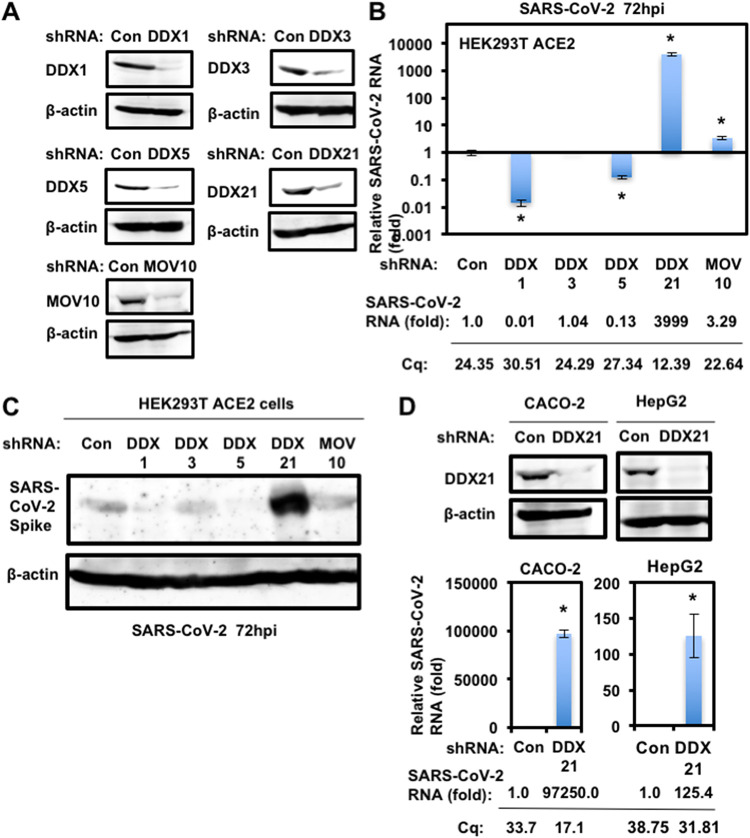
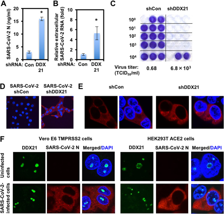
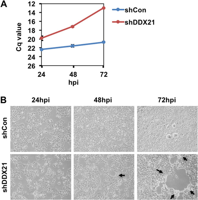
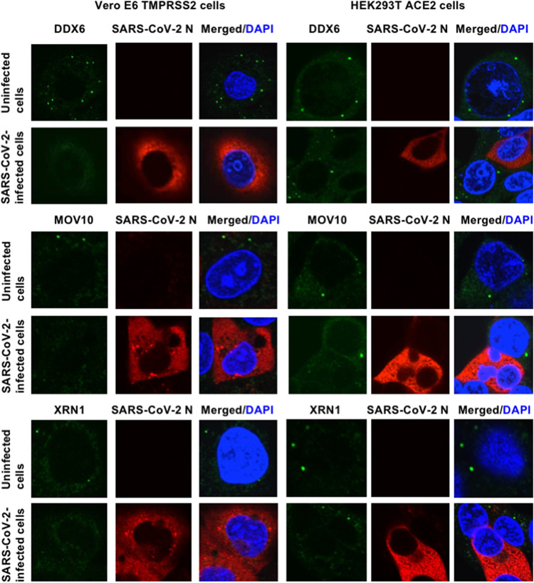
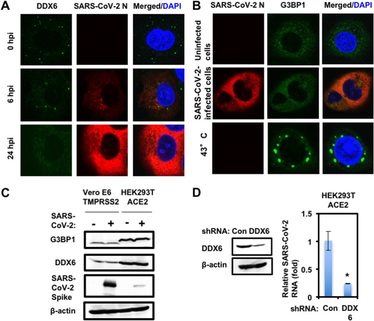
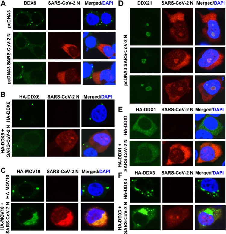
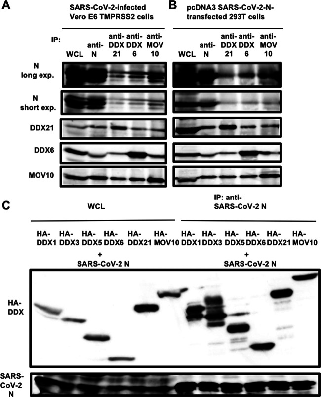
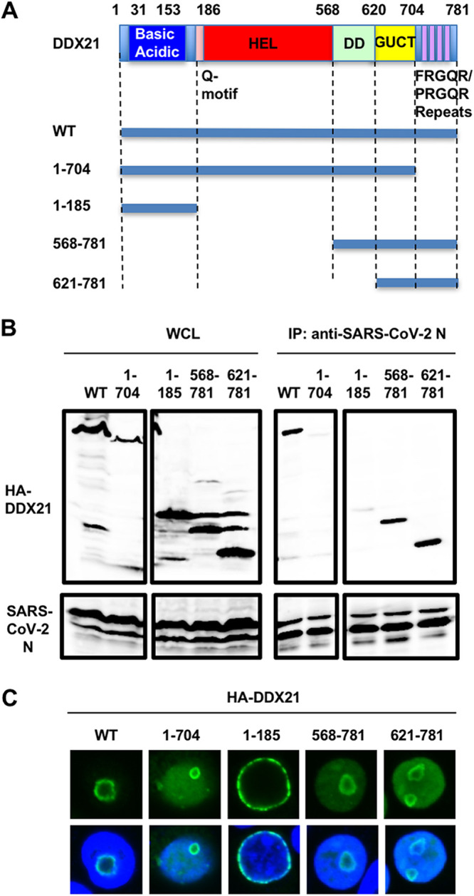
Severe acute respiratory syndrome coronavirus 2 (SARS-CoV-2) has the largest RNA genome, approximately 30 kb, among RNA viruses. The DDX DEAD box RNA helicase is a multifunctional protein involved in all aspects of RNA metabolism. Therefore, host RNA helicases may regulate and maintain such a large viral RNA genome. In this study, I investigated the potential role of several host cellular RNA helicases in SARS-CoV-2 infection. Notably, DDX21 knockdown markedly accumulated intracellular viral RNA and viral production, as well as viral infectivity of SARS-CoV-2, indicating that DDX21 strongly restricts the SARS-CoV-2 infection. In addition, MOV10 RNA helicase also suppressed the SARS-CoV-2 infection. In contrast, DDX1, DDX5, and DDX6 RNA helicases were required for SARS-CoV-2 replication. Indeed, SARS-CoV-2 infection dispersed the P-body formation of DDX6 and MOV10 RNA helicases as well as XRN1 exonuclease, while the viral infection did not induce stress granule formation. Accordingly, the SARS-CoV-2 nucleocapsid (N) protein interacted with DDX1, DDX3, DDX5, DDX6, DDX21, and MOV10 and disrupted the P-body formation, suggesting that SARS-CoV-2 N hijacks DDX6 to carry out viral replication. Conversely, DDX21 and MOV10 restricted SARS-CoV-2 infection through an interaction of SARS-CoV-2 N with host cellular RNA helicases. Altogether, host cellular RNA helicases seem to regulate the SARS-CoV-2 infection. IMPORTANCE SARS-CoV-2 has a large RNA genome, of approximately 30 kb. To regulate and maintain such a large viral RNA genome, host RNA helicases may be involved in SARS-CoV-2 replication. In this study, I have demonstrated that DDX21 and MOV10 RNA helicases limit viral infection and replication. In contrast, DDX1, DDX5, and DDX6 are required for SARS-CoV-2 infection. Interestingly, SARS-CoV-2 infection disrupted P-body formation and attenuated or suppressed stress granule formation. Thus, SARS-CoV-2 seems to hijack host cellular RNA helicases to play a proviral role by facilitating viral infection and replication and by suppressing the host innate immune system.

Keywords: DDX1; DDX21; DDX6; P-body; RNA helicase; SARS-CoV-2; coronavirus; nucleocapsid; nucleolus; stress granule.


Conflict of interest statement

The authors declare no conflict of interest.

We have no conflicts of interest to disclose.

Figures

FIG 1
FIG 1
DDX21 restricts SARS-CoV-2 infection. (A) Inhibition of host cellular RNA helicase protein expression by the shRNA-producing lentiviral vector. The results of Western blot analysis of cellular lysates with anti-DDX1 (A300-521A), anti-DDX3 (A300-474A), anti-DDX6 (A300-460A), anti-DDX21 (A300-627A), anti-MOV10 (A301-571A), or anti-β-actin antibody are shown. (B) The level of intracellular SARS-CoV-2 RNA in the cells at 72 h postinfection at an MOI of 0.5 was monitored by real-time LightCycler RT-PCR. Results from three independent experiments are shown. The level of SARS-CoV-2 RNA in knockdown cells was calculated relative to the level in HEK293T ACE2 cells transduced with a control lentiviral vector (Con). *, P < 0.05 compared to control cells. Cq, quantitation cycle. (C) SARS-CoV-2 spike protein expression levels in knockdown cells. The results of the Western blot analysis of cellular lysates with anti-SARS-CoV-2 Spike (GTX632604 [1A9]) or anti-β-actin antibody in the SARS-CoV-2-infected HEK293T ACE2 cells at 72 h postinfection at an MOI of 0.5 are shown. (D) DDX21 restricts SARS-CoV-2 infection in Caco-2 and HepG2 cells. Inhibition of endogenous DDX21 protein expression by the shRNA-producing lentiviral vector. The results of Western blot analysis of cellular lysates with anti-DDX21 or anti-β-actin antibody in Caco-2 and HepG2 cells are shown. The level of intracellular SARS-CoV-2 RNA in the cells at 72 h postinfection at an MOI of 0.5 was monitored by real-time LightCycler RT-PCR. Results from three independent experiments are shown. The level of SARS-CoV-2 RNA in DDX21 knockdown cells was calculated relative to the level in HEK293T ACE2 cells transduced with a control lentiviral vector (Con). *, P < 0.05 compared to control cells.
FIG 2
FIG 2
Characterization of antiviral effect of DDX21. (A) DDX21 suppresses SARS-CoV-2 production. The levels of extracellular SARS-CoV-2 N protein in the culture supernatants from the DDX21 knockdown HEK293T ACE2 cells 72 h after inoculation of SARS-CoV-2 at an MOI of 0.5 were determined by ELISA. Experiments were done in triplicate, and columns show the mean SARS-CoV-2 N protein levels. *, P < 0.05 compared to control cells. (B) DDX21 inhibits the level of extracellular SARS-CoV-2 RNA. The levels of extracellular SARS-CoV-2 RNA in the culture supernatants from the DDX21 knockdown HEK293T ACE2 cells 72 h after inoculation of SARS-CoV-2 at an MOI of 0.5 were monitored by real-time LightCycler RT-PCR. Results from three independent experiments are shown. The level of SARS-CoV-2 RNA in DDX21 knockdown cells was calculated relative to the level in HEK293T ACE2 cells transduced with a control lentiviral vector (Con). *, P < 0.05 compared to control cells. (C) The virus titer of SARS-CoV-2 in the culture supernatants from the DDX21 knockdown HEK293T ACE2 cells 72 h after inoculation of SARS-CoV-2. Naive Vero E6 TMPRSS2 cells were seeded in 24-well plates at 5 × 104 cells per well and then infected the next day with the indicated serial 10-fold dilutions of culture supernatants. The cells were stained with 0.6% Coomassie brilliant blue in 50% methanol and 10% acetate at 72 h postinfection were monitored for cytopathic effect. The virus titer was determined as TCID50/mL. (D) The infectivity of SARS-CoV-2 in the culture supernatants from the control or DDX21 knockdown HEK293T ACE2 cells 72 h after inoculation of SARS-CoV-2 was compared by immunofluorescence. Naive Vero E6 TMPRSS2 cells were plated on Lab-Tek 2-well chamber slides at 2 × 104 cells per well. The next day, 1 μL of culture supernatants of SARS-CoV-2-infected control or DDX21 knockdown HEK293T ACE2 cells was inoculated. The cells were fixed at 24 h postinfection and stained with anti-SARS-CoV-2 nucleocapsid (ab273434 [6H3]). Cells were then stained with donkey anti-mouse IgG (H+L) Alexa Fluor 594-conjugated secondary antibody. Images were visualized using confocal laser scanning microscopy. Nuclei were stained with DAPI (blue). (E) Subcellular localization of SARS-CoV-2 N protein in control or DDX21 knockdown HEK293T ACE2 cells 24 h after inoculation of SARS-CoV-2. The cells were stained with anti-SARS-CoV-2 nucleocapsid. (F) Subcellular localization of endogenous DDX21 and SARS-CoV-2 N protein in Vero E6 TMPRSS2 or HEK293T ACE2 cells 24 h after inoculation of SARS-CoV-2. The cells were stained with anti-SARS-CoV-2 nucleocapsid and anti-DDX21 (A300-627A) antibodies. Cells were then stained with donkey anti-rabbit IgG (H+L) Alexa Fluor 488-conjugated secondary antibody and donkey anti-mouse IgG (H+L) Alexa Fluor 594-conjugated secondary antibody. The two-color overlay images are also exhibited (Merged).
FIG 3
FIG 3
DDX21 knockdown facilitates SARS-CoV-2 propagation and spread. (A) SARS-CoV-2 RNA multiplication in control (shCon) or DDX21 knockdown HEK293T ACE2 cells (shDDX21) (2 × 105 cells/well) at the indicated time postinfection with SARS-CoV-2 (2.7 × 105 TCID50/μL), as determined by real-time RT-PCR. Cq, quantitation cycle. (B) SARS-CoV-2-infected control (shCon) or DDX21 knockdown HEK293T ACE2 cells (shDDX21) at the indicated time postinfection with SARS-CoV-2, as in panel A. Black arrows indicate syncytium formation.
FIG 4
FIG 4
SARS-CoV-2 disrupts P-body formation. Uninfected Vero E6 TMPRSS2 or HEK293T ACE2 cells and their SARS-CoV-2-infected cells at 24 h postinfection were stained with anti-SARS-CoV-2 nucleocapsid (ab273434 [6H3]) and anti-DDX6 (A300-460A) antibodies. The cells were also stained with anti-SARS-CoV-2 nucleocapsid and either anti-Xrn1 (A300-443A) or anti-MOV10 (A301-571A) antibodies. Cells were then stained with donkey anti-rabbit IgG (H+L) Alexa Fluor 488-conjugated secondary antibody and donkey anti-mouse IgG (H+L) Alexa Fluor 594-conjugated secondary antibody. Images were visualized using confocal laser scanning microscopy. The two-color overlay images are also exhibited (Merged). Nuclei were stained with DAPI (blue).
FIG 5
FIG 5
SARS-CoV-2 hijacks DDX6 for viral replication. (A) Dynamic redistribution of DDX6 in response to SARS-CoV-2 infection. Vero E6 TMPRSS2 cells at the indicated times after inoculation of SARS-CoV-2 were stained with anti-SARS-CoV-2 nucleocapsid (ab273434 [6H3]) and anti-DDX6 (A300-460A) antibodies. (B) SARS-CoV-2 does not induce stress granule formation. Uninfected Vero E6 TMPRSS2 or SARS-CoV-2-infected cells at 24 h postinfection were incubated at 37°C. Uninfected cells were also incubated at 43°C for 45 min. Cells were then stained with anti-SARS-CoV-2 nucleocapsid and anti-G3BP1 (A302-033A) antibodies. (C) Host protein expression levels in response to SARS-CoV-2 infection. The results of the Western blot analysis of cellular lysates with anti-SARS-CoV-2 spike (GTX632604 [1A9]), anti-DDX6, anti-G3BP1, or anti-β-actin antibody in the SARS-CoV-2-infected Vero E6 TMPRSS2 or the HEK293T ACE2 cells at 24 h postinfection at an MOI of 0.5 as well as in the uninfected cells are shown. (D) Requirement of DDX6 for SARS-CoV-2 infection. Inhibition of endogenous DDX6 protein expression by the shRNA-producing lentiviral vector. The results of Western blot analysis of cellular lysates with anti-DDX6 or anti-β-actin antibody are shown. The level of intracellular SARS-CoV-2 RNA in the cells at 72 h postinfection at an MOI of 0.5 was monitored by real-time LightCycler RT-PCR. Results from three independent experiments are shown. The level of SARS-CoV-2 RNA in the DDX6 knockdown cells was calculated relative to the level in HEK293T ACE2 cells transduced with a control lentiviral vector (Con). *, P < 0.05 compared to control cells.
FIG 6
FIG 6
SARS-CoV-2 nucleocapsid (N) protein disrupts the P-body formation of DDX6 and MOV10. (A) Disruption of P-body formation of endogenous DDX6 by ectopically expressed SARS-CoV-2 N. 293T cells transfected with 200 ng of pcDNA3.1-SARS-CoV-2 N (37) were stained with anti-SARS-CoV-2 nucleocapsid (GTX632269 [6H3]) and anti-DDX6 (A300-460A) antibodies. (B) Disruption of P-body formation of HA-tagged DDX6 by ectopically expressed SARS-CoV-2 N. 293T cells cotransfected with 200 ng of pcDNA3-HA-DDX6 (9) and either 200 ng of pcDNA3.1-SARS-CoV-2 N or pcDNA3 were stained with anti-SARS-CoV-2 nucleocapsid and anti-HA (3F10) antibodies. (C) Disruption of P-body formation of HA-tagged MOV10 by SARS-CoV-2 N and colocalization of SARS-CoV-2 N and HA-MOV10. 293T cells cotransfected with 200 ng of pcDNA3-HA-MOV10 and either 200 ng of pcDNA3.1-SARS-CoV-2 N or pcDNA3 were stained with anti-SARS-CoV-2 nucleocapsid and anti-HA antibodies. (D) Colocalization of endogenous DDX21 and ectopically expressed SARS-CoV-2 N in nucleoli. 293T cells transfected with 200 ng of pcDNA3.1-SARS-CoV-2 N were stained with anti-SARS-CoV-2 nucleocapsid and anti-DDX21 (A300-627A) antibodies. (E) Colocalization of HA-tagged DDX1 and SARS-CoV-2 N. 293T cells cotransfected with 200 ng of pcDNA3-HA-DDX1 (9) and either 200 ng of pcDNA3.1-SARS-CoV-2 N or pcDNA3 were stained with anti-SARS-CoV-2 nucleocapsid and anti-HA antibodies. (F) Subcellular localization of HA-tagged DDX3 and SARS-CoV-2 N. 293T cells cotransfected with 200 ng of pHA-DDX3 (8–12) and either 200 ng of pcDNA3.1-SARS-CoV-2 N or pcDNA3 were stained with anti-SARS-CoV-2 nucleocapsid and anti-HA antibodies.
FIG 7
FIG 7
SARS-CoV-2 N binds to host cellular RNA helicases. (A) Vero E6 TMPRSS2 cells (5 × 105 cells/well) were infected with SARS-CoV-2 at an MOI of 0.5. The cell lysates were collected at 24 h postinfection. (B) 293T cells (2 × 105 cells/well) were transfected with 4 μg of pcDNA3.1-SARS-CoV-2 N. The cell lysates were immunoprecipitated with anti-SARS-CoV-2 nucleocapsid (GTX632269 [6H3]), anti-DDX21 (A300-627A), anti-DDX6 (A300-460A), or anti-MOV10 (A301-571A) antibody, followed by immunoblotting analysis using anti-SARS-CoV-2 nucleocapsid, anti-DDX21, anti-DDX6, and anti-MOV10 antibodies, respectively. Both short-exposure and long-exposure images are shown. (C) 293T cells (2 × 106 cells in a 10-cm dish) were cotransfected with 10 μg of pcDNA3-HA-DDX1, pHA-DDX3, pcDNA3-HA-DDX5, pcDNA3-HA-DDX6, pcDNA3-HA-DDX21, or pcDNA3-HA-MOV10 and/or 15 μg of pcDNA3.1-SARS-CoV-2 N. The cell lysates were immunoprecipitated with anti-SARS-CoV-2 nucleocapsid (GTX632269 [6H3]) antibody, followed by immunoblotting analysis using anti-SARS-CoV-2 nucleocapsid and anti-HA (3F10) antibodies, respectively. WCL, whole-cell lysate; IP, immunoprecipitation.
FIG 8
FIG 8
SARS-CoV-2 N binds to DDX1, DDX6, and DDX21 but not MOV10 in an RNA-independent manner. (A) Vero E6 TMPRSS2 cells (5 × 105 cells/well) were infected with SARS-CoV-2 at an MOI of 0.5. The cell lysates were collected at 24 h postinfection. The cell lysates were treated with or without 100 μg of RNase A (Nacalai Tesque) and then immunoprecipitated with anti-SARS-CoV-2 nucleocapsid (GTX632269 [6H3]) antibody, followed by immunoblotting analysis using anti-SARS-CoV-2 nucleocapsid, anti-DDX1 (GTX105205 [N3C2]), anti-DDX21, anti-DDX6, and anti-MOV10 antibodies, respectively. (B) 293T cells (2 × 106 cells in a 10-cm dish) were transfected with 25 μg of pcDNA3.1-SARS-CoV-2 N. The cell lysates were treated with or without 100 μg of RNase A (Nacalai Tesque) and then were immunoprecipitated with anti-SARS-CoV-2 nucleocapsid (GTX632269 [6H3]) antibody, followed by immunoblotting analysis using anti-SARS-CoV-2 nucleocapsid, anti-DDX21, anti-DDX6, and anti-MOV10 antibodies, respectively. (C) 293T cells (2 × 106 cells in a 10-cm dish) were cotransfected with 10 μg of p23-DDX21 WT (FLAG), p23-DDX21 DEV (FLAG), or p23-DDX21 SAT (FLAG) (65) and/or 15 μg of pcDNA3.1-SARS-CoV-2 N. The cell lysates were immunoprecipitated with anti-SARS-CoV-2 nucleocapsid (GTX632269 [6H3]) antibody, followed by immunoblotting analysis using anti-SARS-CoV-2 nucleocapsid and anti-FLAG (M2) antibodies, respectively.
FIG 9
FIG 9
The C-terminal domain on DDX21 is essential for binding with SARS-CoV-2 N. (A) Schematic representation of DDX21 and the deletion mutants used in the present study. The basic acidic domain, Q motif, helicase (HEL), dimerization domain (DD), Gu C terminus (GUCT), and a C-terminal basic tail (FRGQR/PRGQR repeats) are indicated. (B) 293T cells (2 × 106 cells in a 10-cm dish) were cotransfected with 10 μg of pcDNA3-HA-DDX21 WT, 1–704, 1–185, 568–781, and 621–781 and/or 15 μg of pcDNA3.1-SARS-CoV-2 N. The cell lysates were immunoprecipitated with anti-SARS-CoV-2 nucleocapsid (GTX632269 [6H3]) antibody, followed by immunoblotting analysis using anti-SARS-CoV-2 nucleocapsid and anti-HA (3F10) antibodies, respectively. (C) Subcellular localization of DDX21 and the deletion mutants used in this study. 293T cells transfected with 200 ng of pcDNA3-HA-DDX21 (9) or the deletion mutant-expressing plasmid were stained with anti-HA (3F10) antibody. Nuclei were stained with DAPI (blue).
FIG 10
FIG 10
The N-terminal RNA-binding domain of the nucleocapsid protein is conserved among the coronavirus family. (A) Schematic representation of SARS-CoV-2 nucleocapsid. (B) Alignment of the N-terminal RNA-binding and the C-terminal dimerization domains from different coronavirus N proteins: SARS-CoV (P59595, group 2b), SARS-CoV-2 (P0DTC9.1), and avian infectious bronchitis virus (IBV) strain Beaudette (P69596, group 3).

References

    1. Linder P. 2008. mRNA export: RNP remodeling by DEAD-box proteins. Curr Biol 18:R297–R299. 10.1016/j.cub.2008.02.027. - DOI - PubMed
    1. Linder P, Lasko P. 2006. Bent out of shape: RNA unwinding by the DEAD-box helicase Vasa. Cell 125:219–221. 10.1016/j.cell.2006.03.030. - DOI - PubMed
    1. Rocak S, Linder P. 2004. DEAD-box proteins: the driving forces behind RNA metabolism. Nat Rev Mol Cell Biol 5:232–241. 10.1038/nrm1335. - DOI - PubMed
    1. Cordin O, Banroques J, Tanner NK, Linder P. 2006. The DEAD-box protein family of RNA helicases. Gene 367:17–37. 10.1016/j.gene.2005.10.019. - DOI - PubMed
    1. Jankowsky E. 2011. RNA helicases at work: binding and rearranging. Trends Biochem Sci 36:19–29. 10.1016/j.tibs.2010.07.008. - DOI - PMC - PubMed

Publication types