. 2022 Feb 4;8(5):eabl8920.
doi: 10.1126/sciadv.abl8920. Epub 2022 Feb 2.

Dexamethasone sensitizes to ferroptosis by glucocorticoid receptor-induced dipeptidase-1 expression and glutathione depletion

Affiliations

Dexamethasone sensitizes to ferroptosis by glucocorticoid receptor-induced dipeptidase-1 expression and glutathione depletion

Anne von Mässenhausen et al. Sci Adv. .

Abstract

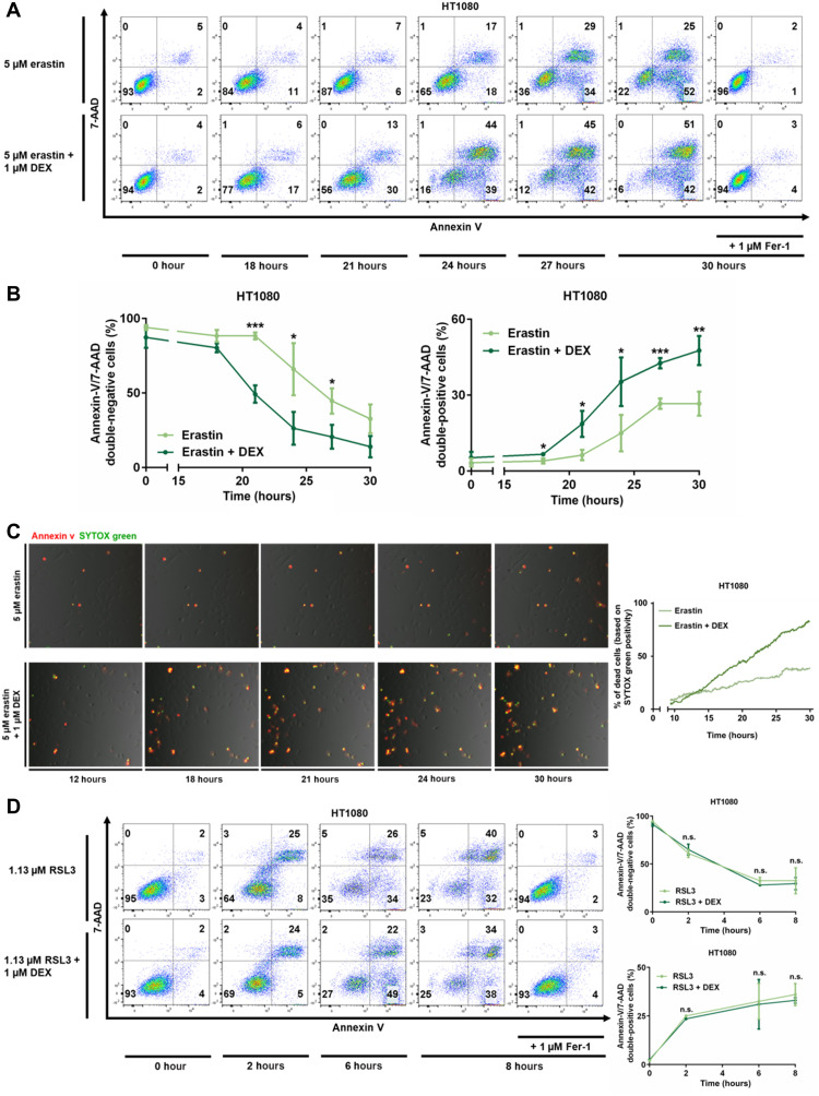
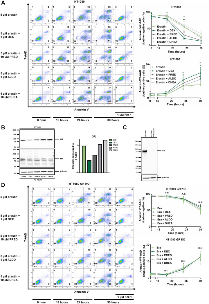
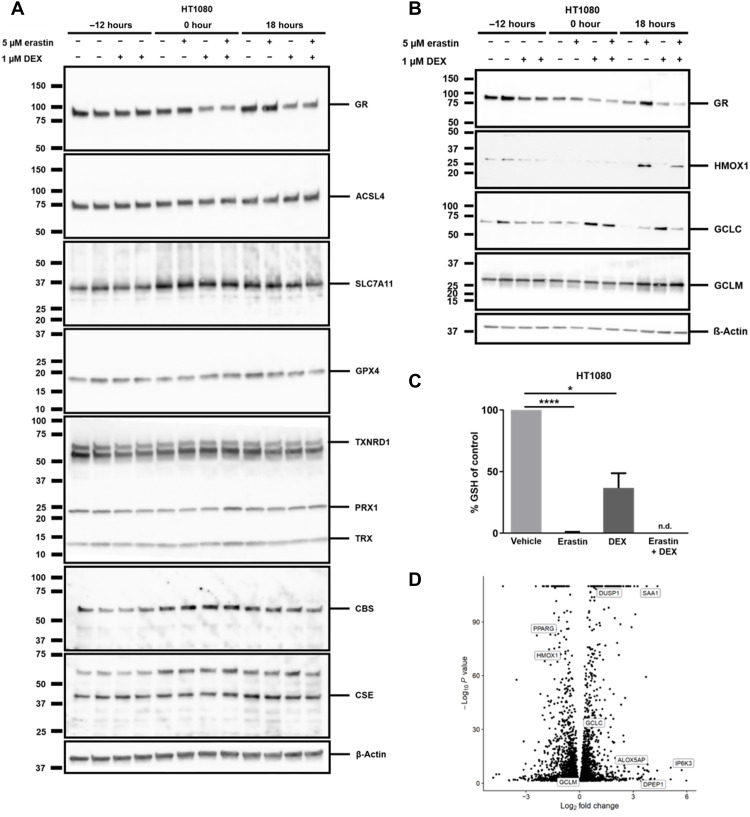
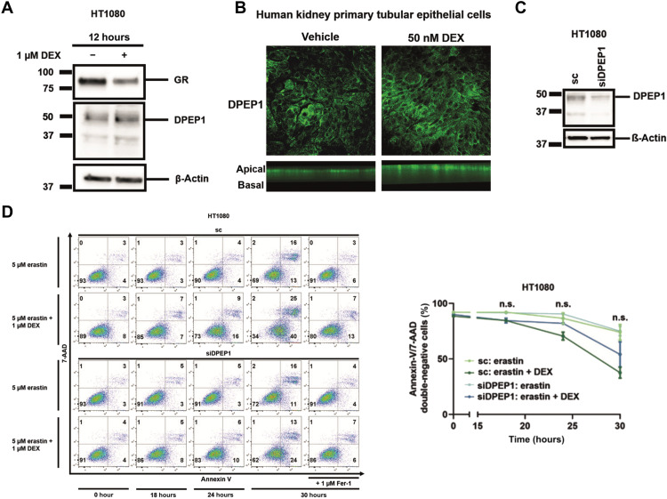
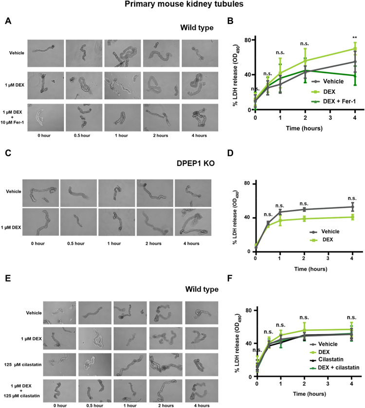
Dexamethasone is widely used as an immunosuppressive therapy and recently as COVID-19 treatment. Here, we demonstrate that dexamethasone sensitizes to ferroptosis, a form of iron-catalyzed necrosis, previously suggested to contribute to diseases such as acute kidney injury, myocardial infarction, and stroke, all of which are triggered by glutathione (GSH) depletion. GSH levels were significantly decreased by dexamethasone. Mechanistically, we identified that dexamethasone up-regulated the GSH metabolism regulating protein dipeptidase-1 (DPEP1) in a glucocorticoid receptor (GR)-dependent manner. DPEP1 knockdown reversed the phenotype of dexamethasone-induced ferroptosis sensitization. Ferroptosis inhibitors, the DPEP1 inhibitor cilastatin, or genetic DPEP1 inactivation reversed the dexamethasone-induced increase in tubular necrosis in freshly isolated renal tubules. Our data indicate that dexamethasone sensitizes to ferroptosis by a GR-mediated increase in DPEP1 expression and GSH depletion. Together, we identified a previously unknown mechanism of glucocorticoid-mediated sensitization to ferroptosis bearing clinical and therapeutic implications.


Figures

Fig. 1.
Fig. 1.. Dexamethasone sensitizes to erastin-induced, but not to RSL3-induced, ferroptosis.
(A) HT1080 cells were treated for indicated times with 5 μM erastin and 1 μM Fer-1 with or without pretreatment of 1 μM dexamethasone for 12 hours. 7-AAD and annexin V were read out by fluorescence-activated cell sorting (FACS). (B) Quantification of data presented in (A) and repetitions of this experiment. (C) Still images of indicated times of a double-chamber time-lapse imaging and quantification of SYTOX-positive cells over time in HT1080 cells pretreated with dexamethasone as indicated. (D) HT1080 cells were induced to undergo ferroptosis by the GSH peroxidase 4 (GPX4) inhibitor RSL3 and assessed as in (A) after indicated times. Experiments were repeated at least three times, and representative images and FACS plots are shown. The graphs show means ± SD. Statistical analysis was performed using Student’s t test for each time point. *P ≤ 0.05, **P ≤ 0.01, ***P ≤ 0.001, n.s., nonsignificant.
Fig. 2.
Fig. 2.. Steroid-induced sensitization to erastin-induced ferroptosis requires the GR 1.
(A) HT1080 cells were treated with erastin, Fer-1, 1 μM dexamethasone (DEX), 10 μM prednisolone (PRED), 1 μM aldosterone (ALDO), and 10 μM dehydroepiandrostendione (DHEA) as indicated. Primary FACS plots and respective quantifications of indicated populations are demonstrated. Note the sensitization to ferroptosis induced by DEX and PRED, but not by ALDO and DHEA. (B) Assessment of GR and mineralocorticoid receptor (MR) down-regulation in response to indicated steroid hormones. β-Actin serves as a loading control. (C) CRISPR-Cas9–mediated knockout of GR from HT1080 cells, confirmed by Western blotting. (D) Experiment performed as described for (A), but using GR-knockout cells. Note the loss of sensitization to ferroptosis. All experiments shown are representative of at least three independent complete repetitions performed. The graphs show means ± SD. Statistical analysis was performed using Student’s t test for each time point. *P ≤ 0.05, **P ≤ 0.01, n.s., nonsignificant.
Fig. 3.
Fig. 3.. An unbiased bulk RNA-seq of parental HT1080 and GR-crKO cells reveals dexamethasone-induced genes involved in ferroptosis.
(A) Protein expression of key players in ferroptosis 12 hours after treatment with or without 1 μM dexamethasone with subsequent treatment of induction of ferroptosis for 18 hours with erastin. β-Actin serves as a loading control. (B) Protein expression of HMOX1, GCLC, and GCLM 12 hours after treatment with or without 1 μM dexamethasone with subsequent treatment of induction of ferroptosis for 18 hours with erastin. β-Actin serves as a loading control. (C) GSH content in HT1080 cells treated with or without 1 μm dexamethasone before inducing ferroptosis with 5 μM erastin for 14 hours. (D) Volcano plot of differently regulated genes upon dexamethasone treatment in HT1080 cells. The bar graph shows means ± SD. Statistical analysis was performed using Student’s t test. *P ≤ 0.05, ****P ≤ 0.0001 (GR: glucocorticoid receptor, ACSL4: acyl-CoA synthetase long chain family member 4, SCL7A11: solute carrier family 7 member 11, GPX4: GSH peroxidase 4, TXNRD1: thioredoxin reductase 1, PRX1: peroxiredoxin 1, TRX: thioredoxin, CBS: cystathionine beta-synthase, CSE: cystathionine gamma lyase, HMOX1: heme oxygenase 1, GCLC: glutamate-cysteine ligase catalytic subunit, GCLM: glutamate-cysteine ligase modifier subunit, GSH: glutathione, n.d.: not detectable).
Fig. 4.
Fig. 4.. Dexamethasone-mediated sensitization to erastin-induced ferroptosis is mediated by DPEP1.
(A) HT1080 cells were treated for 12 hours with dexamethasone before investigating protein expression levels of the GR and DPEP1. (B) Human kidney primary tubular epithelial cells (hTEC) were grown on Transwells and treated for 24 hours with dexamethasone before fluorescently labeling DPEP1. (C) HT1080 cells were treated 48 hours with siRNA against DPEP1, and knockdown efficacy was confirmed by Western blot. (D) HT1080 cells were treated with erastin and dexamethasone as indicated following the 48-hour pretreatment with an siRNA against DPEP1. Primary FACS plots and respective quantifications of indicated populations are demonstrated. The graphs show means ± SD. Statistical analysis was performed using Student’s t test for each time point. sc, scrambled; n.s., nonsignificant.
Fig. 5.
Fig. 5.. Ferroptosis in freshly isolated renal tubules is accelerated by dexamethasone in wild-type but not in DPEP1 knockout mice.
(A) Representative images of freshly isolated murine kidney tubules undergoing spontaneous cell death in the presence of either vehicle or 1 μM dexamethasone in the presence or absence of 10 μM Fer-1. (B) LDH release of respective time points. (C) Representative images of freshly isolated murine kidney tubules from DPEP1 knockout mice undergoing spontaneous cell death in the presence of vehicle or 1 μM dexamethasone. (D) LDH release of respective time points. (E) Representative images of freshly isolated murine kidney tubules undergoing spontaneous cell death in the presence of either vehicle, 1 μM dexamethasone, 125 μm cilastatin, or dexamethasone and cilastatin. (F) LDH release of respective time points. The graphs show means ± SD. Statistical analysis was performed using Student’s t test for each time point. **P ≤ 0.01.

References

    1. Jackson R. K., Irving J. A., Veal G. J., Personalization of dexamethasone therapy in childhood acute lymphoblastic leukaemia. Br. J. Haematol. 173, 13–24 (2016). - PubMed
    1. Teachey D. T., Pui C. H., Comparative features and outcomes between paediatric T-cell and B-cell acute lymphoblastic leukaemia. Lancet Oncol. 20, e142–e154 (2019). - PMC - PubMed
    1. The RECOVERY Collaborative , Dexamethasone in hospitalized patients with Covid-19—Preliminary report. N. Eng. J. Med. covidwho-650223, (2020).
    1. Thomusch O., Wiesener M., Opgenoorth M., Pascher A., Woitas R. P., Witzke O., Jaenigen B., Rentsch M., Wolters H., Rath T., Cingöz T., Benck U., Banas B., Hugo C., Rabbit-ATG or basiliximab induction for rapid steroid withdrawal after renal transplantation (Harmony): An open-label, multicentre, randomised controlled trial. Lancet 388, 3006–3016 (2016). - PubMed
    1. Moshirfar M., Somani A. N., Motlagh M. N., Ronquillo Y. C., Management of cataract in the setting of uveitis: A review of the current literature. Curr. Opin. Ophthalmol. 31, 3–9 (2020). - PubMed

Publication types

MeSH terms