. 2022 Feb 1;38(5):110291.
doi: 10.1016/j.celrep.2021.110291.

Targetable HER3 functions driving tumorigenic signaling in HER2-amplified cancers

Affiliations

Targetable HER3 functions driving tumorigenic signaling in HER2-amplified cancers

Marcia R Campbell et al. Cell Rep. .

Erratum in

Abstract

Effective inactivation of the HER2-HER3 tumor driver has remained elusive because of the challenging attributes of the pseudokinase HER3. We report a structure-function study of constitutive HER2-HER3 signaling to identify opportunities for targeting. The allosteric activation of the HER2 kinase domain (KD) by the HER3 KD is required for tumorigenic signaling and can potentially be targeted by allosteric inhibitors. ATP binding within the catalytically inactive HER3 KD provides structural rigidity that is important for signaling, but this is mimicked, not opposed, by small molecule ATP analogs, reported here in a bosutinib-bound crystal structure. Mutational disruption of ATP binding and molecular dynamics simulation of the apo KD of HER3 identify a conformational coupling of the ATP pocket with a hydrophobic AP-2 pocket, analogous to EGFR, that is critical for tumorigenic signaling and feasible for targeting. The value of these potential target sites is confirmed in tumor growth assays using gene replacement techniques.

Keywords: AP-2 pocket; ERBB2; ERBB3; HER2; HER3; allosteric inhibitor; bosutinib; breast cancer.


Conflict of interest statement

Declaration of interests The authors declare no competing interests.

Figures
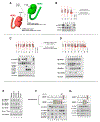
Figure 1.
Figure 1.. HER2 overexpression drives constitutive HER2-HER3 phosphorylation using the canonical allosteric activation mechanism and requires pseudoactive site occupancy in HER3
(A) Schematic depiction of the mutations drawn on cartoon representations of the kinase domains of HER2 and HER3. Amino acid (aa) numbering is based on the nascent reading frame. (B) CHO-K1 cells were cotransfected to overexpress HER2 and to express HER3, such as to generate constitutive HER2 autophosphorylation (lane 1) and HER3 transphosphorylation (lane 5). After 24 h, cell signaling was assayed as shown. Wild-type (WT) constructs were compared with mutants in the indicated combinations. The HER2 N-lobe mutant (I714Q) is a defective allosteric receiver, whereas the HER2 C-lobe mutant (V956R) is an impaired allosteric activator but competent receiver. HER3 is catalytically inactive and thus an obligate allosteric activator, and the HER3 C-lobe mutant (V945R) is an impaired allosteric activator. (C) CHO cells were transfected as in (B) using WT HER2 and HER3 in additional combinations. The HER2 C-lobe mutant (V956R) is an impaired allosteric activator. The HER3 single C-lobe mutant (V945R) is an impaired allosteric activator but retains some activation function. The HER3 triple C-lobe mutant (I938R-V945R-M949R) is an even more impaired allosteric activator. (D) CHO cells were transfected as in (B). The HER2 C-lobe activation function can similarly be further impaired by triple mutation. The double banding of HER3 observed in these CHO-K1 cell transfections is due to differential glycosylation (Figure S1B). (E) CHO cells were cotransfected to overexpress HER2 and express HER3 and were treated with DMSO or either of three small molecules that bind the HER3 KD with high affinity. These were administered at 1 μM for 1 h. The binding KDs for HER3 binding are as follows: bosutinib 0.8 nM, PP-242 120 nM, and dasatinib 18 nM (Davis et al., 2011). The affinities for HER2 KD are >1,400 nM for these drugs. The dephosphorylation of Src is shown as a positive control for bosutinib and dasatinib, which also inhibit Src. (F) CHO-K1 cells were transfected as in (A) using WT or mutant HER2 and HER3 constructs. The HER3 K742M mutant is defective at ATP binding. Lane 8 was treated with 1 μM lapatinib for 1 h. This image was cropped down from 13 lanes for clarity. (G) CHO-K1 cells were transfected as in (C) using WT or mutant HER2 and HER3 constructs.
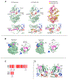
Figure 2.
Figure 2.. Bosutinib reinforces the Src/CDK-like conformation of the HER3 pseudokinase domain
(A) Cartoon representation of crystal structures of HER3/bosutinib (PDB: 6OP9; left) and HER3/AMP-PNP (PDB: 3KEX; middle) and their overlay (right). Carbon atoms of bosutinib are shown in magenta; those of AMP-PNP are shown in yellow. The HER3/bosutinib complex adopts a Src/CDK-like inactive conformation in which helix C (blue) is rotated away from the active site, the activation loop (red) adopts a tethered conformation, and the DFG-aspartate (D852) adopts a DFG-in position. The methoxy substituent of bosutinib’s quinoline ring extends in the same direction as the ribose sugar of AMP-PNP, but the 2,4-dichloro-5-methoxyaniline fragment lies 4–8 Å away from the triphosphate linkage. Weaker electron density corresponding to the N-propoxy-N-methylpiperazine moiety indicated that this region of bosutinib does not interact strongly with HER3. T787 is a gatekeeper residue, and K742 is the “catalytic” lysine. Numbering corresponds to the nascent reading frame (top) or mature protein without the signal peptide (low, in parentheses). (B) The AP-2 pockets in the inactive EGFR kinase (PDB: 3GT8) and the HER3 kinase (PDB: 3KEX) are displayed in magenta and magnified in the panels to the right of full kinase domains shown in surface representation. Hydrophobic residues within the pockets are shown in a sticks and dot representation. Numbering corresponds to the nascent reading frame (top) or mature protein without the signal peptide (low, in parentheses). (C) Conservation of the AP-2 pocket residues in HER receptors. Magenta ovals show HER3 numbering. (D) The spatial relationship between the AP-2 pocket and the nucleotide-binding pocket in the HER3 kinase domain (PDB: 3KEX). K742 interacts with the V786 via hydrogen bond interactions within the β3/β4 strand. Side chains of I741 make direct hydrophobic interactions with V786 and L709.
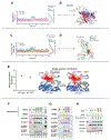
Figure 3.
Figure 3.. Conformation cross-talk between the surface AP-2 pocket of the HER3 KD and the pseudo-active site
(A) RMSD plot from MD simulations of the ATP-bound and apo HER3 pseudokinase states. (B) The superimposed cartoon images of the ATP-bound and apo HER3 final pseudokinase states showcase inward movement of the β3-β4 loop and the collapse of the P loop in the apo state. (C) RMSD plot from MD simulations of the ATP-bound WT, apo WT, and apo K742M HER3 pseudokinase states. (D) The superimposed cartoon images of the ATP-bound WT, apo WT, and apo K742M HER3 final pseudokinase states showcase inward movement of the β3-β4 loop and the collapse of the P loop in the apo state. (E) Left: plot of mutual information in kcal/mol between the transmitter (AP-2 pocket: residues 683, 685, 689, 690, 709, 718, 720, 756, and 767; side chains shown in green in right panel) and receiver (P loop: residues 696–704; side chains shown in yellow in right panel) sites, showing ~2 kcal/mol mutual information in both the absence (WT apo) and presence (WT ATP) of ATP. Error bars reflect standard deviation. Right: illustration of the normalized per-residue contribution to co-information in apo and ATP-bound simulations of HER3; “min” and “max” refer to the minimum and maximum values among all possible residues across all simulations. Residues colored dark red contribute most to communication between the AP-2 pocket and the P loop. (F) CHO-K1 cells were transfected to overexpress HER2 and to express the indicated WT or mutant HER3 constructs. The surface residues F704, L709, and V786 were mutated to impair the binding affinity of the AP-2 pocket. (G) W728 was mutated to collapse the AP-2 pocket. (H) The effect of ligand stimulation was studied on the F704 AP-2 pocket mutant.
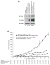
Figure 4.
Figure 4.. HER2-HER3 allosteric transactivation and HER3 AP-2 pocket functions are required for the growth of HER2-amplified tumors in vivo
(A) HCC1569 human HER2-amplified breast cancer cells were engineered to eliminate HER3 expression by CRISPR-Cas targeting (HCC1569-HER3KO). To eliminate the role of clonal growth characteristics in the replacement experiments, we mixed together three separate clones of HCC1569-HER3KO cells to generate a polyclonal HCC1569-HER3KO cell line (lane 2), and this cell line was used as the parental cell line for the various add-back experiments. These were then transduced to re-express WT HER3 (lane 4) or experimental mutant HER3 constructs. Experimental add-backs included a HER3 C-lobe mutant defective at allosteric activation (HER3 I938R/V945R/M949R) and a HER3 with a mutated AP-2 pocket (F704D). The expression of firefly luciferase (lane 3) constitutes a negative control cell type. The add-back HER3 constructs contain C-terminal myc tags. (B) The indicated engineered versions of HER2-amplified HCC1569 tumor cells were inoculated subcutaneously in NSG mice, and tumor volumes were measured over time. The number of surviving mice along the time course of the animal studies is shown for each arm underneath, and the sample size reduction over time in some arms reflects the removal of mice for euthanasia because of large tumor sizes as mandated by guidelines. The error bars reflect SEM.

References

    1. Adams PD, Afonine PV, Bunkoczi G, Chen VB, Davis IW, Echols N, Headd JJ, Hung LW, Kapral GJ, Grosse-Kunstleve RW, et al. (2010). PHENIX: a comprehensive Python-based system for macromolecular structure solution. Acta Crystallogr. D Biol. Crystallogr 66, 213–221. - PMC - PubMed
    1. Amin DN, Sergina N, Ahuja D, McMahon M, Blair JA, Wang D, Hann B, Koch KM, Shokat KM, and Moasser MM (2010). Resiliency and vulnerability in the HER2-HER3 tumorigenic driver. Sci. Transl. Med 2, 16ra17. - PMC - PubMed
    1. Barnes DJ, Palaiologou D, Panousopoulou E, Schultheis B, Yong AS, Wong A, Pattacini L, Goldman JM, and Melo JV (2005). Bcr-Abl expression levels determine the rate of development of resistance to imatinib mesylate in chronic myeloid leukemia. Cancer Res. 65, 8912–8919. - PubMed
    1. Bergdorf M, Baxter S, Rendleman CA, and Shaw DE (2015). Desmond/GPU Performance as of October 2015, D. E. Shaw Research Technical Report DESRES/TR-2015-01.
    1. Best RB, and Hummer G (2009). Optimized molecular dynamics force fields applied to the helix-coil transition of polypeptides. J. Phys. Chem 113, 9004–9015. - PMC - PubMed

Publication types