

Blood-brain barrier leakage in Alzheimer's disease: From discovery to clinical relevance
- PMID: 35108575
- PMCID: PMC9107516
- DOI: 10.1016/j.pharmthera.2022.108119
Blood-brain barrier leakage in Alzheimer's disease: From discovery to clinical relevance
Abstract
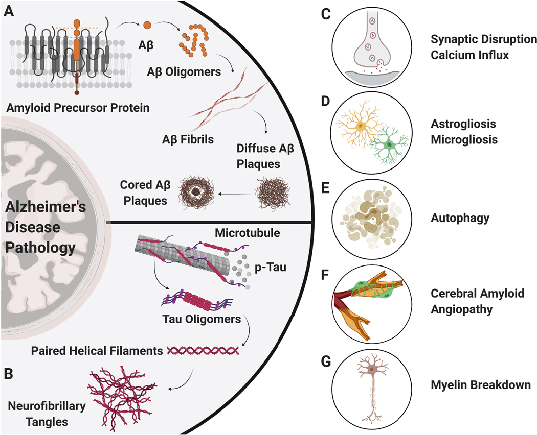
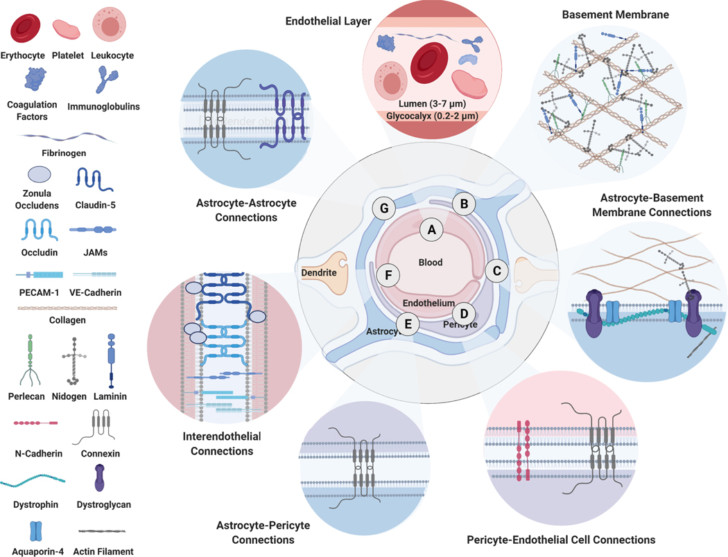
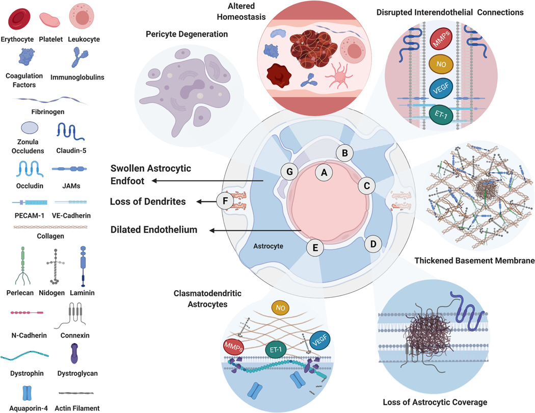
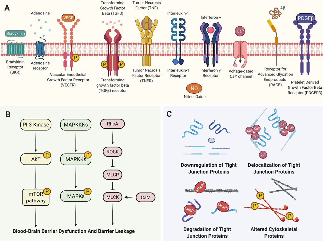
Alzheimer's disease (AD) is the most common form of dementia. AD brain pathology starts decades before the onset of clinical symptoms. One early pathological hallmark is blood-brain barrier dysfunction characterized by barrier leakage and associated with cognitive decline. In this review, we summarize the existing literature on the extent and clinical relevance of barrier leakage in AD. First, we focus on AD animal models and their susceptibility to barrier leakage based on age and genetic background. Second, we re-examine barrier dysfunction in clinical and postmortem studies, summarize changes that lead to barrier leakage in patients and highlight the clinical relevance of barrier leakage in AD. Third, we summarize signaling mechanisms that link barrier leakage to neurodegeneration and cognitive decline in AD. Finally, we discuss clinical relevance and potential therapeutic strategies and provide future perspectives on investigating barrier leakage in AD. Identifying mechanistic steps underlying barrier leakage has the potential to unravel new targets that can be used to develop novel therapeutic strategies to repair barrier leakage and slow cognitive decline in AD and AD-related dementias.
Keywords: Alzheimer’s disease; Barrier dysfunction; Barrier leakage; Blood-brain barrier; Cerebrovasculature; Neurovasculature.
Copyright © 2022 The Author(s). Published by Elsevier Inc. All rights reserved.
Conflict of interest statement
Declaration of Competing Interest The authors declare that there are no conflicts of interest.
Figures

Similar articles
-
Breakdown of the Cerebrovasculature and Blood-Brain Barrier: A Mechanistic Link Between Diabetes Mellitus and Alzheimer's Disease.J Alzheimers Dis. 2016 Sep 6;54(2):445-56. doi: 10.3233/JAD-160284. J Alzheimers Dis. 2016. PMID: 27497477 Review.
-
Inhibition of mTOR protects the blood-brain barrier in models of Alzheimer's disease and vascular cognitive impairment.Am J Physiol Heart Circ Physiol. 2018 Apr 1;314(4):H693-H703. doi: 10.1152/ajpheart.00570.2017. Epub 2017 Dec 22. Am J Physiol Heart Circ Physiol. 2018. PMID: 29351469 Free PMC article.
-
Blood-brain barrier disruption: a pervasive driver and mechanistic link between traumatic brain injury and Alzheimer's disease.Transl Neurodegener. 2025 Mar 26;14(1):16. doi: 10.1186/s40035-025-00478-5. Transl Neurodegener. 2025. PMID: 40140960 Free PMC article. Review.
-
Reversing pathology in a preclinical model of Alzheimer's disease by hacking cerebrovascular neoangiogenesis with advanced cancer therapeutics.EBioMedicine. 2021 Sep;71:103503. doi: 10.1016/j.ebiom.2021.103503. Epub 2021 Sep 15. EBioMedicine. 2021. PMID: 34534764 Free PMC article.
-
Linking Cerebrovascular Dysfunction to Age-Related Hearing Loss and Alzheimer's Disease-Are Systemic Approaches for Diagnosis and Therapy Required?Biomolecules. 2022 Nov 19;12(11):1717. doi: 10.3390/biom12111717. Biomolecules. 2022. PMID: 36421731 Free PMC article. Review.
Cited by
-
Blood-brain barrier permeability is associated with different neuroinflammatory profiles in Alzheimer's disease.Eur J Neurol. 2024 Jan;31(1):e16095. doi: 10.1111/ene.16095. Epub 2023 Oct 12. Eur J Neurol. 2024. PMID: 37823706 Free PMC article.
-
The effects of high plasma levels of Aβ1-42 on mononuclear macrophage in mouse models of Alzheimer's disease.Immun Ageing. 2023 Jul 31;20(1):39. doi: 10.1186/s12979-023-00366-4. Immun Ageing. 2023. PMID: 37525137 Free PMC article.
-
Recent Advancements in Nanomaterials: A Promising Way to Manage Neurodegenerative Disorders.Mol Diagn Ther. 2023 Jul;27(4):457-473. doi: 10.1007/s40291-023-00654-1. Epub 2023 May 22. Mol Diagn Ther. 2023. PMID: 37217723 Review.
-
Celebrating 40 years of the University of Kentucky Alzheimer's Disease Research Center.Alzheimers Dement. 2025 May;21(5):e70181. doi: 10.1002/alz.70181. Alzheimers Dement. 2025. PMID: 40365904 Free PMC article.
-
Acupuncture for neurodegenerative diseases: mechanisms, efficacy, and future research directions.Am J Transl Res. 2025 May 15;17(5):3703-3717. doi: 10.62347/QFJO6227. eCollection 2025. Am J Transl Res. 2025. PMID: 40535632 Free PMC article. Review.
References
-
- Abbott NJ (2013). Blood-brain barrier structure and function and the challenges for CNS drug delivery. J Inherit Metab Dis, 36, 437–449. - PubMed
-
- Adams RA, Passino M, Sachs BD, Nuriel T, & Akassoglou K (2004). Fibrin mechanisms and functions in nervous system pathology. Mol Interv, 4, 163–176. - PubMed
-
- Ahn KC, Learman CR, Dunbar GL, Maiti P, Jang WC, Cha HC, & Song MS (2018). Characterization of Impaired Cerebrovascular Structure in APP/PS1 Mouse Brains. Neuroscience, 385, 246–254. - PubMed
Publication types
MeSH terms
Grants and funding
LinkOut - more resources
Full Text Sources
Medical
