Review
. 2022 Mar;42(3):261-276.
doi: 10.1161/ATVBAHA.121.316930. Epub 2022 Feb 3.

Staphylococcus aureus and Neutrophil Extracellular Traps: The Master Manipulator Meets Its Match in Immunothrombosis

Affiliations
Review

Staphylococcus aureus and Neutrophil Extracellular Traps: The Master Manipulator Meets Its Match in Immunothrombosis

Severien Meyers et al. Arterioscler Thromb Vasc Biol. 2022 Mar.

Abstract

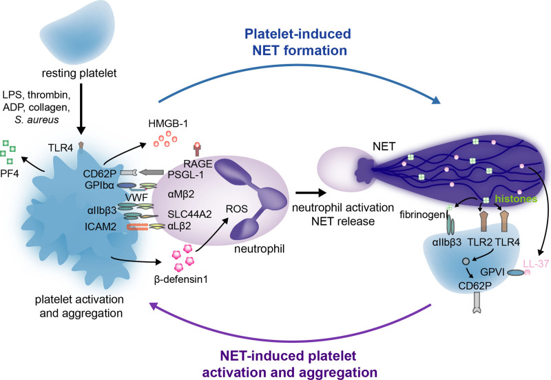
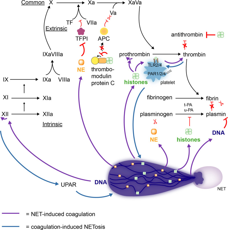
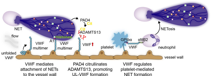
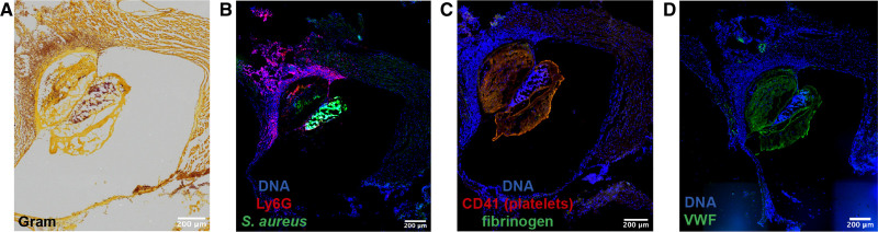
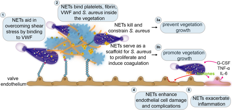
Over the past 10 years, neutrophil extracellular traps (NETs) have become widely accepted as an integral player in immunothrombosis, due to their complex interplay with both pathogens and components of the coagulation system. While the release of NETs is an attempt by neutrophils to trap pathogens and constrain infections, NETs can have bystander effects on the host by inducing uncontrolled thrombosis, inflammation, and tissue damage. From an evolutionary perspective, pathogens have adapted to bypass the host innate immune response. Staphylococcus aureus (S. aureus), in particular, proficiently overcomes NET formation using several virulence factors. Here we review mechanisms of NET formation and how these are intertwined with platelet activation, the release of endothelial von Willebrand factor, and the activation of the coagulation system. We discuss the unique ability of S. aureus to modulate NET formation and alter released NETs, which helps S. aureus to escape from the host's defense mechanisms. We then discuss how platelets and the coagulation system could play a role in NET formation in S. aureus-induced infective endocarditis, and we explain how targeting these complex cellular interactions could reveal novel therapies to treat this disease and other immunothrombotic disorders.

Keywords: Staphylococcus; endocarditis; extracellular trap; neutrophils; platelet activation; virulence factors.


Figures

Figure 1.
Figure 1.
The dynamic interplay between platelets and neutrophil extracellular traps (NETs). While some interactions between platelets and neutrophils trigger activated neutrophils to release NETs, others lead to platelet activation and aggregation. Via various receptors, secreted ligands, and specific components of the coagulation system, platelets can directly or indirectly induce NET formation (left). In return, specific components of NETs, more specifically histones and the cathelicidin LL-37, interact with several platelet receptors that result in platelet activation and aggregation (right). CD62P indicates P-selectin; GPIbα, glycoprotein Ibα; GPVI, glycoprotein VI; HMGB-1, high mobility box group 1; ICAM2, intercellular adhesion molecule 2; LPS, lipopolysaccharide; PF4, platelet factor 4; PSGL-1, P-selectin glycoprotein ligand 1; RAGE, receptor for advanced glycation end product; ROS, reactive oxygen species; SLC44A2, solute carrier family 44 member 2; TLR 2 or 4, toll-like receptor 2 or 4; and VWF, Von Willebrand factor.
Figure 2.
Figure 2.
Neutrophil extracellular traps (NETs) promote fibrin formation by interacting with the coagulation system. NETs stimulate the formation of fibrin by triggering both the extrinsic (TF [tissue factor]) as well as intrinsic (FXII) pathway of the coagulation system (green arrows). In addition, histones are able to prevent the cleavage of FVa by APC (activated protein C). By the autoactivation of prothrombin or the interaction with TLR2 or 4 (toll-like receptor 2 or 4) on platelets, histones directly trigger thrombin generation (green arrows). Besides directly stimulating fibrin formation, NETs prevent its degradation by impairing t-PA (tissue-type plasminogen activator), u-PA (urokinase-type plasminogen activator), antithrombin, and plasmin-mediated lysis (green arrows). Not only are NETs able to interact with various components of the coagulation cascade, but these components (FXII and thrombin) can also prime for NETosis (blue arrows). UPAR indicates urokinase-type plasminogen activator receptor.
Figure 3.
Figure 3.
VWF (Von Willebrand factor) and neutrophil extracellular traps (NETs) interact via various mechanisms. In response to shear stress of flowing blood, ultra-large VWF multimers elongate and expose the A1 domain. Via different mechanisms, NETs and neutrophils interact with these ultra-large VWF multimers. First, the A1 domain on VWF binds NET histones and DNA, thus allowing them to stay in place and damage the vasculature. Second, by releasing PAD4, NETs can promote ADAMTS13 (A disintegrin and metalloprotease with a thrombospondin type 1 motif, member 13) citrullination, a protease that cleaves VWF, and subsequently abolish its activity, promoting ultra-large VWF-platelet string formation. VWF, in turn, can regulate platelet-induced NET formation by acting on platelet GPIbα and neutrophil αMβ2.
Figure 4.
Figure 4.
Staphylococcus aureus both promotes and hampers neutrophil extracellular trap (NET) formation via various virulence factors to survive in the host environment. By interacting with platelet receptors, S. aureus activates and aggregates platelets and possibly promotes platelet-induced NET formation (A). Some S. aureus virulence factors can directly prime IL (interleukin)-6, TNFα (tumor necrosis factor α), reactive oxygen species (ROS), and damage-mediated NETosis (A). While enhancing NETosis could be a strategy of S. aureus to circumvent neutrophil-mediated killing, S. aureus is also equipped to diminish NET formation, destroy already formed NETs, or even to overcome their microbicidal properties (B). αIIbβ3 indicates glycoprotein GPIIb/IIIa; ClfA, clumping factor A; Eap, extracellular adherence protein; EpIP, epidermin leader peptide processing serine protease; FnBPA/B, fibronectin-binding protein A and B; GPIbα, glycoprotein Ib; IL-6/8, interleukin 6 or 8; IsdB, iron-regulated surface determinant B; MPO, myeloperoxidase; NE, neutrophil elastase; NOS, nitric oxide synthase; ROS, reactive oxygen species; SpA, S. aureus protein A; SPIN, staphylococcal peroxidase inhibitor; SSL5, Staphylococcal superantigen-like 5; TNFα, tumor necrosis factor α; VWbp, von Willebrand factor-binding protein; and VWF, Von Willebrand factor.
Figure 5.
Figure 5.
Immunostaining of a cardiac valve vegetation in a model of inflammation-induced IE. IE vegetation was induced upon infection of mice with S. aureus and treatment with local histamine infusion via catheter instillation. A, Brightfield image of a Gram stain with bacteria seen in purple. B–D, Fluorescence images of the cardiac valve vegetation showing: neutrophil-specific marker Ly6G (red) and S. aureus (green) (B); platelet CD41 (red) and fibrinogen (green) (C); VWF (Von Willebrand Factor) in green (D). DNA is identified by Hoechst 33342 staining, depicted in blue. Images were acquired using a Zeiss Axioscan Z1 digital slide scanner or Zeiss Axiovert inverted microscope at the VIB-KU Leuven LiMoNe Bio Imaging Core and the KU Leuven Department of Cardiovascular Sciences Microscopy Core, respectively.
Figure 6.
Figure 6.
The dual role of neutrophil extracellular traps (NETs) in infective endocarditis. Few diseases highlight the crucial interplay between coagulation, bacteria, and immunity better than infective endocarditis, which at its core is an infected blood clot attached to the cardiac valves. (1) Already at the very early stages of this disease, NETs can overcome the turbulent flow of the valves’ environment by binding to the endothelium via VWF (von Willebrand factor). While attached to the endothelium, NETs provide a scaffold for binding of platelets, fibrin, and bacteria. (2) In the already formed vegetation or infected blood clot, NETs also bind platelets, fibrin, and S. aureus. (3) Inside the vegetation, NETs could either prevent its progression by killing and constraining S. aureus (3a) or enhance its formation by further stimulating coagulation or proliferation of S. aureus (3b). The infected blood clot or vegetation typically develops on damaged or inflamed cardiac valves. NETs may also induce further damage (4) and inflammation (5) in the valve endothelium. However, the exact role of NETs in infective endocarditis remains to be established. G-CSF indicates granulocyte colony-stimulating factor; IL-6, interleukin 6; and TNFα, tumor necrosis factor α.

References

    1. Brinkmann V, Reichard U, Goosmann C, Fauler B, Uhlemann Y, Weiss DS, Weinrauch Y, Zychlinsky A. Neutrophil extracellular traps kill bacteria. Science. 2004;303:1532–1535. doi: 10.1126/science.1092385 - PubMed
    1. Tsan MF. Phorbol myristate acetate induced neutrophil autotoxicity. J Cell Physiol. 1980;105:327–334. doi: 10.1002/jcp.1041050215 - PubMed
    1. Tsan MF, Denison RC. Phorbol myristate acetate-induced neutrophil autotoxicity. A comparison with H2O2 toxicity. Inflammation. 1980;4:371–380. doi: 10.1007/BF00916048 - PubMed
    1. Wooldridge LC. XVI. The relation of the white blood corpuscles to the coagulation of the blood. Proc R Soc Lond. 1997;32:413–418.
    1. Al-Salih G, Al-Attar N, Delbosc S, Louedec L, Corvazier E, Loyau S, Michel JB, Pidard D, Duval X, Meilhac O. Role of vegetation-associated protease activity in valve destruction in human infective endocarditis. PLoS One. 2012;7:e45695. doi: 10.1371/journal.pone.0045695 - PMC - PubMed

Publication types

MeSH terms