. 2022 Jan;13(1):1880-1892.
doi: 10.1080/21655979.2021.2018981.

Melatonin alleviates lipopolysaccharide (LPS) / adenosine triphosphate (ATP)-induced pyroptosis in rat alveolar Type II cells (RLE-6TN) through nuclear factor erythroid 2-related factor 2 (Nrf2)-driven reactive oxygen species (ROS) downregulation

Affiliations

Melatonin alleviates lipopolysaccharide (LPS) / adenosine triphosphate (ATP)-induced pyroptosis in rat alveolar Type II cells (RLE-6TN) through nuclear factor erythroid 2-related factor 2 (Nrf2)-driven reactive oxygen species (ROS) downregulation

Tao Zhou et al. Bioengineered. 2022 Jan.

Abstract

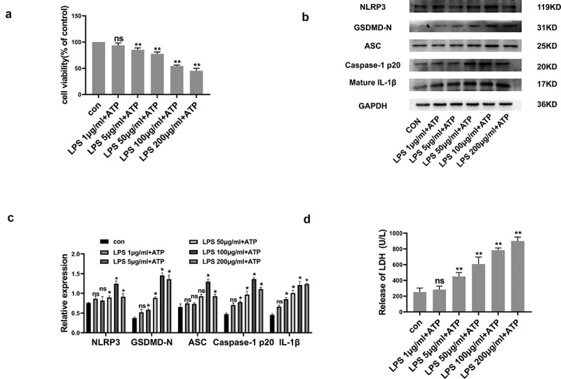
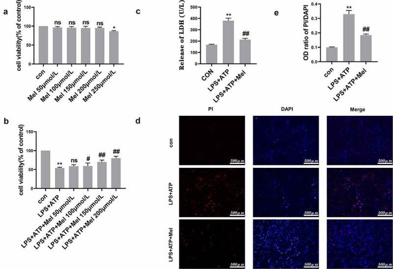
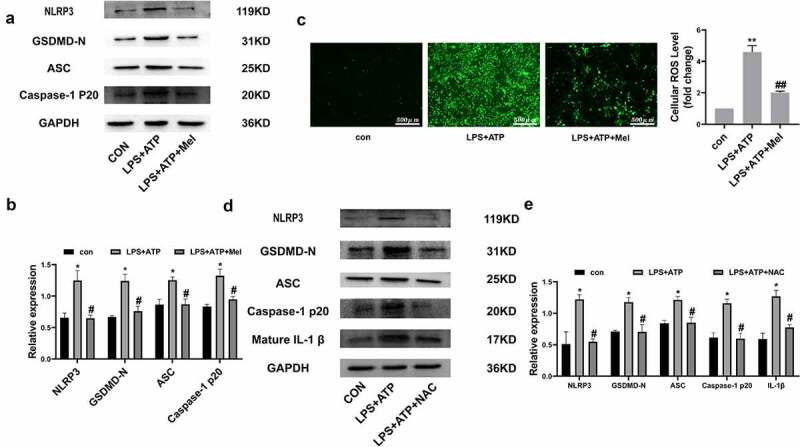
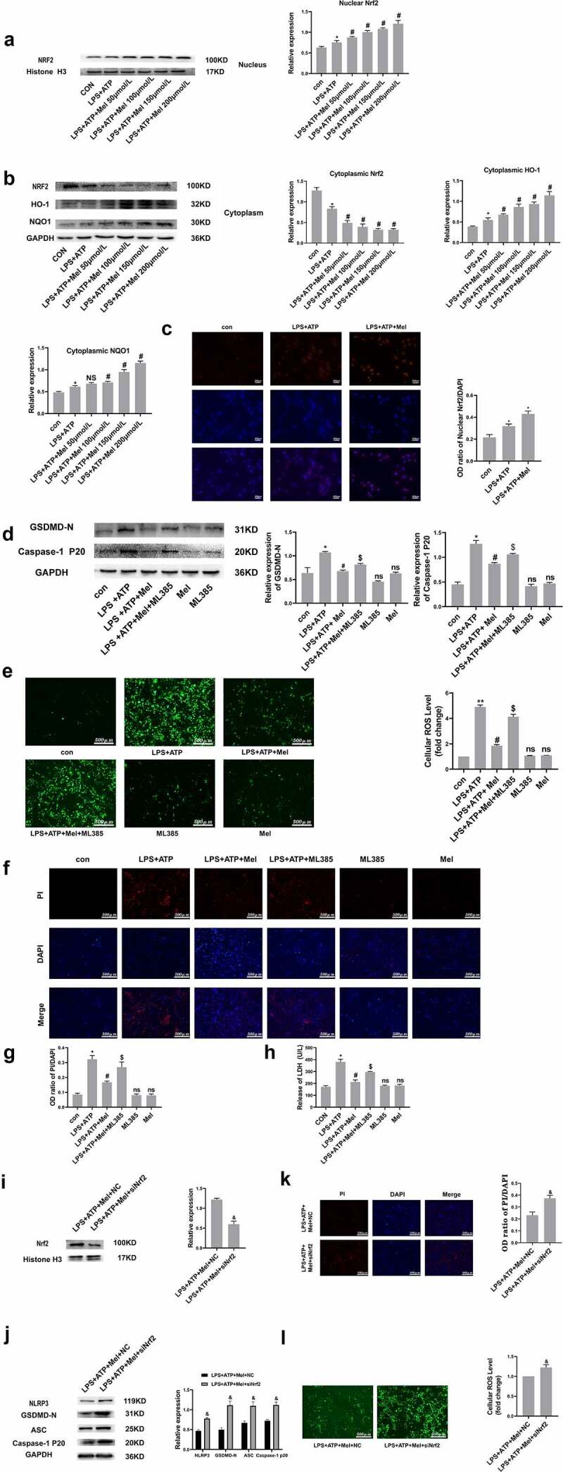
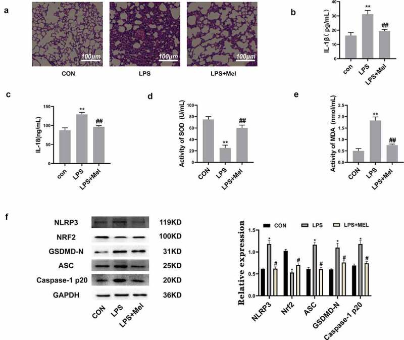
Pyroptosis has pivotal parts within disease development, rendering this attractive mechanism for novel therapeutics. This investigation aimed at analyzing melatonin roles within pyroptosis together with related mechanistics. RLE-6TN cultures were exposed to varying LPS doses for 4.5 h followed by concomitant culturing in the presence of ATP (5 mM) for 0.5 h to induce injury, and the roles of melatonin, N-Acety-L-cysteine (NAC - a ROS scavenger), ML385 (specific Nrf2 inhibitor) were examined. Apoptosis analysis was performed through lactate dehydrogenase (LDH) activity assays, together with propidium iodide (PI) stain-assay. Intracellular ROS were quantified through 2, 7-dichlorodihydrofluorescein diacetate (DCFH-DA). Pyrolysis-associated proteins, such as nucleotide-binding oligomerization domain-like receptor containing pyrin domain 3 (NLRP3), apoptosis-associated speck-like protein containing a CARD (ASC), cysteine aspartate-specific protease-1 P20 (Caspase-1 P20), gasdermin D-N (GSDMD-N), and mature interleukin-1β (IL-1β), were identified through Western blotting. Dataset outcomes demonstrated LPS/ATP induce RLE-6TN cell pyroptosis, while melatonin alleviated this phenomenon, visualized through increased cell survival rate, reduction of LDH discharge and PI+ cellular count. Moreover, melatonin effectively reduced NLRP3 inflammasome triggering in RLE-6TN cells. Meanwhile, this study demonstrated melatonin thwarting over NLRP3 inflammasome triggering was depending on ROS. In addition, this study found that melatonin activated Nrf2/Heme Oxygenase-1 (HO-1) pathway, with pyroptotic-inhibiting function of melatonin was reverted through a bespoke Nrf2-inhibitor and siNrf2. In summary, this study concluded that melatonin prevents RLE-6TN cellular pyroptosis through Nrf2-triggered ROS downregulation.

Keywords: Melatonin; NLRP3 inflammasome; Nrf2; RLE-6TN; ROS; pyroptosis.


Conflict of interest statement

No potential conflict of interest was reported by the author(s).

Figures

Figure 1.
Figure 1.
LPS/ATP reduced cell viability and triggered NLRP3-dependent pyroptosis within RLE-6TN cell cultures. (a) Cell viability measured by CCK-8 assay. LPS/ATP reduced cell viability. (b) Representative Western-blots of NLRP3, GSDMD-N, ASC, Caspase-1 P20 and mature IL-1β in RLE-6TN cells of differing treatment groups. (c) Protein quantitative histogram. (d) Pyroptosis assessed through LDH discharge levels. Datasets presented as mean ± SD (n = 3) for each group. * P < 0.05, ** P < 0.01, ns, P > 0.05 in comparison with controls.
Figure 2.
Figure 2.
Melatonin promoted cell viability and prevented RLE-6TN cells pyroptosis. (a) Cellular viability determined through CCK-8 testing. Melatonin doses ranging from 50 to 200 μmol/L were non-cytotoxic. (b) Melatonin inhibited LPS/ATP-driven pyroptosis with dose-dependent effect at a range of 50–200 μmol/L. (c) Melatonin (200 µmol/L) remarkably decreased LDH discharge in comparison to LPS/ATP group. (d) Fluorescence microscope imaging for DAPI (blue) and PI (red) (scale bar: 500 µm). (e) OD (optical density) ratio of PI/DAPI. Data represented as mean ± SD (n = 3) for each group. * P < 0.05, ** P < 0.01 in comparison to control group. # P < 0.05, ## P < 0.01, ns, P > 0.05 in comparison to LPS/ATP group.
Figure 3.
Figure 3.
Melatonin inhibits ROS-dependent NLRP3 inflammasome triggering. (a) Representative Western-blot images of NLRP3, GSDMD-N, ASC, and Caspase-1 P20 within differing RLE-6TN treatment groups. (b) Protein quantitative histogram. (c) Intracellular ROS (scale bar: 500 µm) and cellular ROS fold-change levels are shown on the right. (d) Representative Western-blot images of NLRP3, GSDMD-N, ASC, Caspase-1 P20, and mature IL-1β. (e) Protein quantitative histogram. Dataset represented as mean ± SD (n = 3) for each group. * P < 0.05, ** P < 0.01, in comparison to control group. # P < 0.05, ## P < 0.01 in comparison to LPS/ATP group.
Figure 4.
Figure 4.
Nrf2 activation is triggered through melatonin. (a and b) Corresponding immunoblots and the relative proteomic concentrations for nuclear Nrf2, cytoplasmic Nrf2, HO-1 and NQO1, and protein quantitative histogram shown on the right and below. (c) Immunofluorescence staining of nuclear Nrf2. (d) Representative immunoblots and relative protein levels of GSDMD-N and Caspase-1 P20, protein quantitative histogram shown on the right. (e) Determination of ROS production (scale bar: 500 µm). (f) Fluorescence microscopy images of DAPI (blue) and PI (red) (scale bar: 500 µm). (g) The OD ratio of PI/DAPI. (h) Determination of LDH release. (i) SiNrf2 downregulated proteomic expression of nuclear Nrf2. (j) SiNrf2 inhibited NLRP3 inflammasome activation. (k) Knockdown of Nrf2 increased the degree of PI-positive cells and (l) ROS production. Datasets represented as mean ± SD (n = 3) per group. * P < 0.05, ** P < 0.01, ns, P > 0.05, compared to the control group. # P < 0.05, NS, P > 0.05, compared to the LPS/ATP group. $ P < 0.05, in comparison to LPS+ATP+Mel group. & P < 0.05, compared to LPS+ATP+Mel+NC group.
Figure 5.
Figure 5.
Melatonin inhibits inflammation and oxidative stress and upregulated Nrf2 in vivo. (a) The HE staining showed melatonin alleviated leukocyte penetration and alveolar interstitial swellings (scale bar: 100 µm). (b) IL-1β levels. (c) IL-18 levels. (d) SOD levels. (e) MDA levels. (f) Corresponding immunoblots and the relative proteomic abundance for NLRP3, Nrf2, ASC, GSDMD-N, Caspase-1 P20, protein quantitative histogram shown on the right. Datasets represented as mean ± SD (n = 6) for each group. ** P < 0.01 in comparison to control group. # # P < 0.01, compared to LPS group.

Similar articles

Cited by

References

    1. Jorgensen I, Miao EA.. Pyroptotic cell death defends against intracellular pathogens. Immunol Rev. 2015;265:130–142. - PMC - PubMed
    1. Li D, Ren W, Jiang Z, et al. Regulation of the NLRP3 inflammasome and macrophage pyroptosis by the p38 MAPK signaling pathway in a mouse model of acute lung injury. Mol Med Rep. 2018;18:4399–4409. - PMC - PubMed
    1. Shao XF, Li B, Shen J, et al. Ghrelin alleviates traumatic brain injury-induced acute lung injury through pyroptosis/NF-κB pathway. Int Immunopharmacol. 2020;79:106175. - PubMed
    1. Lamkanfi M, Dixit VM.. Inflammasomes and their roles in health and disease. Annu Rev Cell Dev Biol. 2012;28:137–161. - PubMed
    1. Wu DD, Pan PH, Liu B, et al. Inhibition of Alveolar macrophage pyroptosis reduces lipopolysaccharide-induced acute lung injury in mice. Chin Med J (Engl). 2015;128:2638–2645. - PMC - PubMed