. 2022 Feb 3;13(1):2.
doi: 10.1186/s43008-022-00088-0.

Epichloë scottii sp. nov., a new endophyte isolated from Melica uniflora is the missing ancestor of Epichloë disjuncta

Affiliations

Epichloë scottii sp. nov., a new endophyte isolated from Melica uniflora is the missing ancestor of Epichloë disjuncta

Torsten Thünen et al. IMA Fungus. .

Abstract

Here we describe a new, haploid and stroma forming species within the genus Epichloë, as Epichloë scottii sp. nov. The fungus was isolated from Melica uniflora growing in Bad Harzburg, Germany. Phylogenetic reconstruction using a combined dataset of the tubB and tefA genes strongly support that E. scottii is a distinct species and the so far unknown ancestor species of the hybrid E. disjuncta. A distribution analysis showed a high infection rate in close vicinity of the initial sampling site and only two more spots with low infection rates. Genetic variations in key genes required for alkaloid production suggested that E. scottii sp. nov. might not be capable of producing any of the major alkaloids including ergot alkaloid, loline, indole-diterpene and peramine. All isolates and individuals found in the distribution analysis were identified as mating-type B explaining the lack of mature stromata during this study. We further release a telomere-to-telomere de novo assembly of all seven chromosomes and the mitogenome of E. scottii sp. nov.

Keywords: Alkaloid profile; BUSCO multigene-phylogeny; Epichloë; Melica uniflora; New species; Oxford nanopore; Telomere-to-telomere de novo genome assembly.


Conflict of interest statement

The authors declare that they have no competing interests.

Figures

Fig. 1
Fig. 1
Sample sites in the nature reserve”Butterberggelände “ in Bad Harzburg, Germany. Site A had a radius of 0.5 m; sites B to H had a radius of 5 m. Stromata bearing samples were discovered at sites A and E. For distribution analysis at each site, 10 specimens with inflorescences and no visible stromata were collected. Numbers in percent indicate the infection rate at the respective site. Scale bar is 500 m. Arrow indicates north. Map was created using blender (https://www.blender.org/) and BlenderGIS PlugIn (https://github.com/domlysz/BlenderGIS). Map data ©2020 Google
Fig. 2
Fig. 2
Stromata on Melica uniflora (indicated by arrows) and growth in culture media. a Partial choking on a stroma bearing specimen of M. uniflora collected at sample site A. b Daughter ramets with stromata of the specimen collected at sample site A grown in the greenhouse. c Stromata on M. uniflora at sample site E. d, e Close-up images of a stroma on a daughter ramet grown in the greenhouse. Scale bars: 3 mm. f, g Isolate growing on PDA, top view (f) and bottom view (g). Bars = 2 cm
Fig. 3
Fig. 3
Photograph and confocal laser scanning micrographs of Epichloë scottiiMelica uniflora association. a Photograph of E. scottii stroma bearing reproductive tiller of M. uniflora. Letters indicate the leaf parts, where samples were taken for CLS microscopy. b–h confocal laser scanning micrographs. Samples were treated with aniline blue diammonium salt to stain fungal and plant cell wall β-glucans (overlay of stain and autofluorescence of cytoplasm depicted in yellow, orange pseudo colors) and wheat germ agglutinin-Alexa Fluor 488 (WGA-AF488) to stain fungal chitin (blue pseudo color) and with propidium iodide (f, g, only) to stain plant and fungal nuclei (yellow pseudo color). (b) epibiotic hyphae of E. scottii on the surface of the stem 2 cm below the base of stroma, maximum projection of 150 µm z-stack. (c) transverse section of unfertilized stroma tissue (st), maximum projection of 35 µm z-stack. d Higher magnification of (c) showing densely colonized vascular bundle (vb) and hyper-proliferative hyphal growth on the cuticle. e epibiotic hyphae of E. scottii on the leaf sheath. f epibiotic hyphae at point of exit of endobiotic hypha (e = expressorium), maximum projection of 9.4 µm z-stack. Only after several hyphal compartments are formed, chitin can be visualized by staining with WGA-AF488, depicted in blue pseudo color. g endobiotic hyphae of E. scottii in leaf sheath epidermis, maximum projection of 144 µm z-stack. h endobiotic hyphae of E. scottii in leaf blade, the channel for blue pseudocolor is overexposed to visualize chitin in septa of endobiotic hyphae, maximum projection of 12 µm z-stack. Septa in F, G, and H are marked with asterisks (*) and nuclei in (f) and (g) with hashes (#). Bars = 20 µm (eh), 50 µm (b, d) 200 µm (c)
Fig. 4
Fig. 4
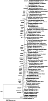
Bayesian inference of the phylogenetic relationship of the fungus described here among Epichloë isolates based on tubB and tefA sequences using SYM + I + G as the nucleotide substitution model. Numbers above nodes are estimates of a posteriori probability (BIpp, ≥ 0.9), and bootstrap values of maximum likelihood (ML) and neighbor-joining (NJ) (≥ 70%), respectively. The topology was rooted with the distantly related species Calviceps purpurea
Fig. 5
Fig. 5
Multi-gene phylogeny of haploid species of the genus Epichloë. A maximum likelihood phylogeny of all haploid species in the genus Epichloë with an available genome reference was built using 2,828 single copy protein orthologs and rooted on two species in the outgroup genus Claviceps. All branches have support values of 1
Fig. 6
Fig. 6
Micrographs of Epichloë scottii on potato dextrose agar. a, b Fungal growth and formation of coiling hyphae. c, d Hyphal anastomosis. e developing hyphae bearing conidiogenous cells and conidia. f Growing hyphae forming ring, conidiogenous cells arising from hyphae and conidia, g details of conidiogenous cells bearing conidia. h conidia. Bars = 10 µm
Fig. 7
Fig. 7
Karyogram of the seven chromosomes of Epichloë scottii. Individual size and GC content is given below each chromosome. Grayscale indicates the density of repeat elements from low (white) to high (back)
Fig. 8
Fig. 8
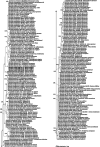
Bayesian inference of the phylogenetic relationship of tubB sequences of the taxa among Epichloë using K80 + I + G as nucleotide substitution model. Numbers above nodes are estimates of a posteriori probability (BIpp, ≥ 0.9). The strains studied here are in Bold. The tree was rooted as midpoint. Host species are provided after each strain

References

    1. Ashrafi S, Helaly S, Schroers H-J, Stadler M, Richert-Poeggeler KR, Dababat AA, Maier W. Ijuhya vitellina sp. Nov., a novel source for chaetoglobosin A, is a destructive parasite of the cereal cyst nematode Heterodera filipjevi. PLoS ONE. 2017;12(7):e0180032. doi: 10.1371/journal.pone.0180032. - DOI - PMC - PubMed
    1. Becker M, Becker Y, Green K, Scott B. The endophytic symbiont Epichloë festucae establishes an epiphyllous net on the surface of Lolium perenne leaves by development of an expressorium, an appressorium-like leaf exit structure. New Phytol. 2016;211(1):240–254. doi: 10.1111/nph.13931. - DOI - PMC - PubMed
    1. Becker Y, Green K, Scott B, Becker M. Artificial inoculation of Epichloë festucae into Lolium perenne, and visualisation of endophytic and epiphyllous fungal growth. Bio-Protoc. 2018 doi: 10.21769/BioProtoc.2990. - DOI - PMC - PubMed
    1. Berry D, Lee K, Winter D, Mace W, Becker Y, Nagabhyru P et al (2021) Cross-species transcriptomics comparing the benign asexual and antagonistic pre-sexual developmental stages of grass-endophytic Epichloë fungi. Under review
    1. Bultman TL, Leuchtmann A. A test of host specialization by insect vectors as a mechanism for reproductive isolation among entomophilous fungal species. Oikos. 2003;103(3):681–687. doi: 10.1034/j.1600-0706.2003.12631.x. - DOI

LinkOut - more resources