. 2022 Feb 8;119(6):e2112059119.
doi: 10.1073/pnas.2112059119.

Mice expressing fluorescent PAR2 reveal that endocytosis mediates colonic inflammation and pain

Affiliations

Mice expressing fluorescent PAR2 reveal that endocytosis mediates colonic inflammation and pain

Rocco Latorre et al. Proc Natl Acad Sci U S A. .

Erratum in

Abstract

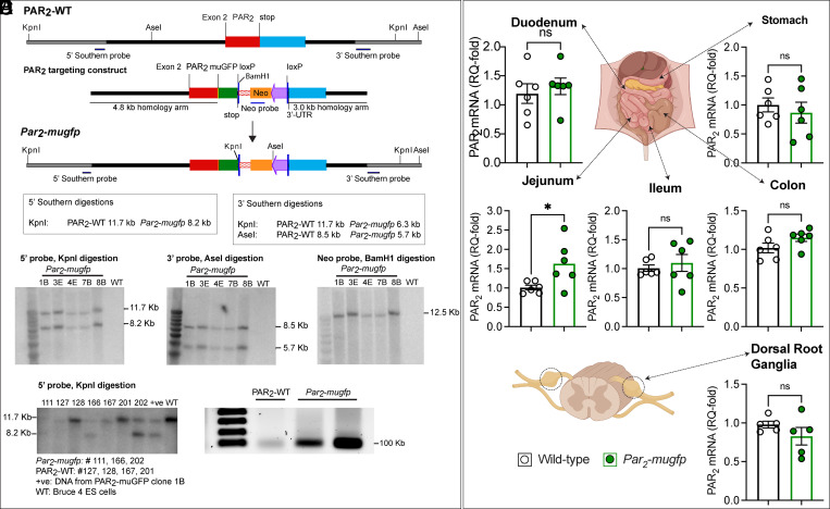
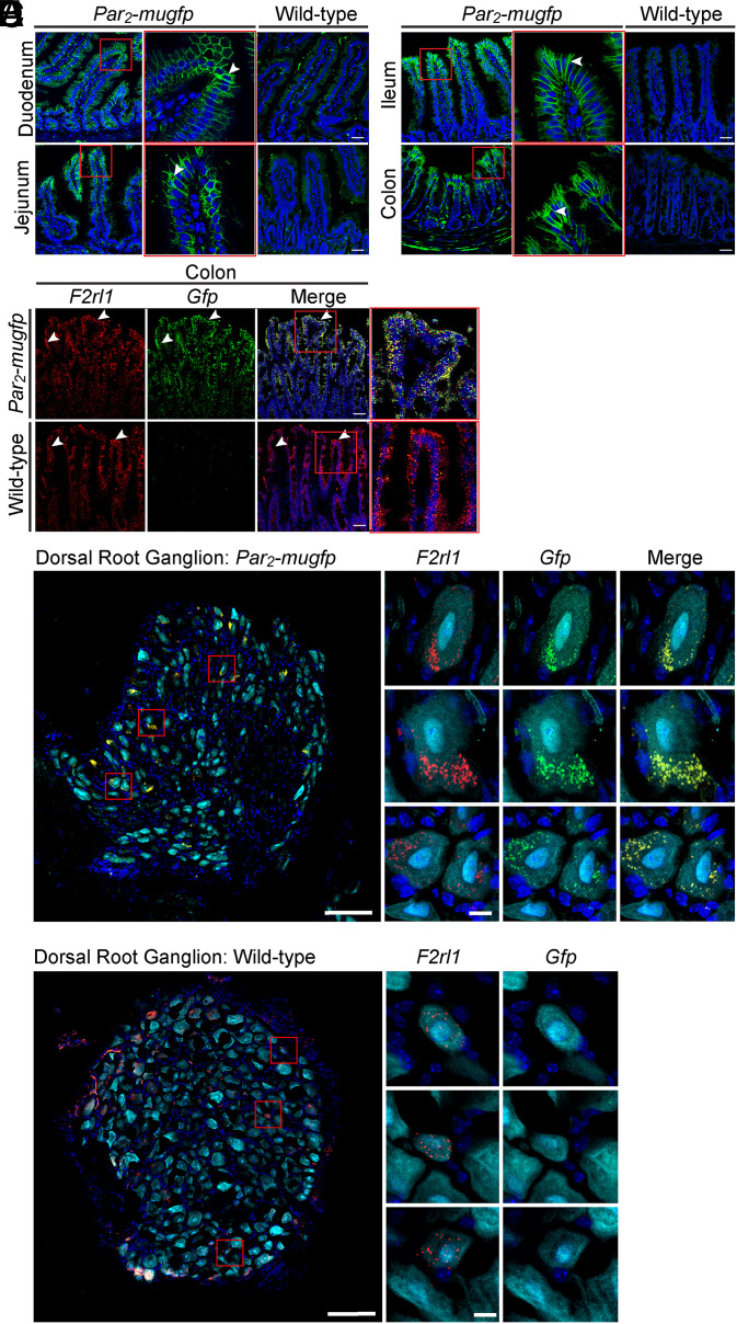
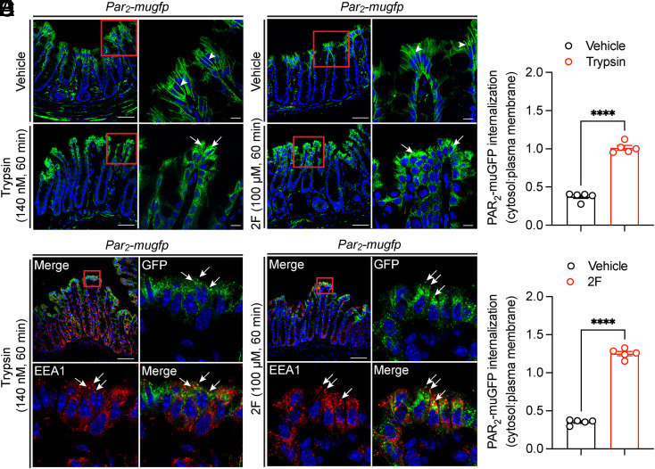
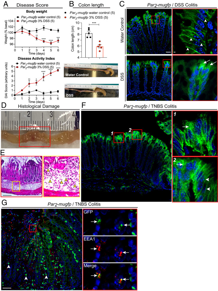
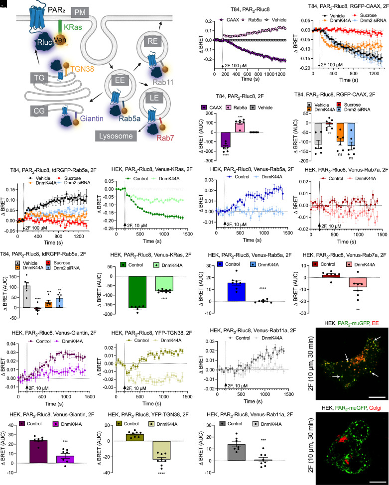
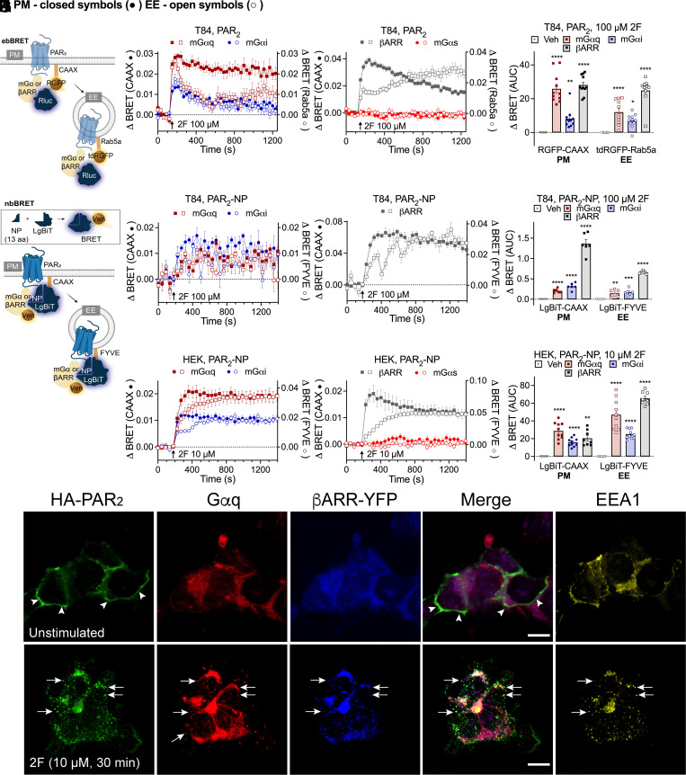
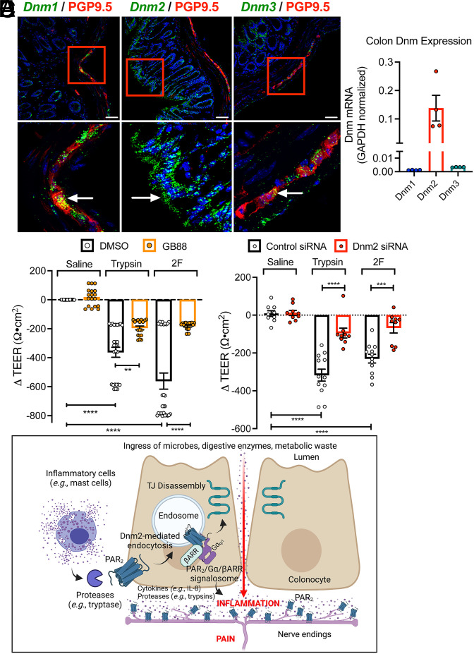
G protein-coupled receptors (GPCRs) regulate many pathophysiological processes and are major therapeutic targets. The impact of disease on the subcellular distribution and function of GPCRs is poorly understood. We investigated trafficking and signaling of protease-activated receptor 2 (PAR2) in colitis. To localize PAR2 and assess redistribution during disease, we generated knockin mice expressing PAR2 fused to monomeric ultrastable green fluorescent protein (muGFP). PAR2-muGFP signaled and trafficked normally. PAR2 messenger RNA was detected at similar levels in Par2-mugfp and wild-type mice. Immunostaining with a GFP antibody and RNAScope in situ hybridization using F2rl1 (PAR2) and Gfp probes revealed that PAR2-muGFP was expressed in epithelial cells of the small and large intestine and in subsets of enteric and dorsal root ganglia neurons. In healthy mice, PAR2-muGFP was prominently localized to the basolateral membrane of colonocytes. In mice with colitis, PAR2-muGFP was depleted from the plasma membrane of colonocytes and redistributed to early endosomes, consistent with generation of proinflammatory proteases that activate PAR2 PAR2 agonists stimulated endocytosis of PAR2 and recruitment of Gαq, Gαi, and β-arrestin to early endosomes of T84 colon carcinoma cells. PAR2 agonists increased paracellular permeability of colonic epithelial cells, induced colonic inflammation and hyperalgesia in mice, and stimulated proinflammatory cytokine release from segments of human colon. Knockdown of dynamin-2 (Dnm2), the major colonocyte isoform, and Dnm inhibition attenuated PAR2 endocytosis, signaling complex assembly and colonic inflammation and hyperalgesia. Thus, PAR2 endocytosis sustains protease-evoked inflammation and nociception and PAR2 in endosomes is a potential therapeutic target for colitis.

Keywords: endocytosis; inflammation; proteases; receptors; signaling.


Conflict of interest statement

Competing interest statement: N.W.B. is a founding scientist of Endosome Therapeutics, Inc. Research in the laboratories of N.W.B., N.A.V., and D.P.P. is funded in part by Takeda Pharmaceuticals International.

Figures

Fig. 1.
Fig. 1.
Generation and characterization of Par2-mugfp mice. (A) Par2-mugfp targeting construct comprising a downstream phosphoglycerine kinase neomycin cassette flanked by loxP sites and a downstream KpnI site. (B) Southern blot confirming correct targeting. (C) Southern blot and (D) RT-PCR blot confirming Par2-mugfp expression in mice. WT, wild-type. (E) Expression of F2rl1 (Par2) mRNA in the digestive tract and DRG of Par2-mugfp and wild-type mice determined by qRT-PCR. n = 6 mice. Mean ± SEM; ns, nonsignificant. *P < 0.05, Student’s t test.
Fig. 2.
Fig. 2.
Localization of PAR2-muGFP in the intestine and DRG by immunofluorescence and RNAScope in situ hybridization. (A and B) Localization of GFP immunoreactivity in duodenum and jejunum (A) and ileum and colon (B) of Par2-mugfp and wild-type mice. (C) RNAScope localization of F2rl1 and Gfp mRNA in colon of Par2-mugfp and wild-type mice. Arrowheads indicate immunoreactivity at the basolateral membrane of epithelial cells and mRNA expression within epithelial cells. (D and E) RNAScope localization of F2rl1 and Gfp mRNA in DRG of Par2-mugfp (D) and wild-type (E) mice. (Scale bars, 10 µm.) Representative images, independent experiments. n = 5 mice.
Fig. 3.
Fig. 3.
Agonist-evoked endocytosis of PAR2-muGFP in the colon. (A and B) Localization of GFP immunoreactivity in isolated segments of colon from Par2-mugfp mice incubated with vehicle, trypsin (A, 140 nM, 60 min) or 2F (B, 100 µM, 60 min). (C and D) Colocalization of GFP and EEA1 in segments treated with trypsin (C) or 2F (D). Arrowheads denote plasma membrane, and arrows denote endosomes. (Scale bar, 10 µm.) Representative images and independent experiments are shown (n = 5 mice). (E and F) Quantification of PAR2-muGFP internalization after vehicle, trypsin (E), or 2F (F) (cytosol–plasma membrane pixel intensity). Mean ± SEM; n = 5 mice. ****P < 0.0001, Student’s t test.
Fig. 4.
Fig. 4.
Colitis-evoked endocytosis of PAR2-muGFP. (AC) DSS colitis in Par2-mugfp mice. (A) Body weight and disease activity index (DAI) after DSS or water (control). (B) Colon length after DSS or water. Mean ± SEM, n = 5 mice. *P < 0.05, **P < 0.01, ***P < 0.001, and ****P < 0.0001 compared to water, two-way ANOVA, Tukey’s test (A), and Student’s t test (B). (C) Redistribution of PAR2-muGFP from the plasma membrane (arrowheads) to endosomes (arrows) after DSS. (DF) TNBS colitis in Par2-mugfp mice. (D) Grossly inflamed colon. (E) Histological sections showing infiltration of neutrophils in the submucosa (yellow arrowheads). (F) Redistribution of PAR2-muGFP from the plasma membrane to endosomes after TNBS. (G) Localization of PAR2-muGFP in infiltrating immune cells (arrowheads) and colocalization of GFP and EEA1 in colonocytes (arrows). (Scale bars, 10 µm.) Representative images and independent experiments are shown (n = 5 mice).
Fig. 5.
Fig. 5.
PAR2 trafficking to endosomes. (A) BRET assays of the proximity between PAR2-Rluc8 and Venus- or YFP-tagged proteins resident to plasma membrane (PM; CAAX or KRas), early endosomes (EE; Rab5a), late endosomes (LE; Rab7a), recycling endosomes (RE; Rab11a), cis-Golgi (CG; giantin), or trans-Golgi (TG; TGN38). (BG) Effects of 2F (100 μM) on translocation of PAR2-Rluc8 from the plasma membrane (RGFP-CAAX) to early endosomes (tdRGFP-Rab5a) of T84 cells treated with vehicle, hypertonic sucrose, or DnmK44A or Dnm2 siRNA. (HS) Effects of 2F (10 μM) on translocation of PAR2-Rluc8 from the plasma membrane [KRas-Venus (H and I)] to early endosomes [Rab5a-Venus (J and K)], late endosomes [Rab7a-Venus (L and M)], cis-Golgi [giantin-Venus (N and O)], trans-Golgi [TGN38-YFP (P and Q)], and recycling endosomes [Rab11-Venus (R and S)] in HEK293T cells. Cells were transfected with DnmK44A or untransfected (control). AUC, area under the curve. Mean ± SEM; n = 6–11 independent experiments. **P < 0.01, ***P < 0.001, and ****P < 0.0001, one-way ANOVA or Tukey’s test. (T and U) Localization of PAR2-muGFP and CellLight markers of early endosomes (T) and the Golgi complex (U) in HEK293T cells. Arrows denote colocalization of PAR2-muGFP and marker of early endosomes. Representative images of n = 5 experiments are shown. (Scale bars, 10 µm.)
Fig. 6.
Fig. 6.
PAR2, Gα and βARR signalosome assembly in endosomes. (A) EbBRET assays of proximity between effector (Gα; βARR) and proteins resident to plasma membrane (PM; CAAX) or early endosomes (EE; Rab5a). (BD) Effects of 2F (100 μM) on the recruitment of mGαsq, mGαsi, (B) βARR2 or mGαs negative control (C) to the plasma membrane (RGFP-CAAX, left axis, closed symbols) or early endosomes (tdRGFP-Rab5a, right axis, open symbols) of T84 cells, and (D) area under the curve (AUC). (E) NbBRET uses luciferase split into two fragments to detect BRET between receptor and effector (PAR2 and mGα or βARR) for proteins resident to the PM (CAAX) or early endosomes (FYVE). (FH) Effects of 2F (100 μM) on recruitment of mGαsq, mGαsi (F) or βARR1 (G) to the plasma membrane (LgBiT-CAAX) or early endosomes (LgBiT-FYVE) of T84 cells, and (H) AUC. (IK) Effects of 2F (10 μM) on recruitment of mGαsq, mGαsi (I), βARR1 or mGαs (J) to the PM (LgBiT-CAAX) or early endosomes (LgBiT-FYVE) of HEK293T cells. (K) AUC. Mean ± SEM. n = 6–11 independent experiments. *P < 0.05, **P < 0.01, ***P < 0.001, and ****P < 0.0001. (D and K) One-way ANOVA with Dunnett’s (D) or Holm–Šidák’s (K) test compared to vehicle. (H) Unpaired t test. (L) Localization of immunoreactive HA-PAR2, Gαq and EEA1 plus YFP-βARR1 and YFP-βARR2 in HEK293T cells. Cells were unstimulated or incubated with 2F (10 µM, 30 min). Arrowheads, plasma membrane. Arrows, colocalization of HA-PAR2, Gαq and YFP-βARR1 + 2 in EEA1-positive early endosomes. (Scale bars, 10 µm.) Representative images from n = 5 experiments are shown.
Fig. 7.
Fig. 7.
PAR2 endosomal signaling and colonic inflammation and pain. (A) Localization of Dnm1-3 in mouse colon by RNAScope in situ hybridization. (Scale bars, 50 µm.) Representative images, independent experiments, n = 3 mice. (B) Expression of Dnm1-3 in mouse colon by qRT-PCR. n = 4 mice. (C) Effects of trypsin (100 nM), 2F (10 µM), or vehicle (saline, control) on TEER of T84 cells after 6 h. Cells were preincubated with GB88 (PAR2 antagonist) or vehicle (DMSO, control). (D) Effects of trypsin (100 nM), 2F (10 µM), or vehicle (saline, control) on TEER of T84 cells after 5 h. Cells were preincubated with Dnm2 or control siRNA. Mean ± SEM. **P < 0.01, ***P < 0.001, and ****P < 0.0001 by one-way ANOVA, Tukey’s test, or Student’s t test. (E) Proposed mechanism by which proteases (e.g., tryptase) from mucosal inflammatory cells (e.g., mast cells) activate PAR2 at the basolateral membrane of colonocytes to evoke PAR2 endocytosis and assembly of a PAR2, Gα, and βARR signalosome in endosomes. Endosomal signaling causes disassembly of tight junctions (TJ) and release of proinflammatory cytokines (e.g., IL-8) and possibly proteases (e.g., trypsins) from colonocytes. The ingress of microbes, proteases, and metabolites from the colonic lumen, and the release of cytokines and proteases from colonocytes, cause inflammation and pain. Proteases activate PAR2 on nociceptive terminals in the colon to cause pain.
Fig. 8.
Fig. 8.
Contribution of Dnm2 to PAR2-evoked inflammation and pain in the colon. (AH) Dnm2 or control (Ctrl) shRNA was administered to Par2-mugfp mice by intracolonic injection. Mice were studied after 48 h. (A) Localization of Dnm2 by RNAScope. Representative images are shown. (Scale bars, 100 µm.) (B) Quantification of Dnm2 in the colon. (C) Localization of GFP immunoreactivity in the colon at 5 h after intracolonic injection of 2F. Arrows denote endosomes; arrowheads denote cell surface. (Scale bars, 10 µm.) (AC) n = 5 mice. (DF) Levels of TNF-α (D), CXCL1 (E), and IL-1β (F) in the colon 5 h after intracolonic injection of 2F. (G and H) Abdominal von Frey filament withdrawal responses 1–5 h after intracolonic injection of 2F. (G) Time course. (H) Area under the curve (AUC) from 0 to 5 h. (DH) n = 6 mice. (I and J) Abdominal von Frey filament withdrawal responses of wild-type, Par2-mugfp, and Par2Nav1.8 mice 1–6 h after intracolonic injection of 2F. (I) Time course. (J) Area under curve from 0 to 6 h. n = 5 mice. (K) Effects of 2F (10 µM) on IL-8 release from explants of human colonic mucosa. Explants were preincubated with dyngo4a or vehicle (DMSO, control). Mean ± SEM; n = 6. (B) **P < 0.01 by Student’s t test. (DJ) *P < 0.05, **P < 0.01, ***P < 0.001, and ****P < 0.0001 by one- and two-way ANOVA or Tukey’s test. (K) **P < 0.01 by Student’s t test.

References

    1. Canals M., Poole D. P., Veldhuis N. A., Schmidt B. L., Bunnett N. W., G-protein-coupled receptors are dynamic regulators of digestion and targets for digestive diseases. Gastroenterology 156, 1600–1616 (2019). - PMC - PubMed
    1. Peterson Y. K., Luttrell L. M., The diverse roles of arrestin scaffolds in G protein-coupled receptor signaling. Pharmacol. Rev. 69, 256–297 (2017). - PMC - PubMed
    1. DeFea K. A., et al. , Beta-arrestin-dependent endocytosis of proteinase-activated receptor 2 is required for intracellular targeting of activated ERK1/2. J. Cell Biol. 148, 1267–1281 (2000). - PMC - PubMed
    1. Irannejad R., et al. , Conformational biosensors reveal GPCR signalling from endosomes. Nature 495, 534–538 (2013). - PMC - PubMed
    1. Jensen D. D., et al. , Neurokinin 1 receptor signaling in endosomes mediates sustained nociception and is a viable therapeutic target for prolonged pain relief. Sci. Transl. Med. 9, eaal3447 (2017). - PMC - PubMed

Publication types