Clinical Trial
. 2022 Apr 14;28(8):1507-1517.
doi: 10.1158/1078-0432.CCR-21-3909.

Phase Ib Trial of the Combination of Imatinib and Binimetinib in Patients with Advanced Gastrointestinal Stromal Tumors

Affiliations
Clinical Trial

Phase Ib Trial of the Combination of Imatinib and Binimetinib in Patients with Advanced Gastrointestinal Stromal Tumors

Ping Chi et al. Clin Cancer Res. .

Abstract

Purpose: This phase Ib trial was designed to evaluate the safety and early efficacy signal of the combination of imatinib and binimetinib in patients with imatinib-resistant advanced gastrointestinal stromal tumors (GISTs).

Patients and methods: This trial used a standard 3 + 3 design to determine the recommended phase II dose (RP2D). Additional patients were enrolled on an expansion cohort at the RP2D enriching for succinate dehydrogenase (SDH)-deficient GISTs to explore potential efficacy.

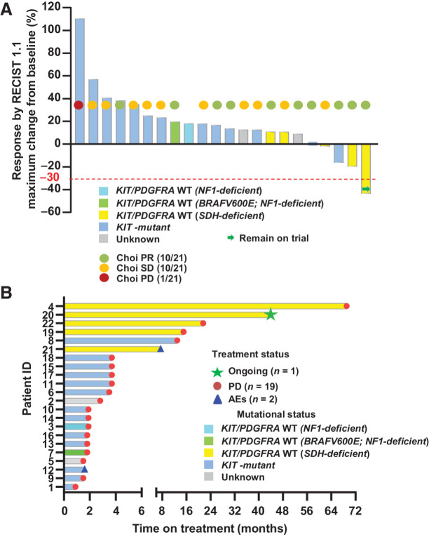
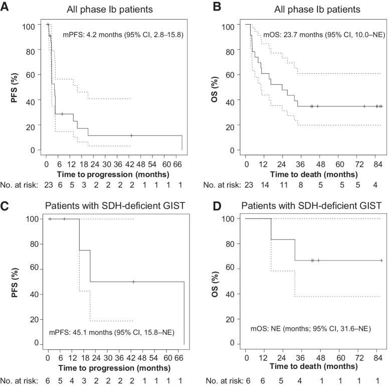
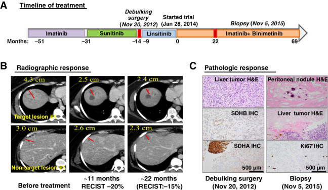
Results: The trial enrolled nine patients in the dose-escalation cohort and 14 in the dose-expansion cohort including six with SDH-deficient GISTs. Imatinib 400 mg daily with binimetinib 45 mg twice daily was established as the RP2D. Dose-limiting toxicity (DLT) was asymptomatic grade 4 creatinine phosphokinase (CPK) elevation. The most common non-DLT grade 3/4 toxicity was asymptomatic CPK elevation (69.6%). Other common ≥grade 2 toxicities included peripheral edema (17.4%), acneiform rash (21.7%), anemia (30.4%), hypophosphatemia (39.1%), and aspartate aminotransferase (AST) increase (17.4%). Two serious adverse events occurred (grade 2 dropped head syndrome and grade 3 central retinal vein occlusion). No unexpected toxicities were observed. Limited clinical activity was observed in KIT-mutant GIST. For SDH-deficient GISTs, one of five had confirmed RECIST1.1 partial response (PR). The median progression-free survival (mPFS) in patients with SDH-deficient GIST was 45.1 months [95% confidence interval (CI), 15.8-not estimable (NE)]; the median overall survival (mOS) was not reached (95% CI, 31.6 months-NE). One patient with a refractory metastatic SDH-deficient GIST had an exceptional pathologic response and durable clinical benefit.

Conclusions: The combination of imatinib and binimetinib is safe with manageable toxicity and has encouraging activity in SDH-deficient but not imatinib-refractory KIT/PDGFRA-mutant GISTs. The observed clinical benefits provide a motivation for a larger trial of the combination strategy in SDH-deficient GISTs.

Trial registration: ClinicalTrials.gov NCT01623349.


Figures

Figure 1. Response rates (RECIST1.1, Choi) and duration of treatment. A, Response rates by RECIST1.1 and Choi criteria. Best objective responses by RECIST1.1 (n = 22), Choi responses (n = 21) around 8 weeks (end of cycle 2, first posttreatment scan) on combination imatinib and binimetinib treatment. The best RECIST1.1 responses are shown as percentage of change from baseline for patients who received the combination of imatinib and binimetinib and with at least one postbaseline scan. The known associated primary driver mutations in KIT, SDH complex, NF1, and BRAF are shown. The best ORR for all phase Ib patients was 4.5% (1/22 confirmed PR), two-sided 95% CI, 0.1 to 22.8. The best ORR for patients with SDH-deficient GIST was 20.0% (1/5 confirmed PR), two-sided 95% CI, 0.5 to 71.6. Choi response rate was 47.6% (95% CI, 25.7–70.2) and 60.0% (95% CI, 14.7–94.7) for all phase Ib patients and patients with SDH-deficient GISTs, respectively. B, Duration of treatment. AE, adverse events; PD, progression of disease; PR, partial response; SD, stable disease.
Figure 1.
Response rates (RECIST1.1, Choi) and duration of treatment. A, Response rates by RECIST1.1 and Choi criteria. Best objective responses by RECIST1.1 (n = 22), Choi responses (n = 21) around 8 weeks (end of cycle 2, first posttreatment scan) on combination imatinib and binimetinib treatment. The best RECIST1.1 responses are shown as percentage of change from baseline for patients who received the combination of imatinib and binimetinib and with at least one postbaseline scan. The known associated primary driver mutations in KIT, SDH complex, NF1, and BRAF are shown. The best ORR for all phase Ib patients was 4.5% (1/22 confirmed PR), two-sided 95% CI, 0.1 to 22.8. The best ORR for patients with SDH-deficient GIST was 20.0% (1/5 confirmed PR), two-sided 95% CI, 0.5 to 71.6. Choi response rate was 47.6% (95% CI, 25.7–70.2) and 60.0% (95% CI, 14.7–94.7) for all phase Ib patients and patients with SDH-deficient GISTs, respectively. B, Duration of treatment. AE, adverse events; PD, progression of disease; PR, partial response; SD, stable disease.
Figure 2. Kaplan–Meier estimates of PFS and OS. PFS by RECIST1.1 (A) and OS (B) of all phase Ib patients. PFS (C) and OS (D) of patients with SDH-deficient GIST. mPFS is based on a Kaplan–Meier estimate of PFS, per investigator assessment. The mPFS was 4.2 months (95% CI, 2.8–15.8 months) and 45.1 months (95% CI, 15.8 months–NE) for all phase Ib patients and patients with SDH-deficient GISTs, respectively. The mOS was 23.7 months (95% CI, 10.0 months–NE), and not estimable (95% CI, 31.6 months–NE) for all phase Ib patients and patients with SDH-deficient GISTs, respectively.
Figure 2.
Kaplan–Meier estimates of PFS and OS. PFS by RECIST1.1 (A) and OS (B) of all phase Ib patients. PFS (C) and OS (D) of patients with SDH-deficient GIST. mPFS is based on a Kaplan–Meier estimate of PFS, per investigator assessment. The mPFS was 4.2 months (95% CI, 2.8–15.8 months) and 45.1 months (95% CI, 15.8 months–NE) for all phase Ib patients and patients with SDH-deficient GISTs, respectively. The mOS was 23.7 months (95% CI, 10.0 months–NE), and not estimable (95% CI, 31.6 months–NE) for all phase Ib patients and patients with SDH-deficient GISTs, respectively.
Figure 3. An example of durable treatment response in a patient with an SDH-deficient GIST. A, Treatment timeline and duration of various treatments the patient received for a metastatic SDH-deficient GIST. B, Representative CT images of the patient's metastatic liver lesions before, approximately 11 months and approximately 22 months after receiving the imatinib plus binimetinib combination therapy. One-dimension measurement in centimeters was provided for RECIST1.1 calculation at different time points. C, Treatment response by histopathologic studies. Representative images of histology and IHC stains for SDHB and SDHA, demonstrating dual SDH-deficiency, in the pretreatment tumor samples (debulking surgery; November 20, 2012) and the histology and the proliferation index marker, Ki67 IHC. The histology from the pretrial treatment liver lesions demonstrated more than 95% viable tumor and less than 5% treatment-associated necrosis. The histology from the on-treatment tumor samples (biopsy; November 5, 2015) showed 100% pathologic response with treatment-associated fibrosis, hyalinization, and dystrophic calcification in a resected metastatic peritoneal nodule and 70% pathologic response with treatment-associated necrosis in the metastatic liver lesion and less than 10% Ki67 IHC in the residual viable tumor. H&E, hematoxylin and eosin.
Figure 3.
An example of durable treatment response in a patient with an SDH-deficient GIST. A, Treatment timeline and duration of various treatments the patient received for a metastatic SDH-deficient GIST. B, Representative CT images of the patient's metastatic liver lesions before, approximately 11 months and approximately 22 months after receiving the imatinib plus binimetinib combination therapy. One-dimension measurement in centimeters was provided for RECIST1.1 calculation at different time points. C, Treatment response by histopathologic studies. Representative images of histology and IHC stains for SDHB and SDHA, demonstrating dual SDH-deficiency, in the pretreatment tumor samples (debulking surgery; November 20, 2012) and the histology and the proliferation index marker, Ki67 IHC. The histology from the pretrial treatment liver lesions demonstrated more than 95% viable tumor and less than 5% treatment-associated necrosis. The histology from the on-treatment tumor samples (biopsy; November 5, 2015) showed 100% pathologic response with treatment-associated fibrosis, hyalinization, and dystrophic calcification in a resected metastatic peritoneal nodule and 70% pathologic response with treatment-associated necrosis in the metastatic liver lesion and less than 10% Ki67 IHC in the residual viable tumor. H&E, hematoxylin and eosin.
Figure 4. Molecular analyses of archived GISTs and the SDH-deficient GIST patient's pre- (debulking surgery; November 20, 2012) and on-treatment (biopsy; November 5, 2015) tumor samples. A, Normalized RNA expression levels of KIT and ETV1 from U133A microarray analyses in MSKCC archived GIST samples harboring KIT (n = 29) or PDGFRA (n = 4) activating mutations, or KIT/PDGFRA WT and SDH-deficient GIST (n = 15). Each dot represents one tumor sample. Error bars: 95% CI. B, Relative RNA expression level of KIT and ETV1 compared with RPL27 (a housekeeping gene) by qRT-PCR in the patient's FFPE pretreatment tumor samples, the ETV1-expressing GIST48 human cell line, and the ETV1-negative tumor samples control. n = 3, technical replicates; error bars: mean ± S.D. Genetic alterations identified by MSK-IMPACT assays (C) and by WES (D) of the pretreatment and on-treatment FFPE tumor samples from the patient with SDH-deficient GIST shown in Fig. 4. The MSK-IMPACT assay included the mean and minimum coverage of the targeted exons and the allele frequency detected for each permutation (C). *, the mutations detected by MSK-IMPACT. SDHA nonsense mutation was not detected by WES due to lack of coverage (D). But KDR missense mutation was identified in both the WES and IMPACT.
Figure 4.
Molecular analyses of archived GISTs and the SDH-deficient GIST patient's pre- (debulking surgery; November 20, 2012) and on-treatment (biopsy; November 5, 2015) tumor samples. A, Normalized RNA expression levels of KIT and ETV1 from U133A microarray analyses in MSKCC archived GIST samples harboring KIT (n = 29) or PDGFRA (n = 4) activating mutations, or KIT/PDGFRA WT and SDH-deficient GIST (n = 15). Each dot represents one tumor sample. Error bars: 95% CI. B, Relative RNA expression level of KIT and ETV1 compared with RPL27 (a housekeeping gene) by qRT-PCR in the patient's FFPE pretreatment tumor samples, the ETV1-expressing GIST48 human cell line, and the ETV1-negative tumor samples control. n = 3, technical replicates; error bars: mean ± S.D. Genetic alterations identified by MSK-IMPACT assays (C) and by WES (D) of the pretreatment and on-treatment FFPE tumor samples from the patient with SDH-deficient GIST shown in Fig. 4. The MSK-IMPACT assay included the mean and minimum coverage of the targeted exons and the allele frequency detected for each permutation (C). *, the mutations detected by MSK-IMPACT. SDHA nonsense mutation was not detected by WES due to lack of coverage (D). But KDR missense mutation was identified in both the WES and IMPACT.

Comment in

  • Clin Cancer Res. 28:1475.

References

    1. Corless CL, Barnett CM, Heinrich MC. Gastrointestinal stromal tumours: origin and molecular oncology. Nat Rev Cancer 2011;11:865–78. - PubMed
    1. Heinrich MC, Corless CL, Duensing A, McGreevey L, Chen CJ, Joseph N, et al. . PDGFRA activating mutations in gastrointestinal stromal tumors. Science 2003;299:708–10. - PubMed
    1. Hirota S, Isozaki K, Moriyama Y, Hashimoto K, Nishida T, Ishiguro S, et al. . Gain-of-function mutations of c-kit in human gastrointestinal stromal tumors. Science 1998;279:577–80. - PubMed
    1. Boikos SA, Pappo AS, Killian JK, LaQuaglia MP, Weldon CB, George S, et al. . Molecular subtypes of KIT/PDGFRA wild-type gastrointestinal stromal tumors: a report from the national institutes of health gastrointestinal stromal tumor clinic. JAMA Oncol 2016;2:922–8. - PMC - PubMed
    1. Killian JK, Kim SY, Miettinen M, Smith C, Merino M, Tsokos M, et al. . Succinate dehydrogenase mutation underlies global epigenomic divergence in gastrointestinal stromal tumor. Cancer Discov 2013;3:648–57. - PMC - PubMed

Publication types

MeSH terms

Associated data