. 2022 Feb 2;13(2):98.
doi: 10.1038/s41419-022-04509-1.

LncRNA FIRRE functions as a tumor promoter by interaction with PTBP1 to stabilize BECN1 mRNA and facilitate autophagy

Affiliations

LncRNA FIRRE functions as a tumor promoter by interaction with PTBP1 to stabilize BECN1 mRNA and facilitate autophagy

Yajie Wang et al. Cell Death Dis. .

Abstract

Long non-coding RNAs (lncRNAs) play critical functions in various cancers. Firre intergenic repeating RNA element (FIRRE), a lncRNA located in the nucleus, was overexpressed in colorectal cancer (CRC). However, the detailed mechanism of FIRRE in CRC remains elusive. Results of RNA sequence and qPCR illustrated overexpression of FIRRE in CRC cell lines and tissues. The aberrant expression of FIRRE was correlated with the migration, invasion, and proliferation in cell lines. In accordance, it was also associated with lymphatic metastasis and distant metastasis in patients with CRC. FIRRE was identified to physically interact with Polypyrimidine tract-binding protein (PTBP1) by RNA pull-down and RNA immunoprecipitation (RIP). Overexpression of FIRRE induced the translocation of PTBP1 from nucleus to cytoplasm, which was displayed by immunofluorescence and western blot. In turn, delocalization of FIRRE from nucleus to cytoplasm is observed after the loss of PTBP1. The RNA-protein complex in the cytoplasm directly bound to BECN1 mRNA, and the binding site was at the 3' end of the mRNA. Cells with FIRRE and PTBP1 depletion alone or in combination were treated by Actinomycin D (ACD). Results of qPCR showed FIRRE stabilized BECN1 mRNA in a PTBP1-medieated manner. In addition, FIRRE contributed to autophagy activity. These findings indicate FIRRE acts as an oncogenic factor in CRC, which induces tumor development through stabilizing BECN1 mRNA and facilitating autophagy in a PTBP1-mediated manner.


Conflict of interest statement

The authors declare no competing interests.

Figures

Fig. 1
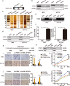
Fig. 1. The expression of FIRRE in colorectal cancer samples.
a HE stain was used to verify the cancer and normal tissues. b Volcano-plots was used to show the differential lncRNAs in samples. The x-coordinate means the fold change of expression difference between two samples. The ordinate shows the statistical value. The dot refers to one gene. Red dots represent genes that are significantly upregulated. Blue dots represent genes that are significantly downregulated. Black dots represent genes that are not significantly different. FIRRE is marked by green box. c The heatmap depicts part of the differential lncRNAs, and FIRRE is marked by red box. d According to the value of FDR, the top 10 upregulated genes in cancer tissues comparing with normal ones were listed. e Expression level of FIRRE was quantitated by qRT-PCR in colon carcinoma cell lines, including RKO, HCT29, HCT116, SW620, and SW480, and in normal intestinal epithelial cell line FHC. f qRT-PCR was conducted to test the expression of FIRRE in cancer tissues and normal tissues. Data are shown in means ± SD from triplicate assays. Bars, ±SD; Statistical analysis: ANOVA test, *P < 0.05, **P < 0.01.*p < 0.05, **p < 0.01, ***p < 0.0001.
Fig. 2
Fig. 2. FIRRE promotes CRC cells migration, invasion, and proliferation.
a qPCR analysis was used to test level of FIRRE in RKO and HCT116 cells transfected with control vector or LV-FIRRE. b The level of FIRRE in RKO and HCT116 cells with Control ASO or FIRRE ASO. c, d The migration assay to explore migration ability of cells with LV-FIRRE or FIRRR ASO. Photos were taken under inverse microscope, Scale bar = 100 µm. *p < 0.05, **p < 0.01. e, f The invasion assay to test the invasion ability of cells with LV-FIRRE or FIRRR ASO, Scale bar = 100 µm. g CCK-8 assays were performed to investigate proliferation of RKO cells with LV-FIRRE or FIRRE ASO as above. Data are presented as mean ± SD for four wells and are representative of three separate times. h CCK-8 assays were performed to investigate proliferation of HCT116 cells with LV-FIRRE or FIRRE ASO. Data are presented as mean ± SD for four wells and are representative of three separate times. i Colony formation assays were utilized to test the colony formation ability in RKO and HCT116 cells after FIRRE overexpression. Data are shown means ± SD in three repeated experiments. j Flow cytometry showed that the S-phase arrest was observed after FIRRE knockdown. Bars, ±SD; Statistical analysis: t-tests *p < 0.05, **p < 0.01, ***p < 0.0001.
Fig. 3
Fig. 3. FIRRE functions as an oncogenic factor in CRC patients.
a The level of FIRRE in CRC patients. p-value is indicated in the plot. b The data show that level of FIRRE was not related with T staging in CRC patients. p-value = 0.541. c The figure shows that higher FIRRE was associated with more advanced N staging. p-value = 0.033. d The figure shows that higher FIRRE was associated with more advanced M staging. p-value = 0.016. e The levels of FIRRE are not associated with the overall TNM staging. p-values are shown in the plot. f FIRRE makes no difference in the Overall Survival of patients with colorectal cancer. Bars, ±SD; Statistical analysis: ANOVA test, t-tests.
Fig. 4
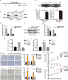
Fig. 4. FIRRE directly interacts with RNA-binding protein PTBP1.
a The antisense and sense of FIRRE were synthesized in vitro through RiboMAX™ Large Scale RNA Production Systems–SP6 and T7 (Promega). b RNA pull-down was conducted with the antisense or sense of FIRRE. Western blot with PTBP1 antibody shows the sense of FIRRE makes more PTBP1 enrichment compared with the antisense. c The silver stain further identified the binding between PTBP1 and FIRRE. PRBP1 was marked with the red arrows. d PTBP1 RIP assay to analyze interactions between PTBP1 and FIRRE in both RKO and HCT116 cells. WB shows the PTBP1 antibody efficiency of immunoprecipitation. e RT-PCR using RIP Primers specific for the FIRRE to validate the interaction between FIRRE and PTBP1. PCR product was observed in the anti-PTBP1 RIP (lane 3) and substantially less was detected in the normal IgG RIP (lane 2). f qRT-PCR to analyze the enrichment of FIRRE in RNA-protein complexes. The FIRRE abundance in anti-PTBP1 group was much more than the IgG group, normalized by the input group. g RRD was synthesized in vitro. RRD was presumed as the binding sites of FIRRE RNA to the PTBP1 protein. h RNA pull-down was conducted with the antisense or sense of RRD. Western blot with PTBP1 antibody shows the sense of RRD makes more PTBP1 enrichment compared with the antisense. i The migration assay to explore the migration ability of cells with FIRRE overexpression and cells with LV-FIRRE and siPTBP1. Photos were taken under inverse microscope, Scale bar = 100 µm. j The invasion assay to test the invasion ability of cells with FIRRE overexpression and cells with LV-FIRRE and siPTBP1, Scale bar = 100 µm. k CCK-8 assays were performed to investigate proliferation of cells with LV-FIRRE or LV-FIRRE and siPTBP1. All experiments were repeated double times or more. Bars, ±SD; Statistical analysis: ANOVA test, t-tests. **p < 0.01, ***p < 0.0001.
Fig. 5
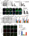
Fig. 5. FIRRE leads to the translocation of PTBP1.
a Immunofluorescence to test the subcellular localization of PTBP1 in cells transfected with FIRRE ASO or lentivirus FIRRE. The picture shows the blue DAPI fluorescence of nuclei and the red staining of anti-PTBP1 antibodies with Cy3-labeled secondary antibodies. b, c, and d Western blot assay of cytosol and nuclear proteins were used to verify the subcellular location of PTBP1 in RKO and HCT116 cells with differential level of FIRRE. GAPDH acted as the marker of cytoplasm, and LaminB1 as the marker of nuclear. b and c, RKO and HCT116 cells with transfection of control vector or lentivirus FIRRE. d RKO and HCT116 cells with Control ASO or FIRRE ASO. e FIRRE localization was determined by RNA-FISH. RKO cells with high expression of FIRRE were transfected with control siRNA (up) or PTBP1 siRNA (down). The nuclear was labeled by DAPI (bule) and the FIRRE RNA probe was stained by Cy3 (red).
Fig. 6
Fig. 6. The RNA-protein complex interacts with and stabilizes BECN1 mRNA.
a Fragment of BECN1 mRNA (named F1) was synthesized in vitro. F1 was presumed as the binding sites of BECN1 mRNA to the PTBP1 protein. b RNA pull-down with F1 RNA probe and control RNA probe was used to identify the interaction of BECN1 and PTBP1. Western blot with PTBP1 antibody shows F1 enriches more PTBP1 compared with the control. c RNA immunoprecipitation (RIP) of PTBP1 interaction with BECN1 RNA in vivo in RKO and HCT116 cells. Protein-RNA complexes captured by anti-PTBP1 or IgG were determined by RT-PCR using specific primers. PCR product was observed in the anti-PTBP1 RIP and substantially less was detected in the normal IgG. d qRT-PCR to analyze the enrichment of FIRRE in RNA-protein complexes. The FIRRE abundance in anti-PTBP1 group was much more than the IgG group, normalized by the input group. e FIRRE depletion induces the decrease of BECN1 mRNA level, which was demonstrated by qRT-PCR. f, g Western blot assay was further used to verify the influence of FIRRE silence on BECN1 mRNA stability in RKO and HCT116 cells. g Quantification of the western blot. h, i RKO(h) and HCT116(i) cells transfected with siPTBP1 or LV-FIRRE alone or in combination were treated with actinomycin D (5 μg/ml). RNA was extracted at 60 mins, and qPCR was used to quantify BECN1 mRNA. All experiments were repeated double times or more. *p < 0.01; **p < 0.001; ***p < 0.0001. j The migration and invasion assay to explore the migration ability of cells with FIRRE overexpression and cells with LV-FIRRE and siBECN1. Photos were taken under inverse microscope, Scale bar = 100 µm. k CCK-8 assays were performed to investigate proliferation of cells with LV-FIRRE or LV-FIRRE and siBECN1. All experiments were repeated double times or more. Bars, ±SD; Statistical analysis: ANOVA test, t-tests. **p < 0.01, ***p < 0.0001.
Fig. 7
Fig. 7. FIRRE activates autophagy.
a Western blots of important autophagy-associated proteins, including SQSMT1 and LC3, in RKO and HCT116 with FIRRE knock-down. Statistical analysis of the WB of SQSMT1 and LC3 is on the right. b The photomicrographs of RKO and HCT116 cells transfected with ASO-FIRRE and stained by indirect immunofluorescence for nuclear (DAPI, blue), and LC3 protein (labeled by Alexa 488, green). Statistical analysis of the average intensity is on the right. c Expression level of BECN1, SQSMT1, and LC3 were detected in cells transfected with siPTBP1 or ASO-FIRRE alone or in combination. d Cells with siPTBP1 or ASO-FIRRE alone or in combination were transfected with lentivirus mCherry-GFP-LC3 to observe the autophagy flux. Then, the presence of GFP-mCherry- (autophagosomes, yellow) and mCherry-positive (autolysosomes, red) dots were obsserved using confocal microscopy. Scale bar = 5 µm. Right panel: quantification of the ratio between red vs. yellow dots to assess an autophagy flux index. All experiments were repeated double times or more. Bars, ±SD; Statistical analysis: ANOVA test, t-tests. *p < 0.01; **p < 0.001; ***p < 0.0001.
Fig. 8
Fig. 8. The mechanism of FIRRE in CRC progress.
FIRRE which is localized in the nucleus binds to PTBP1 protein, promoting the translocation of PTBP1 from nucleus to cytoplasm. The RNA-protein complex in cytoplasm interacts with the 3' end of BECN1 mRNA. This interaction enhances the stability of BECN1 mRNA under treatment of ACD and the activity of autophagy, which contributes to the development of CRC.

Similar articles

Cited by

References

    1. Siegel RL, Miller KD, Goding Sauer A, Fedewa SA, Butterly LF, Anderson JC, et al. Colorectal cancer statistics, 2020. CA Cancer J Clin. 2020;70:145–64. doi: 10.3322/caac.21601. - DOI - PubMed
    1. Zheng RS, Sun KX, Zhang SW, Zeng HM, Zou XN, Chen R, et al. [Report of cancer epidemiology in China, 2015] Zhonghua Zhong Liu Za Zhi. 2019;41:19–28. - PubMed
    1. Han D, Wang M, Ma N, Xu Y, Jiang Y, Gao X. Long noncoding RNAs: novel players in colorectal cancer. Cancer Lett. 2015;361:13–21. doi: 10.1016/j.canlet.2015.03.002. - DOI - PubMed
    1. Schmitt AM, Chang HY. Long noncoding RNAs in cancer pathways. Cancer Cell. 2016;29:452–63. doi: 10.1016/j.ccell.2016.03.010. - DOI - PMC - PubMed
    1. Wang Y, Lu JH, Wu QN, Jin Y, Wang DS, Chen YX, et al. LncRNA LINRIS stabilizes IGF2BP2 and promotes the aerobic glycolysis in colorectal cancer. Mol Cancer. 2019;18:174. doi: 10.1186/s12943-019-1105-0. - DOI - PMC - PubMed

Publication types

MeSH terms