. 2022 Feb 2;13(2):102.
doi: 10.1038/s41419-022-04505-5.

Long non-coding RNA PAARH promotes hepatocellular carcinoma progression and angiogenesis via upregulating HOTTIP and activating HIF-1α/VEGF signaling

Affiliations

Long non-coding RNA PAARH promotes hepatocellular carcinoma progression and angiogenesis via upregulating HOTTIP and activating HIF-1α/VEGF signaling

Huamei Wei et al. Cell Death Dis. .

Abstract

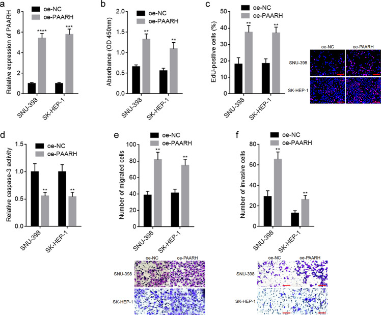
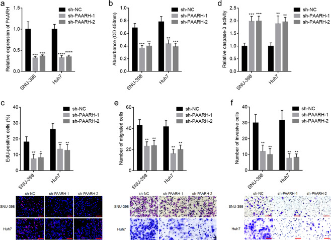
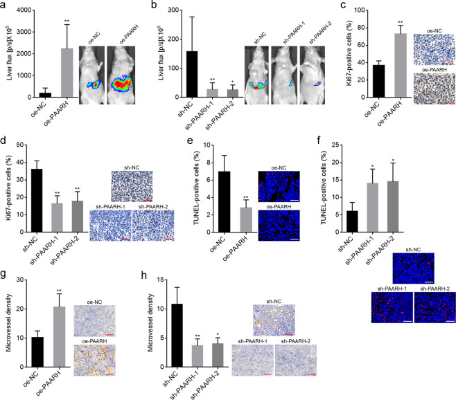
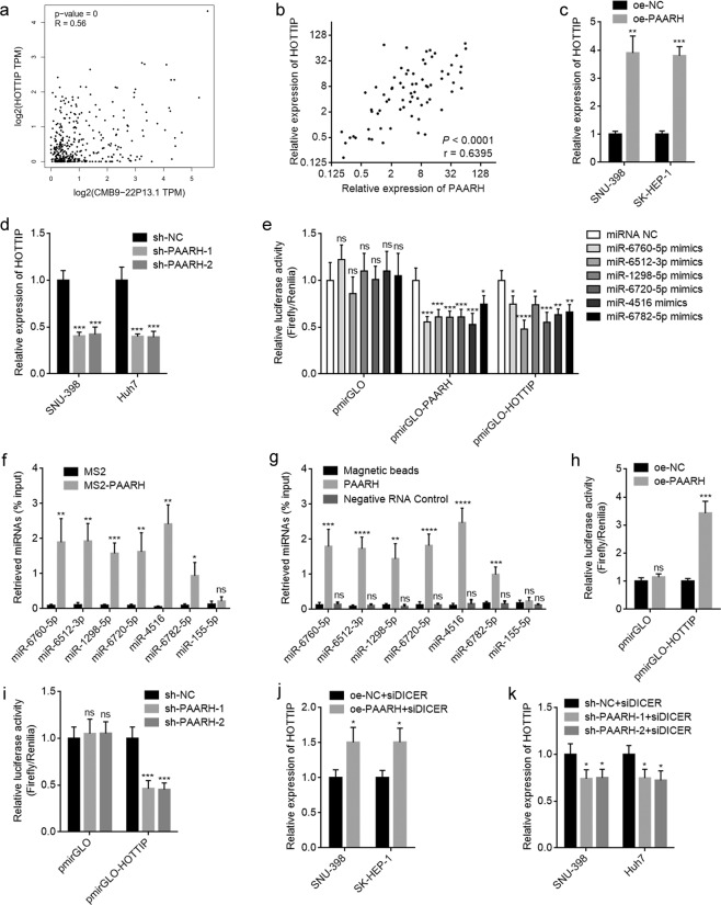
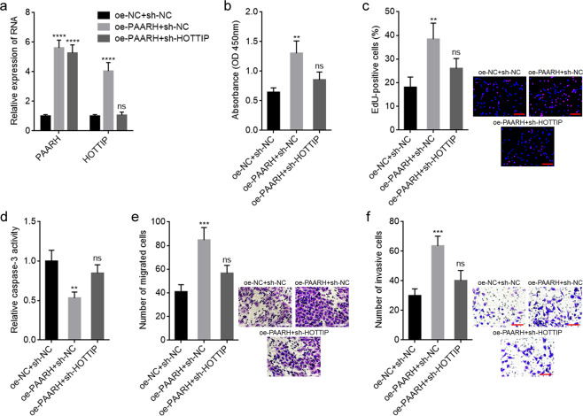
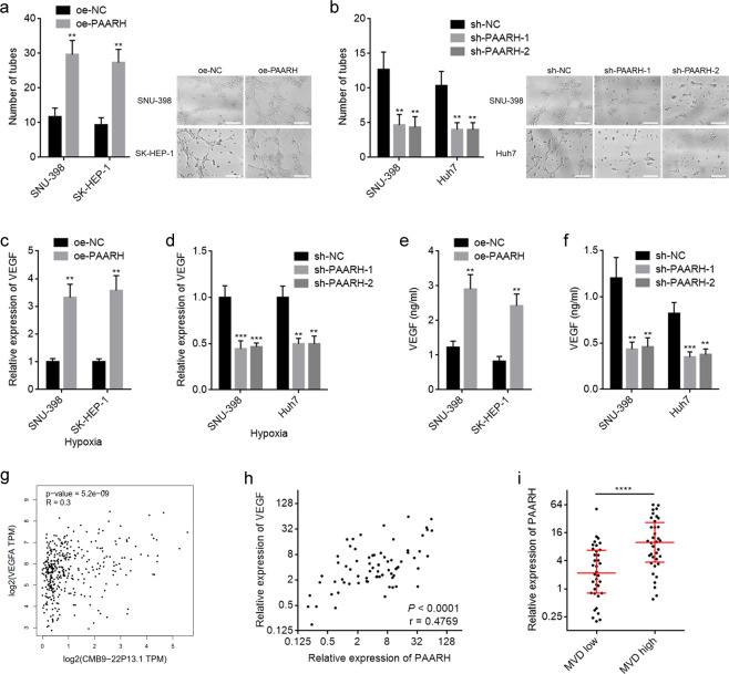
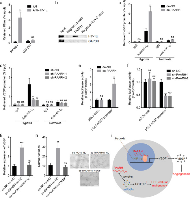
Hepatocellular carcinoma (HCC) is one of the leading lethal malignancies and a hypervascular tumor. Although some long non-coding RNAs (lncRNAs) have been revealed to be involved in HCC. The contributions of lncRNAs to HCC progression and angiogenesis are still largely unknown. In this study, we identified a HCC-related lncRNA, CMB9-22P13.1, which was highly expressed and correlated with advanced stage, vascular invasion, and poor survival in HCC. We named this lncRNA Progression and Angiogenesis Associated RNA in HCC (PAARH). Gain- and loss-of function assays revealed that PAARH facilitated HCC cellular growth, migration, and invasion, repressed HCC cellular apoptosis, and promoted HCC tumor growth and angiogenesis in vivo. PAARH functioned as a competing endogenous RNA to upregulate HOTTIP via sponging miR-6760-5p, miR-6512-3p, miR-1298-5p, miR-6720-5p, miR-4516, and miR-6782-5p. The expression of PAARH was significantly positively associated with HOTTIP in HCC tissues. Functional rescue assays verified that HOTTIP was a critical mediator of the roles of PAARH in modulating HCC cellular growth, apoptosis, migration, and invasion. Furthermore, PAARH was found to physically bind hypoxia inducible factor-1 subunit alpha (HIF-1α), facilitate the recruitment of HIF-1α to VEGF promoter, and activate VEGF expression under hypoxia, which was responsible for the roles of PAARH in promoting angiogenesis. The expression of PAARH was positively associated with VEGF expression and microvessel density in HCC tissues. In conclusion, these findings demonstrated that PAARH promoted HCC progression and angiogenesis via upregulating HOTTIP and activating HIF-1α/VEGF signaling. PAARH represents a potential prognostic biomarker and therapeutic target for HCC.


Conflict of interest statement

The authors declare no competing interests.

Figures

Fig. 1
Fig. 1. PAARH was highly expressed and correlated with poor survival in HCC.
a PAARH (CMB9-22P13.1) expression in 369 HCC tissues and 50 liver tissues according to TCGA LIHC dataset, analyzed by GEPIA. b The correlation between PAARH (CMB9-22P13.1) expression and overall survival according to TCGA LIHC dataset, analyzed by GEPIA. c The correlation between PAARH (CMB9-22P13.1) expression and disease-free survival according to TCGA LIHC dataset, analyzed by GEPIA. d PAARH expression in 72 pairs of HCC tissues and adjacent noncancerous liver tissues was measured by qRT-PCR. ****p < 0.0001 by Wilcoxon matched-pairs signed rank test. e Kaplan–Meier survival analysis of the correlation between PAARH expression and overall survival in our HCC cohort. n = 72, p = 0.0289, HR = 2.019 by log-rank test. f Kaplan–Meier survival analysis of the correlation between PAARH expression and disease-free survival in our HCC cohort. n = 72, p = 0.0264, HR = 1.883 by log-rank test.
Fig. 2
Fig. 2. Ectopic expression of PAARH enhanced HCC cellular malignant phenotype in vitro.
a PAARH expression in SNU-398 and SK-HEP-1 cells with PAARH stable overexpression was measured by qRT-PCR. b Cell growth of SNU-398 and SK-HEP-1 cells with PAARH overexpression was detected using CCK-8 assays. c Cell growth of SNU-398 and SK-HEP-1 cells with PAARH overexpression was detected using EdU incorporation assays. Scale bars, 100 µm. d Cell apoptosis of SNU-398 and SK-HEP-1 cells with PAARH overexpression was detected using caspase-3 activity assays. e Cell migration of SNU-398 and SK-HEP-1 cells with PAARH overexpression was detected using transwell migration assays. Scale bars, 100 µm. f Cell invasion of SNU-398 and SK-HEP-1 cells with PAARH overexpression was detected using transwell invasion assays. Scale bars, 100 µm. Results are shown as mean ± SD based on three independent experiments. **p < 0.01, ***p < 0.001, ****p < 0.0001 by Student’s t test.
Fig. 3
Fig. 3. PAARH silencing reduced HCC cellular malignant phenotype in vitro.
a PAARH expression in SNU-398 and Huh7 cells with PAARH stable silencing was measured by qRT-PCR. b Cell growth of SNU-398 and Huh7 cells with PAARH silencing was detected using CCK-8 assays. c Cell growth of SNU-398 and Huh7 cells with PAARH silencing was detected using EdU incorporation assays. Scale bars, 100 µm. d Cell apoptosis of SNU-398 and Huh7 cells with PAARH silencing was detected using caspase-3 activity assays. e Cell migration of SNU-398 and Huh7 cells with PAARH silencing was detected using transwell migration assays. Scale bars, 100 µm. f Cell invasion of SNU-398 and Huh7 cells with PAARH silencing was detected using transwell invasion assays. Scale bars, 100 µm. Results are shown as mean ± SD based on three independent experiments. *p < 0.05, **p < 0.01, ***p < 0.001, ****p < 0.0001 by one-way ANOVA followed by Dunnett’s multiple comparisons test.
Fig. 4
Fig. 4. PAARH facilitated HCC progression in vivo.
a Bioluminescence imaging of liver tumors in mice at day 14 after inoculation with small pieces of subcutaneous tumors formed by luciferase-labeled SNU-398 cells with PAARH stable overexpression or control. b Bioluminescence imaging of liver tumors in mice at day 14 after inoculation with small pieces of subcutaneous tumors formed by luciferase-labeled SNU-398 cells with PAARH stable silencing or control. c Ki67 IHC staining of liver tumors formed by SNU-398 cells with PAARH stable overexpression or control. Scale bars, 50 µm. d Ki67 IHC staining of liver tumors formed by SNU-398 cells with PAARH stable silencing or control. Scale bars, 50 µm. e TUNEL staining of liver tumors formed by SNU-398 cells with PAARH stable overexpression or control. Scale bars, 50 µm. f TUNEL staining of liver tumors formed by SNU-398 cells with PAARH stable silencing or control. Scale bars, 50 µm. g Microvessel density of liver tumors formed by SNU-398 cells with PAARH stable overexpression or control was detected using CD31 IHC staining. Scale bars, 100 µm. h Microvessel density of liver tumors formed by SNU-398 cells with PAARH stable silencing or control was detected using CD31 IHC staining. Scale bars, 100 µm. Results are shown as mean ± SD based on n = 6 mice in each group. *p < 0.05, **p < 0.01 by Mann–Whitney test (a, c, e, g) or Kruskal–Wallis test followed by Dunn’s multiple comparisons test (b, d, f, h).
Fig. 5
Fig. 5. PAARH increased HOTTIP expression via functioning as a ceRNA.
a The correlation between HOTTIP and PAARH (CMB9-22P13.1) expression in HCC tissues according to TCGA LIHC dataset, analyzed by GEPIA. b The correlation between HOTTIP and PAARH expression in our HCC cohort. n = 72, p < 0.0001, r = 0.6395 by Spearman correlation analysis. c HOTTIP expression levels in SNU-398 and SK-HEP-1 cells with PAARH overexpression or control were measured by qRT-PCR. d HOTTIP expression levels in SNU-398 and Huh7 cells with PAARH stable silencing or control was measured by qRT-PCR. e Luciferase activity in SNU-398 cells co-transfected with miR-6760-5p, miR-6512-3p, miR-1298-5p, miR-6720-5p, miR-4516, or miR-6782-5p and luciferase reporters containing nothing, PAARH or HOTTIP. Results are shown as the relative ratio of firefly luciferase activity to renilla luciferase activity. f MS2 based RIP assays followed by qRT-PCR to detect miRNAs bound to PAARH. g RNA pull-down experiments using RNA end-labeled with desthiobiotin, followed by qRT-PCR to detect miRNAs bound to PAARH. h Luciferase activity in SNU-398 cells with PAARH overexpression or control after transfection of luciferase reporters containing nothing or HOTTIP. Results are shown as the relative ratio of firefly luciferase activity to renilla luciferase activity. i Luciferase activity in SNU-398 cells with PAARH silencing or control after transfection of luciferase reporters containing nothing or HOTTIP. Results are shown as the relative ratio of firefly luciferase activity to renilla luciferase activity. j HOTTIP expression levels in SNU-398 and SK-HEP-1 cells with PAARH overexpression or control after transfection of DICER siRNA pool were measured by qRT-PCR. k HOTTIP expression levels in SNU-398 and Huh7 cells with PAARH stable silencing or control after transfection of DICER siRNA pool was measured by qRT-PCR. For ck, results are shown as mean ± SD based on three independent experiments. *p < 0.05, **p < 0.01, ***p < 0.001, ****p < 0.0001, ns not significant by Student’s t test (c, f, h, j) or one-way ANOVA followed by Dunnett’s multiple comparisons test (d, e, g, i, k).
Fig. 6
Fig. 6. Depletion of HOTTIP reversed the oncogenic roles of PAARH in HCC.
a PAARH and HOTTIP expression in SNU-398 cells with PAARH overexpression and concurrent HOTTIP depletion. b Cell growth of SNU-398 cells with PAARH overexpression and HOTTIP depletion was detected using CCK-8 assays. c Cell growth of SNU-398 cells with PAARH overexpression and HOTTIP depletion was detected using EdU incorporation assays. Scale bars, 100 µm. d Cell apoptosis of SNU-398 cells with PAARH overexpression and HOTTIP depletion was detected using caspase-3 activity assays. e Cell migration of SNU-398 cells with PAARH overexpression and HOTTIP depletion was detected using transwell migration assays. Scale bars, 100 µm. f Cell invasion of SNU-398 cells with PAARH overexpression and HOTTIP depletion was detected using transwell invasion assays. Scale bars, 100 µm. Results are shown as mean ± SD based on three independent experiments. **p < 0.01, ***p < 0.001, ****p < 0.0001, ns not significant, by one-way ANOVA followed by Dunnett’s multiple comparisons test.
Fig. 7
Fig. 7. PAARH promoted angiogenesis through upregulating VEGF.
a The number of endothelial tubes formed by HUVEC treated with conditioned medium from SNU-398 and SK-HEP-1 cells with PAARH overexpression or control under hypoxia for 24 h. Scale bars, 200 µm. b The number of endothelial tubes formed by HUVEC treated with conditioned medium from SNU-398 and Huh7 cells with PAARH silencing or control under hypoxia for 24 h. Scale bars, 200 µm. c VEGF expression in SNU-398 and SK-HEP-1 cells with PAARH overexpression or control under hypoxia for 12 h was measured by qRT-PCR. d VEGF expression in SNU-398 and Huh7 cells with PAARH silencing or control under hypoxia for 12 h was measured by qRT-PCR. e ELISA measurement of VEGF protein level in conditioned medium from SNU-398 and SK-HEP-1 cells with PAARH overexpression or control under hypoxia for 24 h. f ELISA measurement of VEGF protein level in conditioned medium from SNU-398 and Huh7 cells with PAARH silencing or control under hypoxia for 24 h. g The correlation between VEGF and PAARH (CMB9-22P13.1) expression in HCC tissues according to TCGA LIHC dataset, analyzed by GEPIA. h The correlation between VEGF and PAARH expression in our HCC cohort. n = 72, p < 0.0001, r = 0.4769 by Spearman correlation analysis. i PAARH expression levels in HCC tissues with high or low microvessel density (MVD), indicated by CD31 IHC staining. ****p < 0.0001 by Mann–Whitney test. For af, results are shown as mean ± SD based on three independent experiments. **p < 0.01, ***p < 0.001 by Student’s t test (a, c, e) or one-way ANOVA followed by Dunnett’s multiple comparisons test (b, d, f).
Fig. 8
Fig. 8. PAARH physically bound to HIF-1α, recruited HIF-1α to VEGF promoter, and activated VEGF transcription.
a RIP assays followed by qRT-PCR to detect RNAs bound to HIF-1α. b RNA pull-down experiments using RNA end-labeled with desthiobiotin, followed by western blot to detect proteins bound to PAARH. c SNU-398 cells with PAARH overexpression or control were incubated under hypoxia or normoxia for 24 h, followed by ChIP with anti-HIF-1α antibody or nonrelated IgG. Precipitated DNAs were measured by qRT-PCR for VEGF promoter. d SNU-398 cells with PAARH silencing or control were incubated under hypoxia or normoxia for 24 h, followed by ChIP with anti-HIF-1α antibody or nonrelated IgG. Precipitated DNAs were measured by qRT-PCR for VEGF promoter. e Luciferase activity in SNU-398 cells with PAARH overexpression or control after transfection of luciferase reporters containing VEGF promoter or nothing. Results are shown as the relative ratio of firefly luciferase activity to renilla luciferase activity. f Luciferase activity in SNU-398 cells with PAARH silencing or control after transfection of luciferase reporters containing VEGF promoter or nothing. Results are shown as the relative ratio of firefly luciferase activity to renilla luciferase activity. g VEGF expression in SNU-398 cells with PAARH overexpression and concurrent HIF-1α depletion under hypoxia for 12 h was measured by qRT-PCR. h The number of endothelial tubes formed by HUVEC treated with conditioned medium from SNU-398 cells with PAARH overexpression and concurrent VEGF depletion under hypoxia for 24 h. Scale bars, 200 µm. i Schematic model of the roles of PAARH in regulating HCC cellular malignancy and angiogenesis. Results are shown as mean ± SD based on three independent experiments. **p < 0.01, ***p < 0.001, ns not significant, by Student’s t test (a, c, e) or one-way ANOVA followed by Dunnett’s multiple comparisons test (d, fh).

Similar articles

Cited by

References

    1. Sung H, Ferlay J, Siegel RL, Laversanne M, Soerjomataram I, Jemal A, et al. Global Cancer Statistics 2020: GLOBOCAN Estimates of Incidence and Mortality Worldwide for 36 Cancers in 185 Countries. CA Cancer J Clin. 2021;71:209–49. - PubMed
    1. Villanueva A. Hepatocellular Carcinoma. N Engl J Med. 2019;380:1450–62. - PubMed
    1. Fountzilas C, Gupta M, Lee S, Krishnamurthi S, Estfan B, Wang K, et al. A multicentre phase 1b/2 study of tivozanib in patients with advanced inoperable hepatocellular carcinoma. Br J Cancer. 2020;122:963–70. - PMC - PubMed
    1. Briggs A, Daniele B, Dick K, Evans TRJ, Galle PR, Hubner RA, et al. Covariate-adjusted analysis of the Phase 3 REFLECT study of lenvatinib versus sorafenib in the treatment of unresectable hepatocellular carcinoma. Br J Cancer. 2020;122:1754–9. - PMC - PubMed
    1. Liu WR, Tian MX, Tang Z, Fang Y, Zhou YF, Song SS, et al. Nine-factor-based immunohistochemistry classifier predicts recurrence for early-stage hepatocellular carcinoma after curative resection. Br J Cancer. 2020;123:92–100. - PMC - PubMed

Publication types

MeSH terms

Substances