

Deep learning-based segmentation of the thorax in mouse micro-CT scans
- PMID: 35110676
- PMCID: PMC8810936
- DOI: 10.1038/s41598-022-05868-7
Deep learning-based segmentation of the thorax in mouse micro-CT scans
Abstract
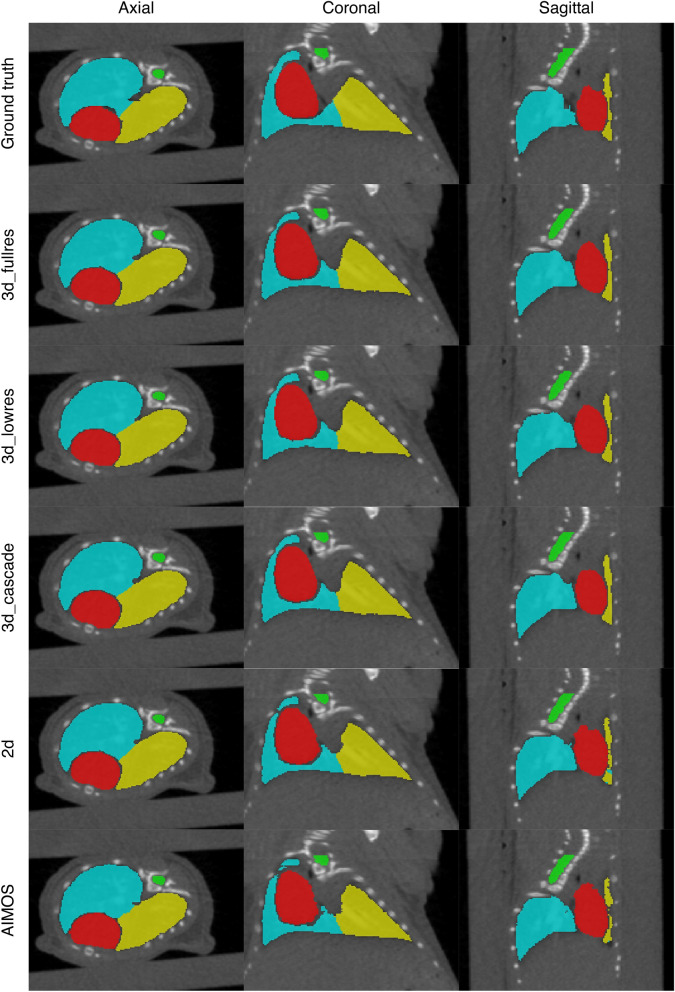
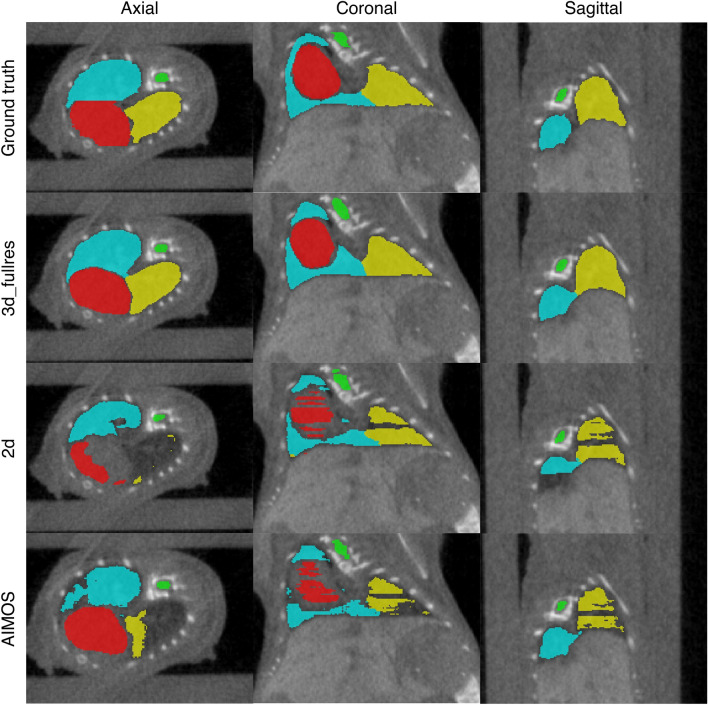
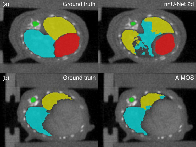
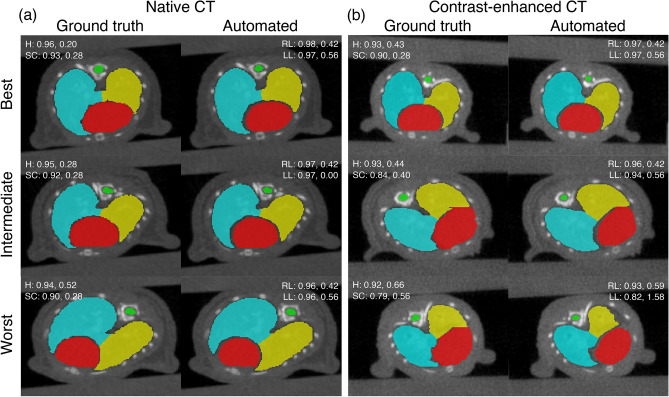
For image-guided small animal irradiations, the whole workflow of imaging, organ contouring, irradiation planning, and delivery is typically performed in a single session requiring continuous administration of anaesthetic agents. Automating contouring leads to a faster workflow, which limits exposure to anaesthesia and thereby, reducing its impact on experimental results and on animal wellbeing. Here, we trained the 2D and 3D U-Net architectures of no-new-Net (nnU-Net) for autocontouring of the thorax in mouse micro-CT images. We trained the models only on native CTs and evaluated their performance using an independent testing dataset (i.e., native CTs not included in the training and validation). Unlike previous studies, we also tested the model performance on an external dataset (i.e., contrast-enhanced CTs) to see how well they predict on CTs completely different from what they were trained on. We also assessed the interobserver variability using the generalized conformity index ([Formula: see text]) among three observers, providing a stronger human baseline for evaluating automated contours than previous studies. Lastly, we showed the benefit on the contouring time compared to manual contouring. The results show that 3D models of nnU-Net achieve superior segmentation accuracy and are more robust to unseen data than 2D models. For all target organs, the mean surface distance (MSD) and the Hausdorff distance (95p HD) of the best performing model for this task (nnU-Net 3d_fullres) are within 0.16 mm and 0.60 mm, respectively. These values are below the minimum required contouring accuracy of 1 mm for small animal irradiations, and improve significantly upon state-of-the-art 2D U-Net-based AIMOS method. Moreover, the conformity indices of the 3d_fullres model also compare favourably to the interobserver variability for all target organs, whereas the 2D models perform poorly in this regard. Importantly, the 3d_fullres model offers 98% reduction in contouring time.
© 2022. The Author(s).
Conflict of interest statement
The authors declare no competing interests.
Figures
References
Publication types
MeSH terms
LinkOut - more resources
Full Text Sources
Research Materials
