. 2022 Feb;602(7896):343-348.
doi: 10.1038/s41586-021-04392-4. Epub 2022 Feb 2.

Structure of a B12-dependent radical SAM enzyme in carbapenem biosynthesis

Affiliations

Structure of a B12-dependent radical SAM enzyme in carbapenem biosynthesis

Hayley L Knox et al. Nature. 2022 Feb.

Abstract

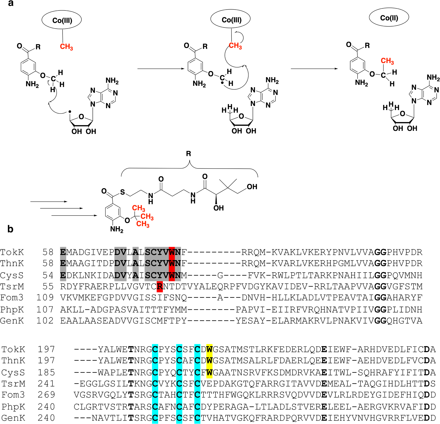
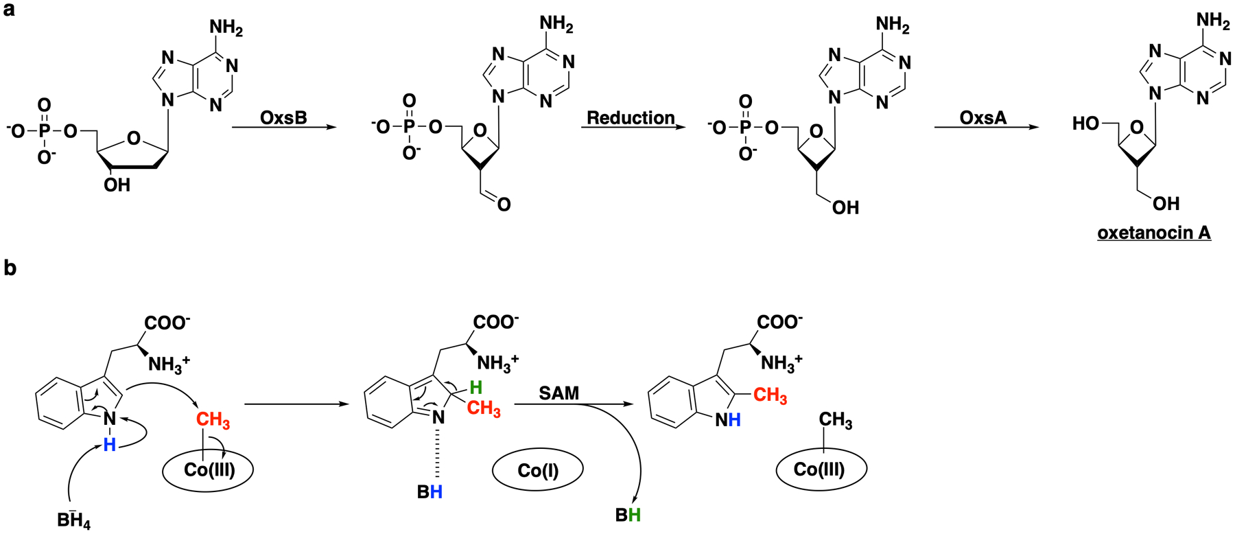
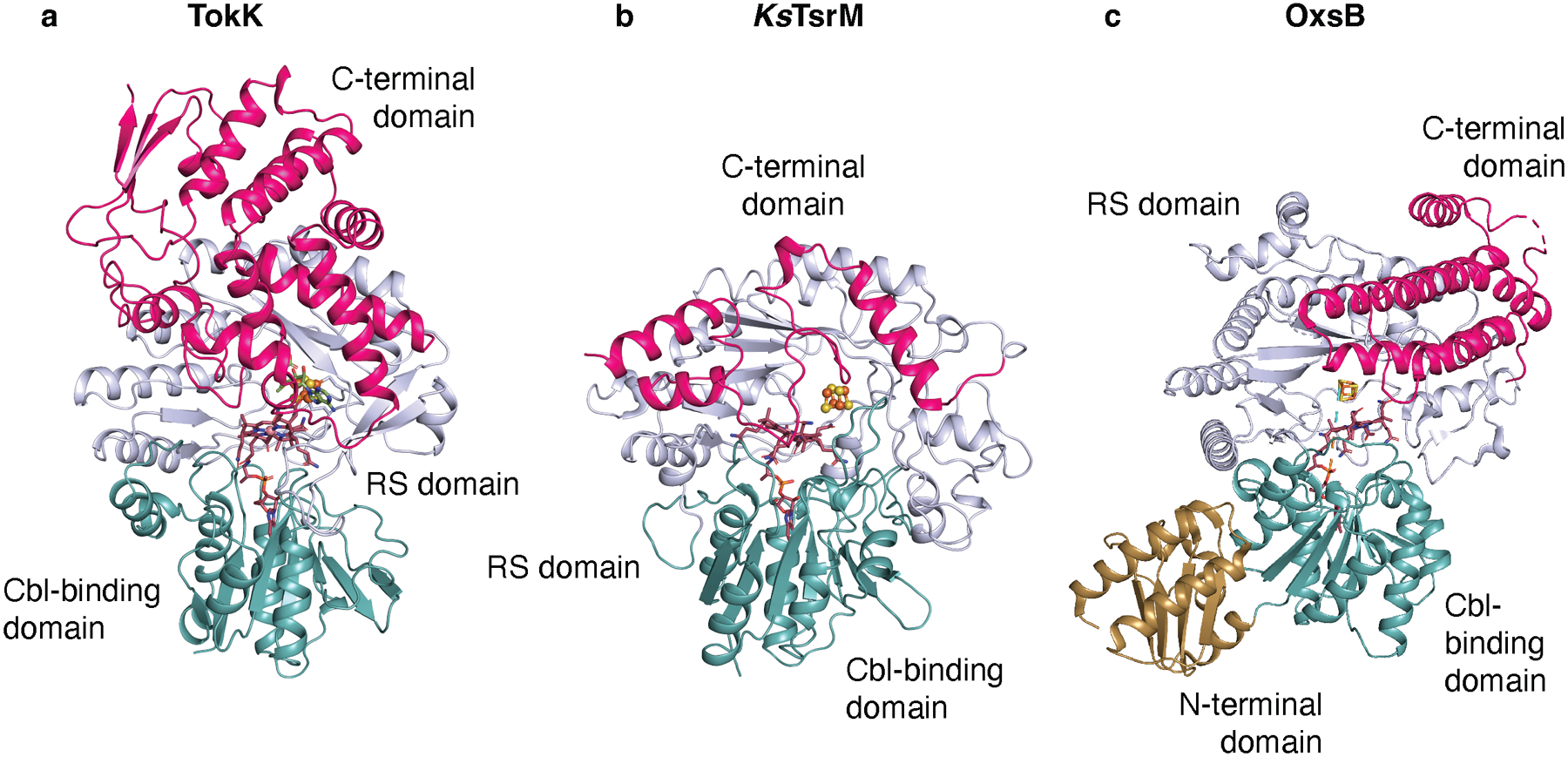
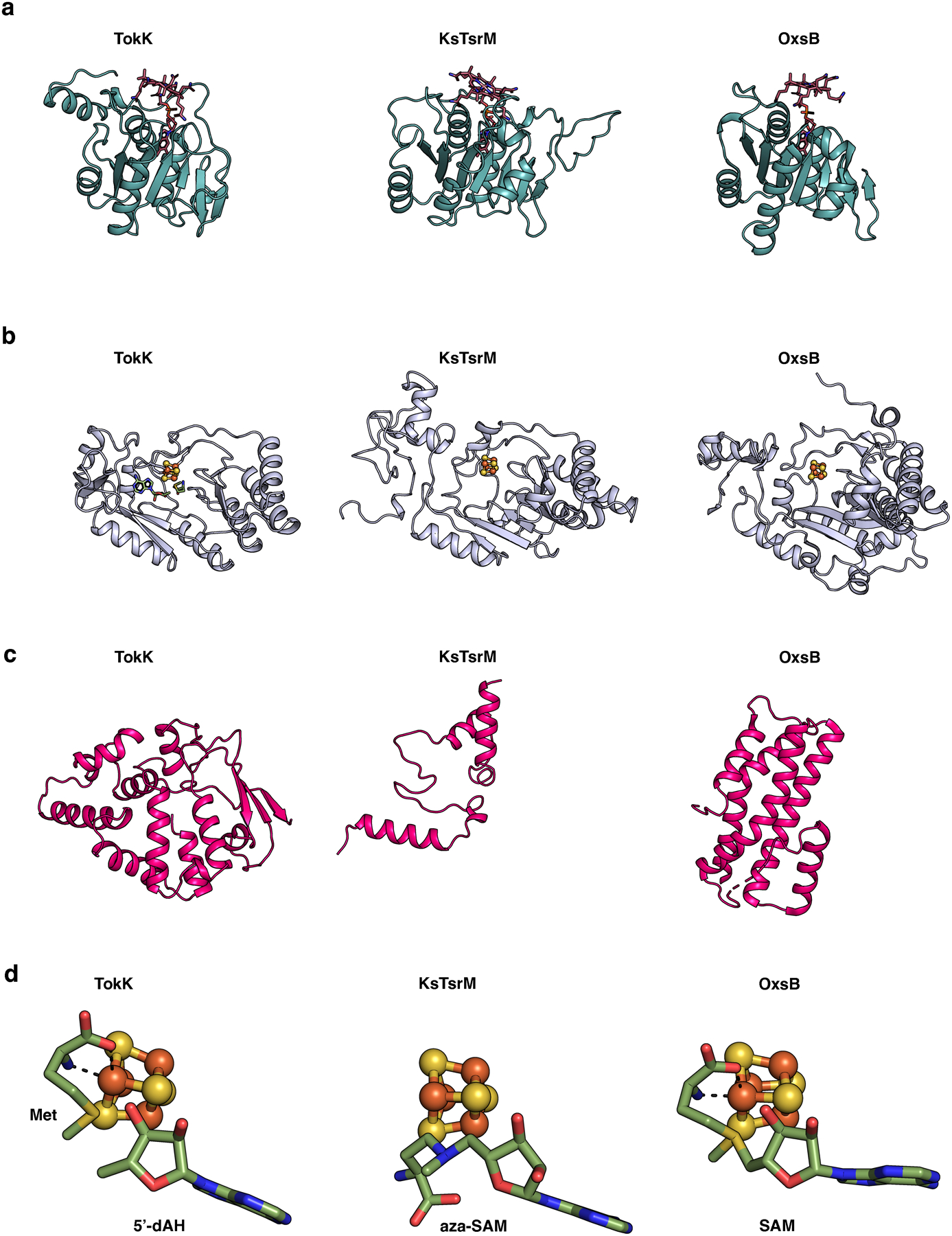
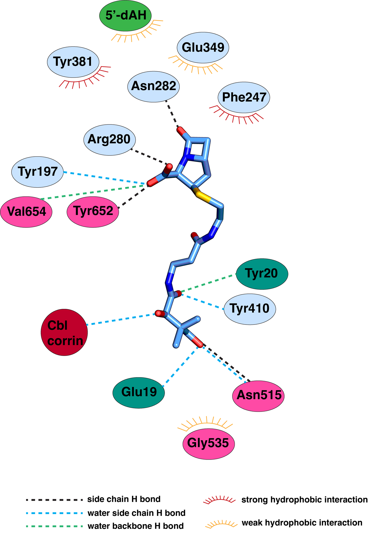
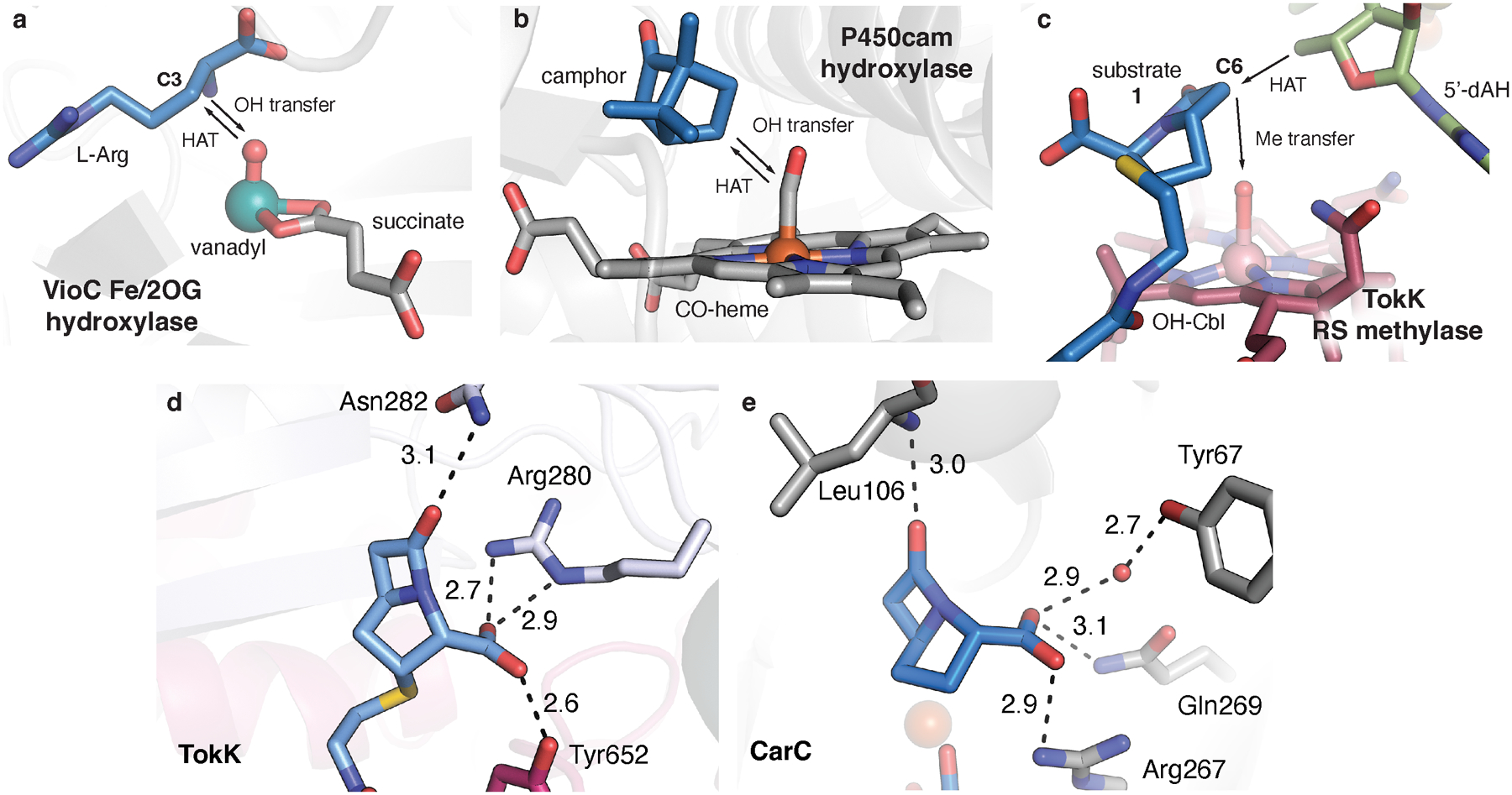
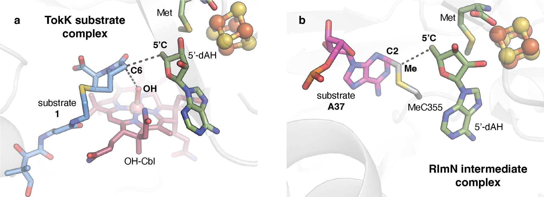
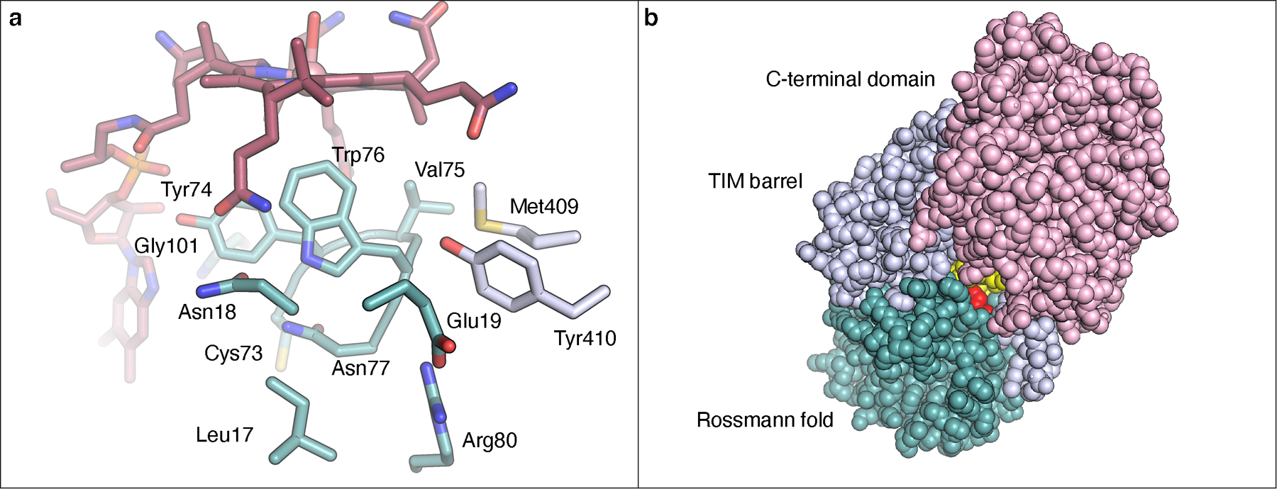
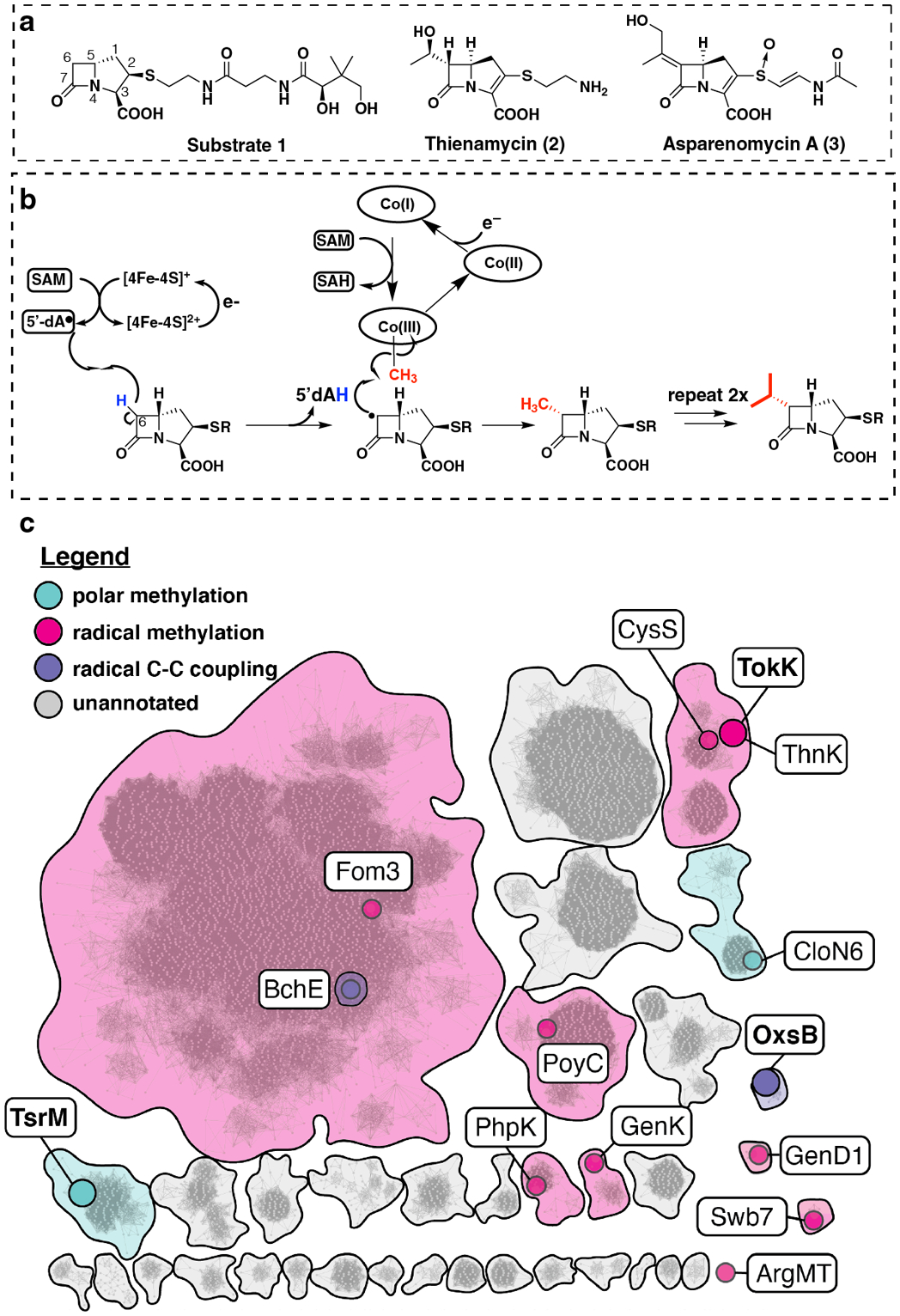
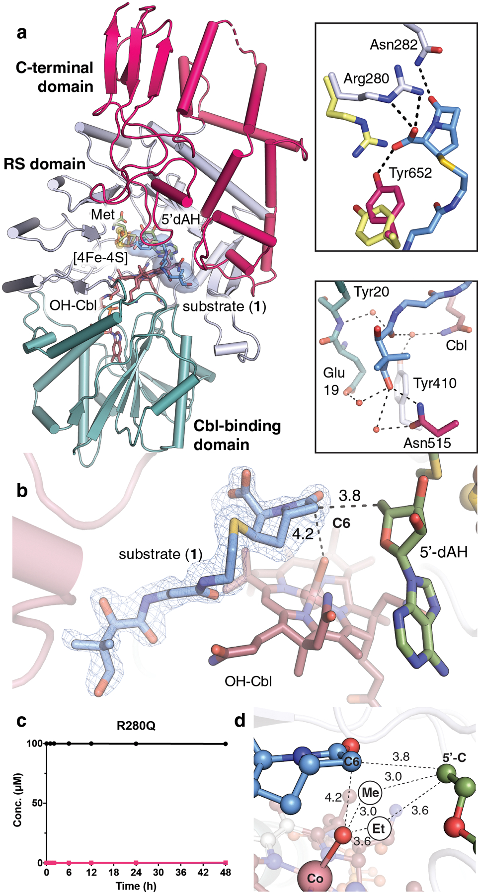
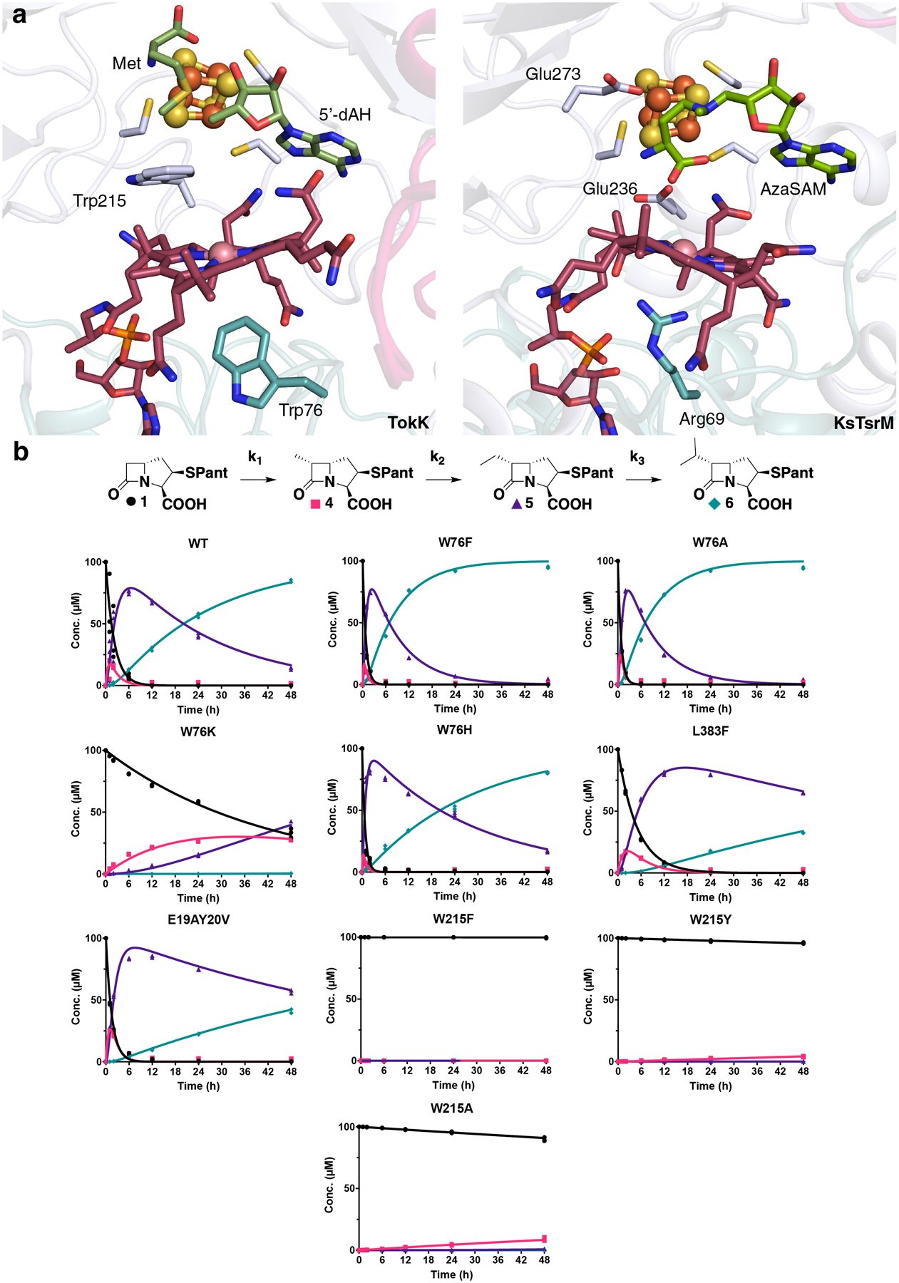
Carbapenems are antibiotics of last resort in the clinic. Owing to their potency and broad-spectrum activity, they are an important part of the antibiotic arsenal. The vital role of carbapenems is exemplified by the approval acquired by Merck from the US Food and Drug Administration (FDA) for the use of an imipenem combination therapy to treat the increased levels of hospital-acquired and ventilator-associated bacterial pneumonia that have occurred during the COVID-19 pandemic1. The C6 hydroxyethyl side chain distinguishes the clinically used carbapenems from the other classes of β-lactam antibiotics and is responsible for their low susceptibility to inactivation by occluding water from the β-lactamase active site2. The construction of the C6 hydroxyethyl side chain is mediated by cobalamin- or B12-dependent radical S-adenosylmethionine (SAM) enzymes3. These radical SAM methylases (RSMTs) assemble the alkyl backbone by sequential methylation reactions, and thereby underlie the therapeutic usefulness of clinically used carbapenems. Here we present X-ray crystal structures of TokK, a B12-dependent RSMT that catalyses three-sequential methylations during the biosynthesis of asparenomycin A. These structures, which contain the two metallocofactors of the enzyme and were determined in the presence and absence of a carbapenam substrate, provide a visualization of a B12-dependent RSMT that uses the radical mechanism that is shared by most of these enzymes. The structures provide insight into the stereochemistry of initial C6 methylation and suggest that substrate positioning governs the rate of each methylation event.


Conflict of interest statement

The authors declare no competing interests

Figures

Extended Data Fig. 1.
Extended Data Fig. 1.. Comparison to CysS, another class B sequential methylase.
CysS performs sequential radical methylations like TokK and ThnK (a). Partial alignment of TokK (Uniprot ID: A0A6B9HEI0), ThnK (Uniprot ID: F8JND9), CysS (Uniprot ID: A0A0H4NV78), TsrM (Uniprot ID: C0JRZ9), Fom3 (Uniprot ID: Q56184), PhpK (Uniprot ID: A0A0M3N271), and GenK (Uniprot ID: Q70KE5) (b). Cysteines that coordinate the iron-sulfur cluster are highlighted in blue. Trp76 (highlighted in red) and Trp215 (highlighted in yellow) in TokK is conserved in ThnK and CysS, but not in other known cobalamin-binding RS methylases. Areas of conservation for TokK, ThnK, and CysS around Trp76 are highlighted in grey. The bottom axial amino acid residue for TsrM (Arg69) is highlighted in red. Completely conserved residues are bolded.
Extended Data Fig. 2.
Extended Data Fig. 2.. Reactions performed by OxsB and TsrM, notable Cbl-binding radical SAM enzymes.
(a) Proposed pathway for the biosynthesis of oxetanocin A by OxsB and OxsA. Currently the aldehyde reduction step is unknown. (b) Proposed non-radical reaction performed by TsrM. The carboxylate in SAM is implicated in playing a dual role in catalysis, both as the source of the methyl donor and as the base to prime the substrate for nucleophilic attack.
Extended Data Fig. 3.
Extended Data Fig. 3.. Structural comparisons of Cbl-dependent RS enzymes.
The overall structures of TokK (a), KsTsrM (PDB ID: 6WTE) (b), and OxsB (PDB ID: 5UL3) (c) are shown as ribbon diagrams and colored by domain (Cbl-binding domain, teal; RS domain, light blue; and C-terminal domain, pink). OxsB has a fourth domain, an N-terminal domain of an unknown function, shown in gold. All three enzymes share very similar Cbl-binding domains with a characteristic Rossmann fold. However, as shown in Extended Data Fig. 4, the RS domain and C-terminal domain differ in each system.
Extended Data Fig. 4.
Extended Data Fig. 4.. Comparison of the Cbl-binding, RS, and C-terminal domains of TokK, KsTsrM, and OxsB.
The domains are colored as in Extended Data Fig. 3. (a) The Rossmann fold, in teal, is highly similar among TokK (PDB ID: 7KDY), KsTsrM (PDB ID: 6WTF), and OxsB (PDB ID: 5UL4). (b) The core of each of the RS domains is a (β/⍺)6 motif; however, there are distinct differences. The RS domain of OxsB is more compact than those of TokK or KsTsrM, and all three have unique extra secondary structure features. (c) The C-terminal domains for TokK, KsTsrM, and OxsB are vastly different in architecture. (d) Comparison of the binding of Met and 5’-dAH, aza-SAM, and SAM for TokK, KsTsrM, and OxsB structures, respectively. Only the relevant binding of SAM to the cluster is shown for OxsB. OxsB has two binding positions of SAM, one to the cluster and one in what is proposed to be an intermediate state towards methylating the Cbl.
Extended Data Fig. 5.
Domain architecture of TokK. (a) The three domains of TokK are portrayed in a ribbon diagram. The N-terminal Cbl-binding domain is shown in teal, the RS domain is shown in light blue, and the C-terminal domain is shown in pink. (b) A topology diagram of TokK with domains colored as in panel A. The cobalamin is portrayed in sticks, and the lower axial Trp side chain is shown as a red dot. The iron-sulfur cluster is shown in orange and yellow spheres, and the ligating Cys residues are shown as small yellow spheres. Zoomed in views of the C-terminal domain (c), RS domain (d), and Cbl-binding domain (e) are shown as ribbon diagrams.
Extended Data Fig. 6.
Extended Data Fig. 6.. Diagram of carbapenam interactions in the TokK complex with substrate.
Carbapenam substrate 1 shown in blue sticks and colored by atom type. Polar and hydrophobic interactions were mapped with the LigPlot program and indicated with dashed lines or a starburst symbol.
Extended Data Fig. 7.
Extended Data Fig. 7.. Comparison of TokK structure with those of enzymes using alternative radical-generating mechanisms.
Residues that interact with the polar substituents in the β-lactam ring are shown in stick format for TokK (a) and the epimerase CarC (PDB ID: 4OJ8) (b). While the two enzymes are structurally distinct, they use a similar number and type of functional groups to anchor the β-lactam by using direct and water-mediated contacts to the C7 carbonyl and C3 carboxylate substituents. Additionally, comparison of the active site with those of hydroxylases reveals differences in the orientation of hydrogen atom transfer (HAT) intermediates and −OH or −CH3 functionalization moieties. Fe(II)- and 2-oxo-glutarate-dependent (Fe/2OG) oxygenases use a ferryl [Fe(IV)-oxo)] intermediate to abstract an H-atom from an unactivated substrate carbon. The resulting Fe(III)-OH complex then couples with the substrate radical to yield a hydroxylated product. A vanadyl [V(IV)-oxo] mimic of the ferryl intermediate in l-Arg C3 hydroxylase, VioC, reveals that HAT and −OH transfer must occur from the same side of the substrate target carbon (c). The distance between the reactive oxo group and the substrate target carbon (indicated by the arrows) is 3.1 Å in VioC. A structure of the heme-dependent hydroxylase, P450cam, shows a similar phenomenon. A CO-bound mimic of the Fe(IV)-porphyrin radical intermediate, compound I, demands the same arrangement of HAT and −OH transfer components relative to the hydroxylation target on the camphor substrate (d). The distance between a mimic of the reactive group and the substrate target carbon (indicated by the arrows) is 3.1 Å in P450cam. By contrast, RS methylases use separate HAT reagents (5’-dA•) and methyl group donors (Me-Cbl), allowing for more diverse stereochemical outcomes in C-H functionalization reactions. (e) The structure of TokK in complex with carbapenam substrate, 1, shows a ~120° angle between the HAT acceptor (5’C of 5’-dAH), the substrate target carbon (C6), and the Cbl top ligand (−OH of OH-Cbl, a surrogate for Me-Cbl).
Extended Data Fig. 8.
Extended Data Fig. 8.. A comparison of the substrate-binding sites in two structurally characterized RS methylases.
The substrate complexes of TokK (a) and RlmN (PDB ID: 5HR7) (b) are shown. RlmN is an RS methylase that uses a radical-based mechanism to methylate an sp2-hybridized carbon (C2) of an adenine base in transfer or ribosomal RNA. By contrast to TokK and other Cbl-dependent RS enzymes, RlmN uses a 5’-dA• to activate a post-translationally modified methyl-Cys residue on a C-terminal loop in the active site to modify its aromatic substrate via radical addition. Comparison of a structure of RlmN in a cross-linked methylCys-tRNA intermediate state to the TokK substrate complex shows that, despite the differences in reaction mechanism, these systems use a similar orientation of radical initiator (5’-dA•), substrate target carbon (C6, C2), and methyl donor (OH, Me). In the TokK substrate complex, the −OH ligand of the Cbl cofactor serves as a surrogate for the position of the methyl donor.
Extended Data Fig. 9.
Extended Data Fig. 9.. Solvent accessibility of the bottom axial face of the Cbl in TokK.
(a) Nearest residues (~6 Å) to the bottom face of the Cbl. (b) View of TokK using space-filling model to show a channel to the active site. As can be seen, only a very small portion of the Cbl (colored yellow) is solvent accessible, with most of the Cbl being buried within the Rossmann fold. Trp76 is colored red.
Fig 1.
Fig 1.. Cobalamin-dependent radical-mediated methylations in carbapenem biosynthesis.
(a) Structure of (2R) pantetheinylated carbapenam precursor substrate 1 modified by ThnK and TokK. Related carbapenem natural products containing C6-alkyl substituents include thienamycin and asparenomycin A. (b) Proposed mechanism describing the three sequential methylations catalyzed by TokK. (c) An abbreviated sequence similarity network (SSN) of cobalamin-binding RS enzymes, highlighting selected sequence clusters and nodes. The full network (Fig. S1) was generated from ~11,000 annotated cobalamin-dependent RS enzymes with an alignment score of 65. Each node represents a single sequence or a set of sequences with >40% sequence identity. Sequence clusters and nodes are colored by predicted reaction mechanism (see legend in figure panel). Nodes containing functionally annotated sequences are indicated in color. Structurally characterized enzymes are represented by enlarged nodes and labeled in boldface.
Fig. 2.
Fig. 2.. TokK binds its carbapenam substrate at the interface of three domains.
(a) The overall structure of TokK (chain A) is illustrated as a ribbon diagram and colored by domain. The Cbl-binding Rossmann fold is shown in teal with the cobalamin cofactor in stick format and colored by atom type. The RS domain is shown in light blue. A [4Fe-4S] cluster is shown in orange and yellow spheres. 5’-dAH and Met coproducts are shown in stick format. The C-terminal domain is shown in pink. Carbapenam substrate (1) is shown in light blue sticks, colored by atom type. (b) An Fo-Fc omit electron density map is shown for (1) (blue mesh, contoured at 3.0σ). Substrates, cofactors, and coproducts are shown in stick format. Distances between reactive groups are given in units of Å. (c) Product formation for the R280Q TokK variant. Substrate is shown in black spheres and the first methylated product is shown in pink squares. All activity assays were conducted with substrate 1, shown in Fig. 1a. (d) Projections of the additional methyl groups added to C6 of (1) and their respective distances (in Å) from the 5’ carbon of 5’-dAH and the hydroxyl moiety of OHCbl, the latter of which serves as a surrogate of the active MeCbl cofactor. Spheres labeled Me and Et represent the suggested positions of the newly installed carbon atoms in the mono-methylated and dimethylated products.
Fig. 3.
Fig. 3.. The Cbl and substrate binding sites influence overall activity and the relative rates of each TokK methylation step.
(a) Comparative analysis of the side chains proximal to the Co ion in two cobalamin-binding RS enzymes, TokK and KsTsrM (PDB ID: 6WTF). Selected amino acid side chains are shown in stick format and the Co ion is shown as a pink sphere. (b) Schematic diagram of the three sequential methylations performed by TokK. 48 h time course experiments performed in triplicate (each replicate represented as a symbol) tracking substrate (black spheres), methyl (pink squares), ethyl (purple triangles), and isopropyl (blue diamonds) product formation. Each product was estimated using COPASI (irreversible mass action model using the reaction scheme shown above) and simulated using Virtual Cell (shown as lines) for wild-type, W76F, W76A, W76K, W76H, L383F, E19AY20V, W215F, W215Y, and W215A.

References

    1. US Food and Drug Administration, Supplemental New Drug Approval: RECARBRIO (imipenem, cilastatin, and relebactam) for injection, available at https://www.fda.gov/RegulatoryInformation/Guidances/default.htm (2020).
    1. Fisher JF & Mobashery S Three decades of the class A beta-lactamase acyl-enzyme. Curr Protein Pept Sci 10, 401–407, doi: 10.2174/138920309789351967 (2009). - DOI - PMC - PubMed
    1. Sinner EK, Marous DR, Townsend CA Evolution of Methods for the Study of Cobalamin-Dependent Radical SAM Enzymes. ACS Bio & Med Chem Au 0, 10.1021/acsbiomedchemau.1021c00032, doi: 10.1021/acsbiomedchemau.1c00032 (2021). - DOI - PMC - PubMed
    1. Li R, Lloyd EP, Moshos KA & Townsend CA Identification and characterization of the carbapenem MM 4550 and its gene cluster in Streptomyces argenteolus ATCC 11009. Chembiochem 15, 320–331, doi: 10.1002/cbic.201300319 (2014). - DOI - PMC - PubMed
    1. Nunez LE, Mendez C, Brana AF, Blanco G & Salas JA The biosynthetic gene cluster for the beta-lactam carbapenem thienamycin in Streptomyces cattleya. Chem Biol 10, 301–311, doi: 10.1016/s1074-5521(03)00069-3 (2003). - DOI - PubMed

Publication types

MeSH terms