Review
. 2022 Jan 17:9:775102.
doi: 10.3389/fcell.2021.775102. eCollection 2021.

Development, Diversity, and Neurogenic Capacity of Enteric Glia

Affiliations
Review

Development, Diversity, and Neurogenic Capacity of Enteric Glia

Werend Boesmans et al. Front Cell Dev Biol. .

Abstract

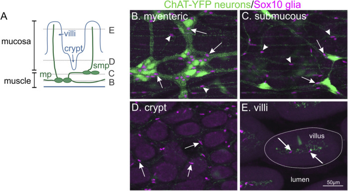
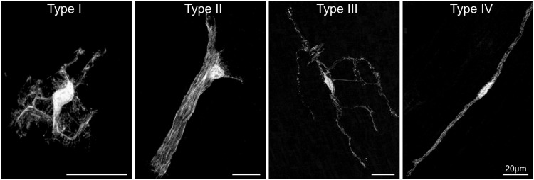
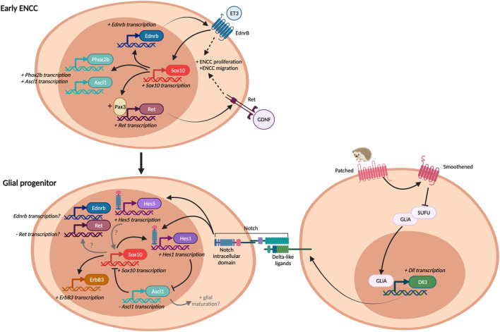
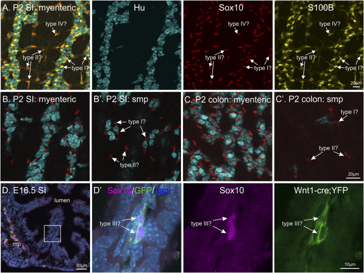
Enteric glia are a fascinating population of cells. Initially identified in the gut wall as the "support" cells of the enteric nervous system, studies over the past 20 years have unveiled a vast array of functions carried out by enteric glia. They mediate enteric nervous system signalling and play a vital role in the local regulation of gut functions. Enteric glial cells interact with other gastrointestinal cell types such as those of the epithelium and immune system to preserve homeostasis, and are perceptive to luminal content. Their functional versatility and phenotypic heterogeneity are mirrored by an extensive level of plasticity, illustrated by their reactivity in conditions associated with enteric nervous system dysfunction and disease. As one of the hallmarks of their plasticity and extending their operative relationship with enteric neurons, enteric glia also display neurogenic potential. In this review, we focus on the development of enteric glial cells, and the mechanisms behind their heterogeneity in the adult gut. In addition, we discuss what is currently known about the role of enteric glia as neural precursors in the enteric nervous system.

Keywords: enteric nervous system; glial cells; gliogenesis; neural crest; neurogenesis.


Conflict of interest statement

The authors declare that the research was conducted in the absence of any commercial or financial relationships that could be construed as a potential conflict of interest.

Figures

FIGURE 1
FIGURE 1
Enteric glial cells are located throughout all different concentric layers of the gut. (A): Diagram of the different cell layers in the small intestine. (B–E): Whole-mount immunohistochemistry on the small intestine of ChAT-YFP mouse, where excitatory enteric neurons are labelled with GFP. Ce3D clearing was performed to visualise all tissue layers. The location of Sox10-immunoreactive enteric glial nuclei (magenta, arrows) can be observed with neuronal cell bodies in the myenteric plexus (mp, B), submucous plexus (smp, C) and also associated with neuronal fibres in the muscle layers (arrowheads). In addition, enteric glia are found throughout the mucosa surrounding the crypts (D) and in villi (E).
FIGURE 2
FIGURE 2
The morphological enteric glial cell subtypes. Individual enteric glial cells with GFP labelling throughout their cell body to visualise their morphology. Sparse labelling of individual cells was achieved using the Sox10-cre;MADM transgenic mouse system. Adapted from (Boesmans et al., 2015).
FIGURE 3
FIGURE 3
Pathways in gliogenesis. Some pathways are commonly employed both in early ENCC development and enteric glial differentiation, but may play different roles in each situation, depending on the presence of other factors. Adapted and expanded from (Liu and Ngan, 2014).
FIGURE 4
FIGURE 4
Differentiation of enteric glial subtypes during ENS development? (A–C): Whole-mount postnatal day 2 (P2) murine gut with immunohistochemical staining against HuC/D (blue), Sox10 (red) and S100B (yellow). By P2, enteric glia are present in different locations in the various gut layers: in the myenteric and submucous (smp, arrows) plexus in both the small intestine (SI) and colon, and also surrounding neurons within ganglia. Should these enteric glia be classified as type I, II, III or IV based on their locations? (D): Transverse cryosection of E16.5 small intestine from Wnt1-cre;R26R-YFP mice, where all neural crest-derived cells express YFP. Immunohistochemical labelling against Sox10 (magenta), HuC/D (red), YFP (green) and dapi (blue) was performed. Inset on left image is magnified in the following panel. Sox10+ cells can be observed in some villi (arrows), these could be classified as either type III enteric glia or progenitor cells.
FIGURE 5
FIGURE 5
Enteric glia are present in the gut in the absence of enteric neurons: the aganglionic zone of Hirschsprung Disease (HSCR) tissue. Whole-mount immunohistochemistry of Ednrb-KO mouse (A–F) and human HSCR patient tissue (H-H′). (A): Diagram of mouse colon including the ENS wavefront and showing the regions with “normal” ganglia, the transition zone (TZ), and the aganglionic zone (AZ). (B–F): Representative images from each of these regions, following immunohistochemistry against S100B (green), HuC/D (red) and neuronal class III β−tubulin (Tuj1, blue). The majority of S100B + glia in the aganglionic region follow nerve bundles, both large and small (arrows). Sometimes, individual Hu + neurons are present in the aganglionic zone (open arrows). NB. These are not “skip segments” as there are no clear ganglia, instead, they are just a handful of individual neuronal cell bodies. (G-G’): Patient HSCR samples labelled against Sox10 (red) and Tuj1 (blue). In the aganglionic zone, Sox10 + glia are also associated with nerve processes. The same proximal—distal orientation has been maintained in all tissues.

References

    1. Almond S., Lindley R. M., Kenny S. E., Connell M. G., Edgar D. H. (2007). Characterisation and Transplantation of Enteric Nervous System Progenitor Cells. Gut 56, 489–496. 10.1136/gut.2006.094565 - DOI - PMC - PubMed
    1. Amiel J., Sproat-Emison E., Garcia-Barcelo M., Lantieri F., Burzynski G., Borrego S., et al. (2008). Hirschsprung Disease, Associated Syndromes and Genetics: a Review. J. Med. Genet. 45, 1–14. 10.1136/jmg.2007.053959 - DOI - PubMed
    1. Anderson R. B., Stewart A. L., Young H. M. (2006). Phenotypes of Neural-Crest-Derived Cells in Vagal and Sacral Pathways. Cell Tissue Res 323, 11–25. 10.1007/s00441-005-0047-6 - DOI - PubMed
    1. Andrew A., Kramer B., Rawdon B. B. (1982). The Embryonic Origin of Endocrine Cells of the Gastrointestinal Tract. Gen. Comp. Endocrinol. 47, 249–265. 10.1016/0016-6480(82)90228-3 - DOI - PubMed
    1. Avetisyan M., Schill E. M., Heuckeroth R. O. (2015). Building a Second Brain in the Bowel. J. Clin. Invest. 125, 899–907. 10.1172/jci76307 - DOI - PMC - PubMed