. 2022 Jan 17:8:780750.
doi: 10.3389/fmed.2021.780750. eCollection 2021.

Inflammation-Induced Coagulopathy Substantially Differs Between COVID-19 and Septic Shock: A Prospective Observational Study

Affiliations

Inflammation-Induced Coagulopathy Substantially Differs Between COVID-19 and Septic Shock: A Prospective Observational Study

Mélanie Dechamps et al. Front Med (Lausanne). .

Abstract

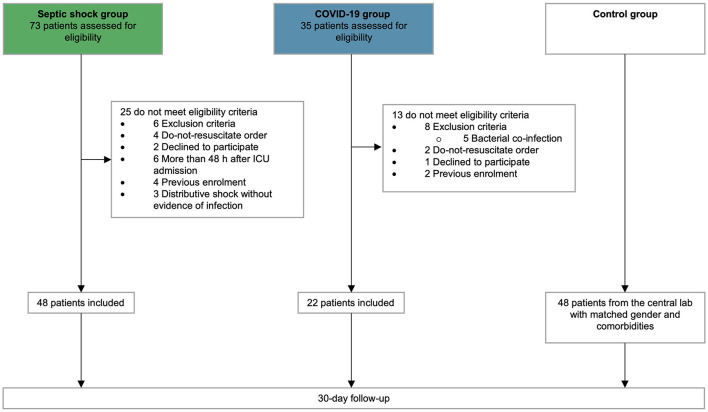
Critical COVID-19, like septic shock, is related to a dysregulated systemic inflammatory reaction and is associated with a high incidence of thrombosis and microthrombosis. Improving the understanding of the underlying pathophysiology of critical COVID-19 could help in finding new therapeutic targets already explored in the treatment of septic shock. The current study prospectively compared 48 patients with septic shock and 22 patients with critical COVID-19 regarding their clinical characteristics and outcomes, as well as key plasmatic soluble biomarkers of inflammation, coagulation, endothelial activation, platelet activation, and NETosis. Forty-eight patients with matched age, gender, and co-morbidities were used as controls. Critical COVID-19 patients exhibited less organ failure but a prolonged ICU length-of-stay due to a prolonged respiratory failure. Inflammatory reaction of critical COVID-19 was distinguished by very high levels of interleukin (IL)-1β and T lymphocyte activation (including IL-7 and CD40L), whereas septic shock displays higher levels of IL-6, IL-8, and a more significant elevation of myeloid response biomarkers, including Triggering Receptor Expressed on Myeloid cells-1 (TREM-1) and IL-1ra. Subsequent inflammation-induced coagulopathy of COVID-19 also differed from sepsis-induced coagulopathy (SIC) and was characterized by a marked increase in soluble tissue factor (TF) but less platelets, antithrombin, and fibrinogen consumption, and less fibrinolysis alteration. In conclusion, COVID-19 inflammation-induced coagulopathy substantially differs from SIC. Modulating TF release and activity should be evaluated in critical COVID-19 patients.

Keywords: COVID-19; NETosis; coagulopathy; endothelium; inflammation; platelet; septic shock.


Conflict of interest statement

JD is the CEO and founder of QUALIblood s.a., a Belgian Contract Research Organization. LM and HH were employed by the company QUALIblood s.a. and authors MDer and LJ were employed by the company Inotrem s.a. The remaining authors declare that the research was conducted in the absence of any commercial or financial relationships that could be construed as a potential conflict of interest. The authors declare that this study received funding from Foundation Saint-Luc. The funder was not involved in the study design, collection, analysis, interpretation of data, the writing of this article or the decision to submit it for publication.

Figures

Figure 1
Figure 1
Flow chart. Flow chart of patients screening and inclusion.
Figure 2
Figure 2
Endothelial activation and coagulation. Scatter graphs of soluble biomarkers of (A–F) endothelial activation and (G–L) coagulation. Individual values (open circle), mean (colored rectangle), and standard deviation are presented on the graphs. *p < 0.05 between septic shock and control patients. #p < 0.05 between COVID-19 and control patients. $p < 0.05 between septic shock and COVID-19 patients. ICAM-1, intercellular adhesion molecule-1; INR, international normalized ratio; PAI-1, plasminogen activator inhibitor-1; TAT, thrombin–antithrombin complex; TF, tissue factor; TFPI, tissue factor pathway inhibitor; tPA, tissue plasminogen activator; vWF, von Willebrand factor.
Figure 3
Figure 3
Platelet activation and NETosis. Scatter graphs of soluble biomarkers of (A,B) platelet activation and (C,D) NETosis. Individual values (open circle), mean (colored rectangle), and standard deviation are presented on the graphs. *p < 0.05 between septic shock and control patients. #p < 0.05 between COVID-19 and control patients. $p < 0.05 between septic shock and COVID-19 patients. (E) Representative microscopy pictures of endothelial cells (CD31, green), Cit-H3 (orange), neutrophils (NE, yellow), platelets (CD42b, light blue), nucleus (DAPI, dark blue), and merged staining, from control, septic shock, and COVID-19 lungs. sCD62P, soluble p-selectin; Cit-H3, citrullinated histone 3; NE, neutrophil elastase; NET, neutrophil extracellular traps; MPO, myeloperoxidase; sTLT-1, soluble triggering receptor expressed on myeloid cells (TREM) like transcript-1.
Figure 4
Figure 4
Inflammation and immune response. Scatter graphs of (A–E) ubiquitous proinflammatory cytokines, (F–I) myeloid inflammatory cytokines, and (J–S) lymphoid inflammatory cytokines. Individual values (open circle), mean (colored rectangle), and standard deviation are presented on the graphs. *p < 0.05 between septic shock and control patients. #p < 0.05 between COVID-19 and control patients. $p < 0.05 between septic shock and COVID-19 patients. CD40L, CD40 ligand; CRP, C-reactive protein; IFNγ, interferon gamma; IL, interleukin; IL-1ra, IL-1 receptor antagonist; IP-10, interferon gamma-induced protein 10; MCP-1, monocyte chemoattractant protein 1; MIP-1α, Macrophage inflammatory protein-1α; sTREM-1, soluble triggering receptor expressed on myeloid cells 1.
Figure 5
Figure 5
Principal component analysis of study cohort. Principal component analysis showing (A) representation of COVID-19 and septic shock population in the two dimensions (score plot). Variables are separated in biplots of (B) ubiquitous, (C) myeloid, (D) lymphoid biomarkers, (E) endothelial activation, (F) coagulation activation, and (G) platelet and NETosis activation with correlation. Scores and loadings are presented in a scatterplot of one principal component (PC) against another. The loadings are represented in a circle of correlations: the closer the arrow of a loading is to the circle, the more the variable is well-represented in the space of the two plotted PCs and contributed to the building of these PCs. This graph also indicates the positive or negative links between the variables and groupings of variables. Arrow colors correspond to the between-group comparison as mentioned in the scatter graphs. CD40L, CD40 ligand; 1Cit-H3, citrullinated histone H3; CRP, C-reactive protein; ICAM-1, intercellular adhesion molecule-1; IFNγ, interferon gamma; IL, interleukin; IL-1ra, IL-1 receptor antagonist; INR, international normalized ration; IP-10, interferon gamma-induced protein 10; MCP-1, monocyte chemoattractant protein 1; MIP-1α, macrophage inflammatory protein-1α; PCA, principal component analysis; MPO, myeloperoxidase; NE, neutrophil elastase; PAI-1, plasminogen activator inhibitor-1; TAT, thrombin–antithrombin complex; TF, tissue factor; TFPI, tissue factor pathway inhibitor; sTLT-1, soluble TREM like transcript-1; tPA, tissue plasminogen activator; sTREM-1, soluble triggering receptor expressed on myeloid cells 1; TT, thrombin time; VCAM-1, vascular cell adhesion protein 1; vWF, von Willebrand factor; WBC, white blood cells.

References

    1. Huang C, Wang Y, Li X, Ren L, Zhao J, Hu Y, et al. . Clinical features of patients infected with 2019 novel coronavirus in Wuhan, China. Lancet. (2020) 395:497–506. 10.1016/S0140-6736(20)30183-5 - DOI - PMC - PubMed
    1. Fajgenbaum DC, June CH. Cytokine storm. N Engl J Med. (2020) 383:2255–73. 10.1056/NEJMra2026131 - DOI - PMC - PubMed
    1. Klok FA, Kruip M, van der Meer NJM, Arbous MS, Gommers D, Kant KM, et al. . Incidence of thrombotic complications in critically ill ICU patients with COVID-19. Thromb Res. (2020) 191:145–7. 10.1016/j.thromres.2020.04.013 - DOI - PMC - PubMed
    1. Helms J, Tacquard C, Severac F, Leonard-Lorant I, Ohana M, Delabranche X, et al. . High risk of thrombosis in patients with severe SARS-CoV-2 infection: a multicenter prospective cohort study. Intens Care Med. (2020) 46:1089–98. 10.1007/s00134-020-06062-x - DOI - PMC - PubMed
    1. Ackermann M, Verleden SE, Kuehnel M, Haverich A, Welte T, Laenger F, et al. . Pulmonary vascular endothelialitis, thrombosis, and angiogenesis in Covid-19. N Engl J Med. (2020) 383:120–8. 10.1056/NEJMoa2015432 - DOI - PMC - PubMed

LinkOut - more resources