Review
. 2022 Jan 17:8:768106.
doi: 10.3389/fmolb.2021.768106. eCollection 2021.

Computational Methods for Single-Cell Imaging and Omics Data Integration

Affiliations
Review

Computational Methods for Single-Cell Imaging and Omics Data Integration

Ebony Rose Watson et al. Front Mol Biosci. .

Abstract

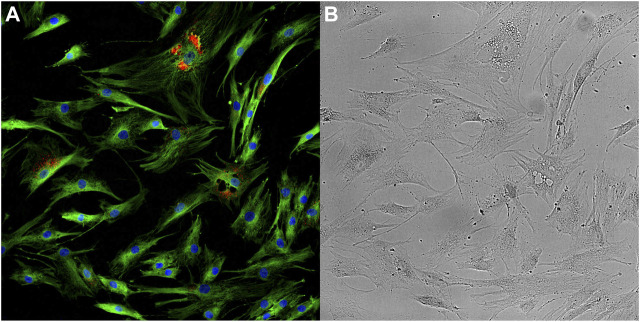
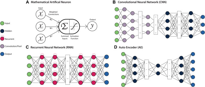
Integrating single cell omics and single cell imaging allows for a more effective characterisation of the underlying mechanisms that drive a phenotype at the tissue level, creating a comprehensive profile at the cellular level. Although the use of imaging data is well established in biomedical research, its primary application has been to observe phenotypes at the tissue or organ level, often using medical imaging techniques such as MRI, CT, and PET. These imaging technologies complement omics-based data in biomedical research because they are helpful for identifying associations between genotype and phenotype, along with functional changes occurring at the tissue level. Single cell imaging can act as an intermediary between these levels. Meanwhile new technologies continue to arrive that can be used to interrogate the genome of single cells and its related omics datasets. As these two areas, single cell imaging and single cell omics, each advance independently with the development of novel techniques, the opportunity to integrate these data types becomes more and more attractive. This review outlines some of the technologies and methods currently available for generating, processing, and analysing single-cell omics- and imaging data, and how they could be integrated to further our understanding of complex biological phenomena like ageing. We include an emphasis on machine learning algorithms because of their ability to identify complex patterns in large multidimensional data.

Keywords: ageing; data integration; machine learning; single cell imaging; single cell omics.


Conflict of interest statement

The authors declare that the research was conducted in the absence of any commercial or financial relationships that could be construed as a potential conflict of interest.

Figures

FIGURE 1
FIGURE 1
Optical microscopy images taken of ageing mesenchymal stem cells. Fluorescence image (A) provides information on the abundance and distribution of DNA (blue), α-Tubulin (green) and Senescence-associated beta-galactosidase (red). Brightfield image (B) provides information on the cellular and sub-cellular morphology. Images have been enhanced for visualisation.
FIGURE 2
FIGURE 2
Diagram depicting multi-modal data integration strategies according to the correlation, sequential and integrative categorisations. Triangles (green) and circles (blue) represent datasets from distinct biological data modalities. (A) For correlation-based integration strategies, distinct data modalities are processed and analysed independently, and correlations between the data are identified from the results. (B) In sequential integration strategies the results of the analysis on one data modality are refined by the integration of additional data modalities in subsequent analyses. (C) In the integrative analysis approach, each data modality undergoes feature transformation independently, which are subsequently combined and analysed.
FIGURE 3
FIGURE 3
Diagram depicting multi-modal data integration strategies according to the concatenation-, transformation- and model-based categorisation. Triangles (green), circles (blue) and squares (orange) represent datasets from distinct biological data modalities. (A) In concatenation-based integration, multi-modal data is joined at the raw or processed level before being passed to an ensuing model for analysis. (B) In transformation-based strategies, each data modality undergoes modelling to transform features separately, which are subsequently integrated and passed to a final model for analysis (C) In model-based integration, each data modality undergoes modelling and analysis independently, and model outputs are integrated to generate the final result.
FIGURE 4
FIGURE 4
Deep Artificial Neural Network (ANN) Architectures. Left: a key for several types of neurons used in ANN architectures. (A) Mathematical model of a neuron. The weighted (Wi) sum of all inputs (Xi) to the neuron is computed and passed to the activation function, which produces the neurons output. This output is propagated as an input to neurons in subsequent layers of the network. (B) A Convolutional Neural Network (CNN) is a feed-forward ANN architecture containing convolutional and pooling layers, which allow local patterns to be learned and detected in a spatially invariant manner. (C) A Recurrent Neural Network (RNN) is a recursive ANN architecture containing neurons with an internal memory state, which retain information about prior inputs to the model. (D) An Autoencoder (AE) is a feed-forward ANN architecture that is comprised of an encoder module that learns a latent representation of the input, and a decoder module that reconstructs the original input data from the encoded representation.

References

    1. Abdelaal T., Michielsen L., Cats D., Hoogduin D., Mei H., Reinders M. J. T., et al. (2019). A Comparison of Automatic Cell Identification Methods for Single-Cell RNA Sequencing Data. Genome Biol. 20 (1), 194. 10.1186/s13059-019-1795-z - DOI - PMC - PubMed
    1. Abràmoff M. D., Magalhães P. J., Ram S. J. (2004). Image Processing with ImageJ. Biophotonics Int. 11 (7), 36–42.
    1. Acar E., Papalexakis E. E., Gürdeniz G., Rasmussen M. A., Lawaetz A. J., Nilsson M., et al. (2014). Structure-revealing Data Fusion. BMC Bioinformatics 15 (1), 239. 10.1186/1471-2105-15-239 - DOI - PMC - PubMed
    1. Aibar S., González-Blas C. B., Moerman T., Huynh-Thu V. A., Imrichova H., Hulselmans G., et al. (2017). SCENIC: Single-Cell Regulatory Network Inference and Clustering. Nat. Methods 14 (11), 1083–1086. 10.1038/nmeth.4463 - DOI - PMC - PubMed
    1. Algar W. R., Hildebrandt N., Vogel S. S., Medintz I. L. (2019). FRET as a Biomolecular Research Tool - Understanding its Potential while Avoiding Pitfalls. Nat. Methods 16 (9), 815–829. 10.1038/s41592-019-0530-8 - DOI - PubMed