

Inter-observer variability of organ contouring for preclinical studies with cone beam Computed Tomography imaging
- PMID: 35111981
- PMCID: PMC8790504
- DOI: 10.1016/j.phro.2022.01.002
Inter-observer variability of organ contouring for preclinical studies with cone beam Computed Tomography imaging
Abstract
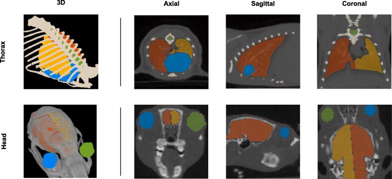
Background and purpose: In preclinical radiation studies, there is great interest in quantifying the radiation response of healthy tissues. Manual contouring has significant impact on the treatment-planning because of variation introduced by human interpretation. This results in inconsistencies when assessing normal tissue volumes. Evaluation of these discrepancies can provide a better understanding on the limitations of the current preclinical radiation workflow. In the present work, interobserver variability (IOV) in manual contouring of rodent normal tissues on cone-beam Computed Tomography, in head and thorax regions was evaluated.
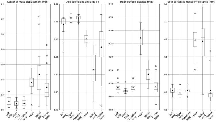
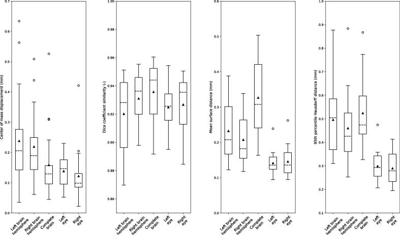
Materials and methods: Two animal technicians performed manually (assisted) contouring of normal tissues located within the thorax and head regions of rodents, 20 cases per body site. Mean surface distance (MSD), displacement of center of mass (ΔCoM), DICE similarity coefficient (DSC) and the 95th percentile Hausdorff distance (HD95) were calculated between the contours of the two observers to evaluate the IOV.
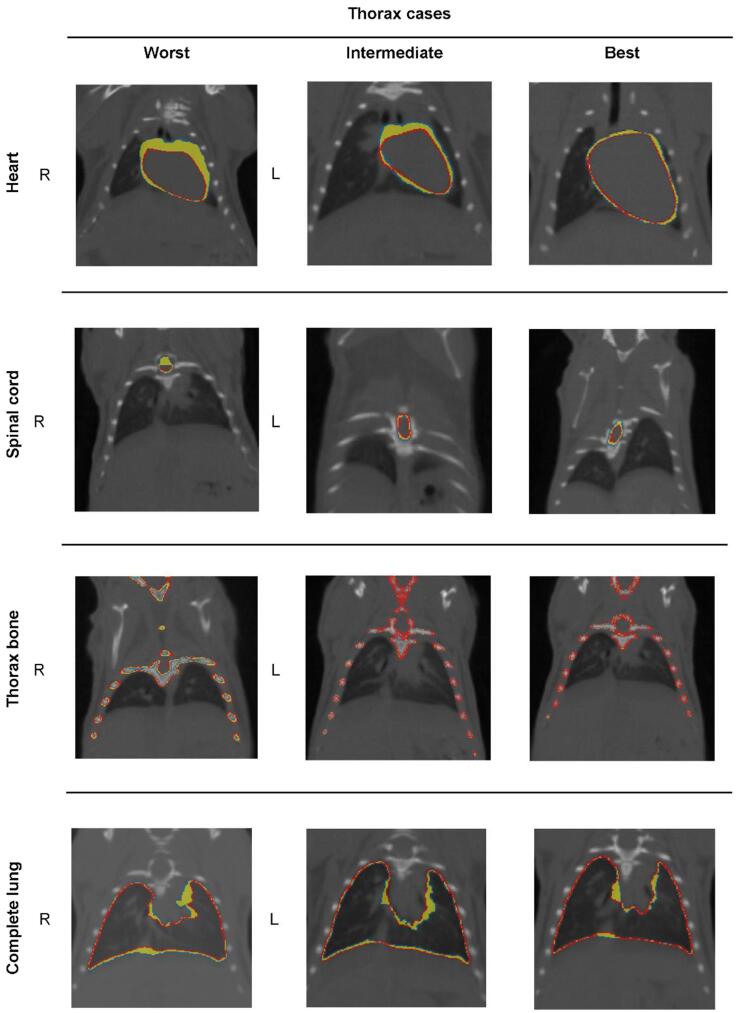
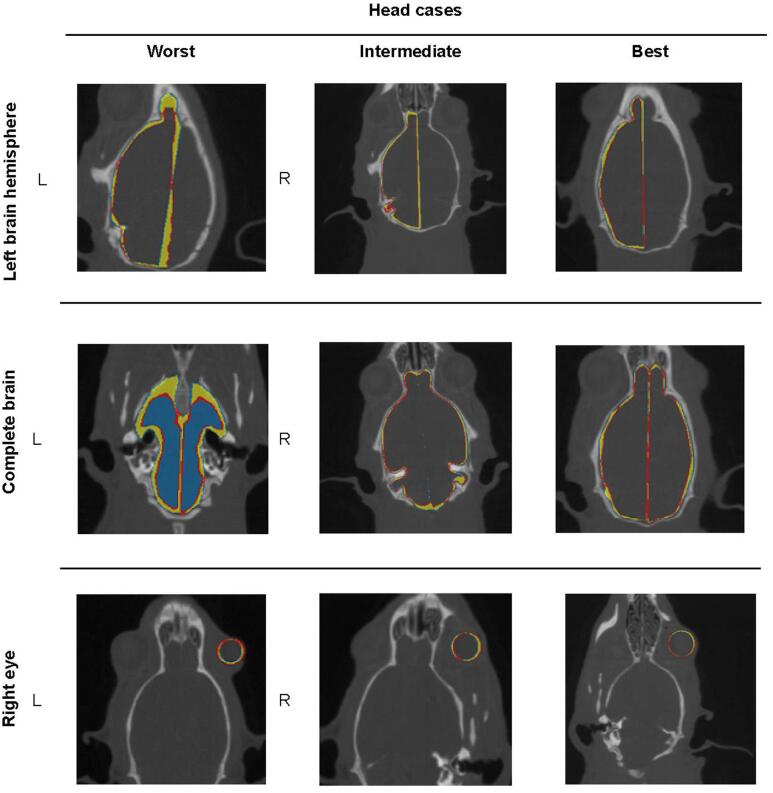
Results: For the thorax organs, right lung had the lowest IOV (ΔCoM: 0.08 ± 0.04 mm, DSC: 0.96 ± 0.01, MSD:0.07 ± 0.01 mm, HD95:0.20 ± 0.03 mm) while spinal cord, the highest IOV (ΔCoM:0.5 ± 0.3 mm, DSC:0.81 ± 0.05, MSD:0.14 ± 0.03 mm, HD95:0.8 ± 0.2 mm). Regarding head organs, right eye demonstrated the lowest IOV (ΔCoM:0.12 ± 0.08 mm, DSC: 0.93 ± 0.02, MSD: 0.15 ± 0.04 mm, HD95: 0.29 ± 0.07 mm) while complete brain, the highest IOV (ΔCoM: 0.2 ± 0.1 mm, DSC: 0.94 ± 0.02, MSD: 0.3 ± 0.1 mm, HD95: 0.5 ± 0.1 mm).
Conclusions: Our findings reveal small IOV, within the sub-mm range, for thorax and head normal tissues in rodents. The set of contours can serve as a basis for developing an automated delineation method for e.g., treatment planning.
Keywords: CT; Interobserver variability; Organ contouring; Preclinical.
© 2022 The Authors.
Conflict of interest statement
The authors declare that they have no known competing financial interests or personal relationships that could have appeared to influence the work reported in this paper.
Figures
References
-
- Iglesias VS, Hoof SJ van, Vaniqui A, Schyns LE, Lieuwes N, Yaromina A, et al. An orthotopic non-small cell lung cancer model for image-guided small animal radiotherapy platforms. 2018;92. https://doi.org/10.1259/BJR.20180476. - PMC - PubMed
-
- Verhaegen F, Granton P, Tryggestad E. Small animal radiotherapy research platforms. Phys Med Biol 2011;56. https://doi.org/10.1088/0031-9155/56/12/R01. - PubMed
LinkOut - more resources
Full Text Sources
