. 2022 May;52(5):825-834.
doi: 10.1002/eji.202149324. Epub 2022 Feb 15.

Targeting Bcl6 in the TREX1 D18N murine model ameliorates autoimmunity by modulating T-follicular helper cells and germinal center B cells

Affiliations

Targeting Bcl6 in the TREX1 D18N murine model ameliorates autoimmunity by modulating T-follicular helper cells and germinal center B cells

Rajkumar Venkatadri et al. Eur J Immunol. 2022 May.

Abstract

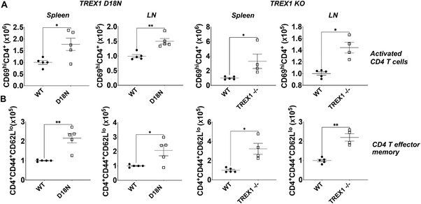
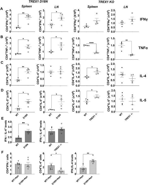
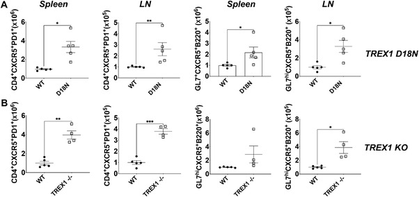
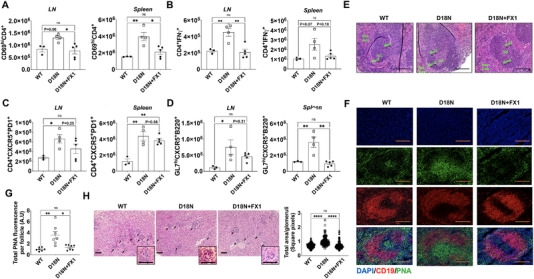
The Three Prime Repair EXonuclease I (TREX1) is critical for degrading post-apoptosis DNA. Mice expressing catalytically inactive TREX1 (TREX1 D18N) develop lupus-like autoimmunity due to chronic sensing of undegraded TREX1 DNA substrates, production of the inflammatory cytokines, and the inappropriate activation of innate and adaptive immunity. This study aimed to investigate Thelper (Th) dysregulation in the TREX1 D18N model system as a potential mechanism for lupus-like autoimmunity. Comparison of immune cells in secondary lymphoid organs, spleen and peripheral lymph nodes (LNs) between TREX1 D18N mice and the TREX1 null mice revealed that the TREX1 D18N mice exhibit a Th1 bias. Additionally, the T-follicular helper cells (Tfh) and the germinal celter (GC) B cells were also elevated in the TREX1 D18N mice. Targeting Bcl6, a lineage-defining transcription factor for Tfh and GC B cells, with a commercially available Bcl6 inhibitor, FX1, attenuated Tfh, GC, and Th1 responses, and rescued TREX1 D18N mice from autoimmunity. The study presents Tfh and GC B-cell responses as potential targets in autoimmunity and that Bcl6 inhibitors may offer therapeutic approach in TREX1-associated or other lupus-like diseases.

Keywords: Bcl6; TREX1; Tfh; Th1; autoimmunity; inflammation.


Conflict of interest statement

RS is an inventor for US patents 6897041 and 9840545 licensed to Slate Bio Inc., holds equity and is a consultant for Slate Bio Inc., unrelated to this work. The research was conducted in the absence of any commercial or financial relationships that could pose a potential conflict of interest.

Figures

Figure 1
Figure 1
Elevated levels of CD4 T helper cells and T effector memory (Tem) in TREX1 deficiency. Spleen and pooled peripheral lymph nodes (cervical, axillary, brachial, and inguinal lymph nodes) were obtained from female (8–12 weeks old) TREX1 D18N and TREX1 knockout mice and levels of (A) activated CD4+ T cells (CD4+CD69hi) and (B) Tem status (CD4+CD44+CD62Llo) were analyzed by flow cytometry. Absolute number data are presented as mean ± SEM relative to WT mice (normalized absolute numbers) and a total of four to five mice per group over three independent experiments, with the dots representing individual mice. *p < 0.01, **p < 0.001 as calculated by two‐tailed unpaired Student's t‐test and Mann–Whitney test. Also see Supporting information Fig. S1 for data in percentages.
Figure 2
Figure 2
Status of T‐helper cytokines in TREX1 deficiency. Spleen and lymph nodes (LN) from female TREX1 D18N and TREX1 knockout mice were stimulated with PMA/Ionomycin for 5 h and levels of Th1 cytokines IFN‐γ (A), TNF‐α (B), and Th2 cytokines IL‐4 (C) and IL‐5 (D) were analyzed by flow cytometry. (E) The ratio of IFN‐γ to IL‐4 producing CD4 T cells indicated a significant Th1 bias as shown in the spleen of TREX1 D18N mice, but did not reach statistical significance in TREX1 KO animals. (F) In vitro T‐cell stimulation assay using BM‐derived macrophages as described in the Materials and Methods showed that TREX D18N macrophages induced similar levels of %IFN‐γ producing T cells, but much fewer %IL‐4 producing cells. A ratio of IFN‐γ/IL‐4 confirmed the greater Th1 bias induced by the TREX1 D18N macrophages. Data presented as mean ± SEM relative to WT mice (normalized absolute numbers) and a total of four to five mice per group over three independent experiments, with the dots representing individual mice, and three independent experiments for in vitro assays. *p < 0.01, **p < 0.001 as calculated by two‐tailed unpaired Student's t‐test. Also see Supporting information Fig. S2 for data in percentages.
Figure 3
Figure 3
Elevated T‐follicular helper (Tfh) cell response and germinal center reaction in TREX1 deficiency. Levels of Tfh cells (CD4+CXCR5+PD1+) and GL7hiCXCR5+B220+ B‐cells in (A) TREX1 D18N and (B) TREX1 KO female mice analyzed by flow cytometry. Data presented as mean ± SEM relative to WT mice (normalized absolute numbers) and a total of four to five mice per group over three independent experiments, with the dots representing individual mice. *p < 0.01, **p < 0.001, ***p < 0.0001 as calculated by two‐tailed unpaired Student's t‐test. Also see Supporting information Fig. S3 for data in percentages.
Figure 4
Figure 4
Bcl6 inhibition rescues from autoimmunity in TREX1 D18N mutant mice. Mice were treated as described in Materials and Methods and analyzed by flow cytometry for (A) Activated (CD69+) CD4; (B) IFN‐γ producing CD4 T cells; (C) CD4+CXCR5+PD1+ T follicular helper cells; and (D) GL7hiCXCR5+B220+ B cells; (E) Representative H&E stained sections of the spleens. Spleens from WT, D18N mutant DMSO control, and D18N mutant FX1‐treated animals were stained with H&E and screened for germinal centers (GC), Mantle zone (ManZ), Marginal zone (MarZ), and Red pulp, Scale bars 50 μm. (F) Immunofluorescence for PNA/CD19/DAPI, Scale bars 50 μm and (G) Quantification of total PNA fluorescence per splenic follicle by ImageJ. (H) Representative H & E stained sections of the kidneys and quantification of total area per glomeruli by ImageJ, Scale bars 50 μm (Insert shows glomeruli). Arrows point to individual glomeruli. Data are shown as mean ± SEM and show a total of three to five mice per group over two independent experiments and shown as relative to D18N animals. *p < 0.01, **p < 0.001, ***p < 0.0001 as calculated by multiple comparison by one‐way ANOVA. Also see Supporting information Fig. S4 for data in percentages and GL‐7 staining for GC B cells.

References

    1. Lu, Q. and Lemke, G. , Homeostatic regulation of the immune system by receptor tyrosine kinases of the Tyro 3 family. Science 2001. 293: 306–311. - PubMed
    1. Cohen, P. L. , Caricchio, R. , Abraham, V. , Camenisch, T. D. , Jennette, J. C. , Roubey, R. A. , Earp, H. S. et al., Delayed apoptotic cell clearance and lupus‐like autoimmunity in mice lacking the c‐mer membrane tyrosine kinase. J. Exp. Med. 2002. 196: 135–140. - PMC - PubMed
    1. Hanayama, R. , Tanaka, M. , Miyasaka, K. , Aozasa, K. , Koike, M. , Uchiyama, Y. and Nagata, S. , Autoimmune disease and impaired uptake of apoptotic cells in MFG‐E8‐deficient mice. Science 2004. 304: 1147–1150. - PubMed
    1. Perrino, F. W. , Miller, H. and Ealey, K. A. , Identification of a 3'→5'‐exonuclease that removes cytosine arabinoside monophosphate from 3' termini of DNA. J. Biol. Chem. 1994. 269: 16357–16363. - PubMed
    1. Mazur, D. J. and Perrino, F. W. , Identification and expression of the TREX1 and TREX2 cDNA sequences encoding mammalian 3'→5' exonucleases. J. Biol. Chem. 1999. 274: 19655–19660. - PubMed