

Swallowing assessment in patients with dysphagia: Validity and reliability of a pocket-sized ultrasound system
- PMID: 35112768
- PMCID: PMC9305130
- DOI: 10.1111/1460-6984.12703
Swallowing assessment in patients with dysphagia: Validity and reliability of a pocket-sized ultrasound system
Abstract
Background: The use of ultrasound as an adjunct to clinical swallowing evaluation provides quantitative physiological and morphological data. As a low-risk procedure, ultrasound imaging can be performed outside of a medical setting. This is particularly important for patients living in rural areas with restricted access to a hospital. Technical advances have produced pocket-sized ultrasound technology that is more affordable, and therefore within the fiscal reach of most allied health services.
Aims: To explore the validity and reliability of pocket-sized ultrasound technology in dysphagia assessment.
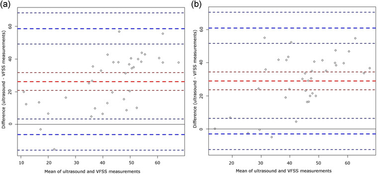
Methods & procedures: Data were acquired from 43 patients with dysphagia using the Clarius ultrasound device. Ultrasound and videofluoroscopic measures of hyoid and laryngeal displacement during liquid and puree swallowing were collected concurrently to quantify correlation and agreement between identical measures derived from the two instruments. Reliability of ultrasound was assessed for measures of hyoid and laryngeal displacement, tongue thickness, and size of the submental muscles in eight patients. Reliability was evaluated for the entire process of data acquisition including scanning and online measurement using an iPad in a clinical setting and for offline measurement on a computer screen to explore environmental influences on reliability.
Outcomes & results: Results revealed poor correlation between the measures of interest across instruments. Reliability of the entire process of data acquisition in a clinical setting was insufficient while reliability was more promising for offline measurements.
Conclusions & implications: The clinical use of pocket-sized ultrasound devices, such as the Clarius system, for swallowing evaluation is not indicated at this time. Enhanced validity and reliability of the entire process of data acquisition are needed prior to clinical translation of such technology.
What this paper adds: What is already known on the subject The use of ultrasound allows for radiation-free, non-invasive swallowing assessment. Some data suggest that ultrasound is valid and reliable in the evaluation of swallowing using standard-sized equipment. Insufficient validity and reliability have been reported for pocket-sized ultrasound technology in the assessment of healthy swallowing. What this paper adds to existing knowledge This research is the first to provide validity and reliability data of the pocket-sized Clarius technology in the evaluation of swallowing in patients with dysphagia. Insufficient validity and reliability of online data acquisition in a clinical environment were found. Reliability for offline measurement was more promising. What are the potential or actual clinical implications of this work? The clinical use of pocket-sized ultrasound devices, such as the Clarius system, for swallowing assessment is not indicated at this time.
Keywords: deglutition; dysphagia; reliability; ultrasound; validity; videofluoroscopy.
© 2022 The Authors. International Journal of Language & Communication Disorders published by John Wiley & Sons Ltd on behalf of Royal College of Speech and Language Therapists.
Conflict of interest statement
The authors declare no conflict of interest.
Figures


References
-
- Allen, M. (2017) Correlation, Pearson. The SAGE encyclopedia of communication research methods. Thousand Oaks, CA: SAGE Publications.
-
- Bartlett, J.W. & Frost, C. (2008) Reliability, repeatability and reproducibility: analysis of measurement errors in continuous variables. Ultrasound in Obstetrics and Gynecology, 31, 466–475. - PubMed
-
- Bates, D. , Mächler, M. , Bolker, B. & Walker, S. (2015) Fitting linear mixed‐effects models using {lme4}. Journal of Statistical Software, 67, 1–48.
-
- Daniels, S.K. & Huckabee, M.L. (2014) Dysphagia following stroke, San Diego: Plural Publishing.
Publication types
MeSH terms
LinkOut - more resources
Full Text Sources
Medical
Research Materials
