Clinical Trial
. 2022 Mar 1;140(3):243-249.
doi: 10.1001/jamaophthalmol.2021.6067.

Association of Pegcetacoplan With Progression of Incomplete Retinal Pigment Epithelium and Outer Retinal Atrophy in Age-Related Macular Degeneration: A Post Hoc Analysis of the FILLY Randomized Clinical Trial

Affiliations
Clinical Trial

Association of Pegcetacoplan With Progression of Incomplete Retinal Pigment Epithelium and Outer Retinal Atrophy in Age-Related Macular Degeneration: A Post Hoc Analysis of the FILLY Randomized Clinical Trial

Muneeswar Gupta Nittala et al. JAMA Ophthalmol. .

Abstract

Importance: Change in areas of incomplete retinal pigment epithelium (RPE) and outer retinal atrophy (iRORA) within eyes with geographic atrophy (GA) might reflect similar changes among eyes with drusen but no GA.

Objective: To evaluate the potential association of pegcetacoplan with progression of iRORA in eyes with GA secondary to AMD.

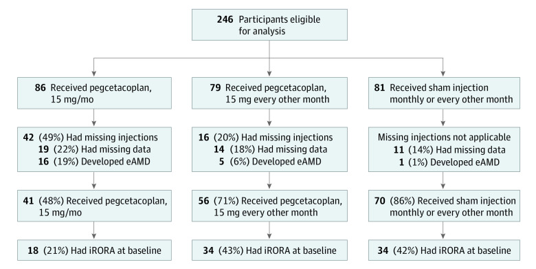
Design, setting, and participants: This post hoc analysis of the phase 2 multicenter, randomized, single-masked, sham-controlled FILLY trial of intravitreal pegcetacoplan for 12 months took place from February 2 to July 7, 2020. Participants comprised 167 patients with GA secondary to AMD who received pegcetacoplan monthly (n = 41) or every other month (n = 56) or a sham injection (n = 70) in the FILLY trial, completed the month 12 study visit, and did not develop exudative AMD.

Interventions: Intravitreal pegcetacoplan, 15 mg, or sham injection, monthly or every other month for 12 months.

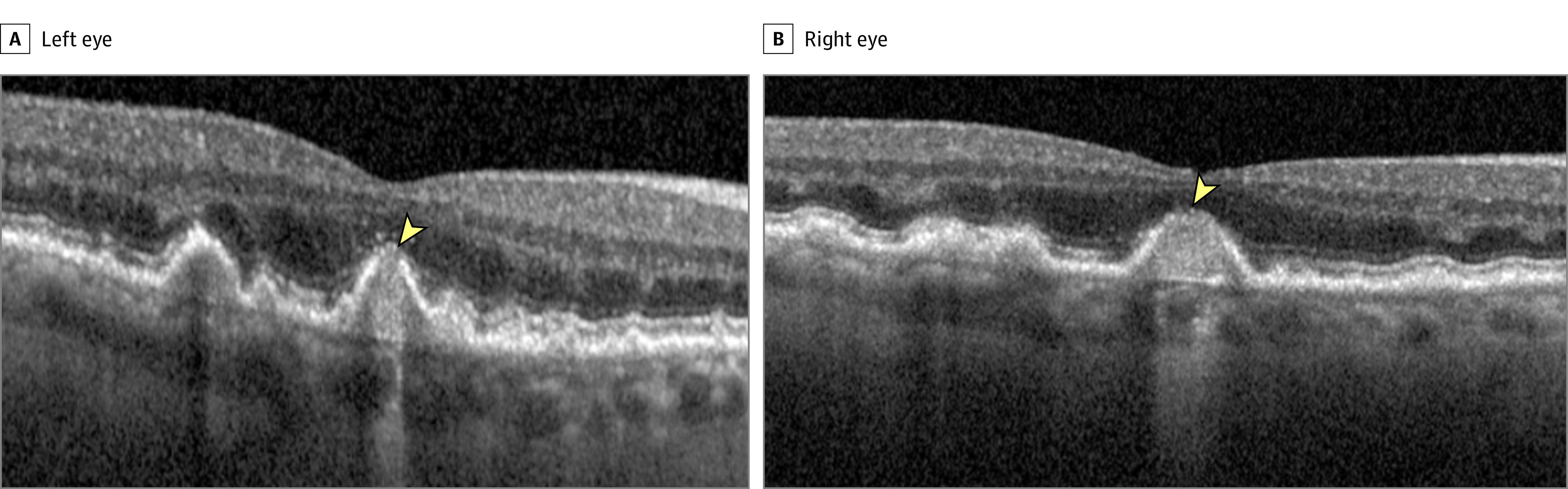
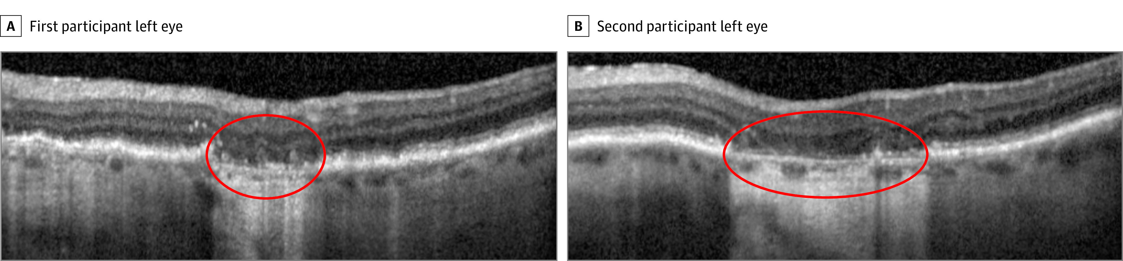
Main outcomes and measures: Masked readers analyzed spectral-domain optical coherence tomography scans in regions beyond a perimeter of 500 μm from the GA border according to the Classification of Atrophy Meetings criteria. Primary outcome measures were progression from iRORA to complete RPE and outer retina atrophy (cRORA) from baseline to 6 and 12 months.

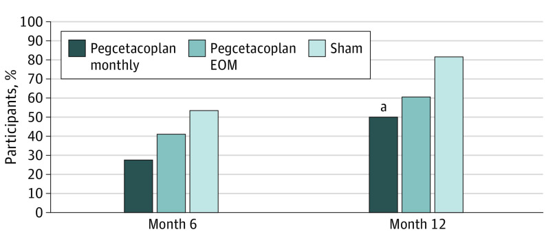
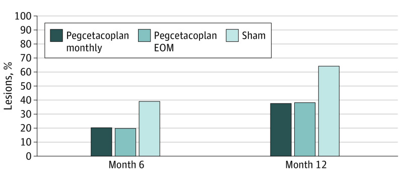
Results: Among the 167 patients in the study, at baseline, iRORA was present in 45.0% of study eyes (18 of 40) in the pegcetacoplan monthly group, 61.8% of study eyes (34 of 55) in the pegcetacoplan every other month group, and 50.7% of study eyes (34 of 67) in the sham group. At 12 months, progression from iRORA to cRORA occurred in 50.0% of study eyes (9 of 18) in the pegcetacoplan monthly group (P = .02 vs sham), 60.6% of study eyes (20 of 33) in the pegcetacoplan every other month group (P = .06 vs sham), and 81.8% of study eyes (27 of 33) in the sham group. Compared with sham treatment, the relative risk of progression at 12 months from iRORA to cRORA was 0.61 (95% CI, 0.37-1.00) for eyes in the pegcetacoplan monthly group and 0.74 (95% CI, 0.54-1.02) for eyes in the pegcetacoplan every other month group.

Conclusions and relevance: Eyes receiving intravitreal pegcetacoplan had lower rates of progression from iRORA to cRORA compared with controls, suggesting a potential role for pegcetacoplan therapy earlier in the progression of AMD prior to the development of GA.

Trial registration: ClinicalTrials.gov Identifier: NCT02503332.


Conflict of interest statement

Conflict of Interest Disclosures: Dr Metlapally reported having a patent pending relevant to this manuscript. Dr Ip reported receiving personal fees from Novartis, Genentech, Allergan, Regeneron, RegenexBio, Apellis, Aerie, Alimera, Amgen, Cell Lineage Therapeutics, Clearside, and OccuRX outside the submitted work. Dr Chakravarthy reported receiving payment or honoraria for lectures, presentations, speaker’s bureaus, manuscript writing, or educational events from Apellis, Iveric Bio, Novartis, and Hoffmann-La Roche; and participating as chair on a Data Safety Monitoring Board for trials sponsored by Bayer outside the submitted work. Dr Holz reported receiving grants and personal fees from Apellis and Heidelberg Engineering during the conduct of the study; grants from Genentech/Roche, Novartis, Bayer, Pixium Vision, Optos, Iveric Bio, and Gyroscope Therapeutics; and personal fees from Genentech/Roche, Novartis, Bayer, and Iveric Bio outside the submitted work. Dr Staurenghi reported receiving personal fees from Apellis and Iveric Bio during the conduct of the study; receiving personal fees and grants from Novartis, Bayer, and Centervue; personal fees from Boehringer Ingelheim, Heidelberg Engineering, Genentech/ Roche, Topcon, and ORA; and grants from Carl Zeiss Meditec, Canon, Nidek, and Optovue outside the submitted work. Dr Waheed reported receiving personal fees from Apellis Pharmaceuticals and Nidek; personal fees and equity from Gyroscope Therapeutics; and nonfinancial support from Carl Zeiss Meditec, Optovue, and Heidelberg Software outside the submitted work. Mr Koester reported receiving personal fees from and being a shareholder in Apellis Pharmaceuticals during the conduct of the study. Dr Sadda reported receiving grants from Apellis Pharmaceuticals during the conduct of the study; personal fees from Apellis Pharmaceuticals, Iveric Bio, Amgen, Allergan/AbbVie, Regeneron, Novartis, Roche/Genentech, 4DMT, Janssen, Nanoscope, Optos, Centervue, and Heidelberg; and nonfinancial support from Carl Zeiss Meditec, Nidek, Topcon, Optos, and Heidelberg outside the submitted work. No other disclosures were reported.

Figures

Figure 1.
Figure 1.. Participant Flow Diagram
eAMD indicates exudative age-related macular degeneration; iRORA, incomplete retinal pigment epithelium and outer retina atrophy.
Figure 2.
Figure 2.. Incomplete Retinal Pigment Epithelium (RPE) and Outer Retina Atrophy (iRORA) at Baseline in Eyes of 2 Participants
The arrowhead indicates presence of iRORA. RPE disruption and hypertransmission are evident, with some loss of the outer retinal bands at the apex of the lesion. The region of RPE disruption is less than 250 μm in both cases. The case in the left eye is a borderline example; the hypertransmission is 250 μm or more, but there appears to be some partial RPE preservation in the temporal section of the region of hypertransmission, and the zone of RPE disruption is less than 250 μm.
Figure 3.
Figure 3.. Complete Retinal Pigment Epithelium and Outer Retina Atrophy (cRORA) at Follow-up in Eyes of 2 Participants
The area within the red oval indicates the presence of cRORA, characterized by loss of the retinal pigment epithelium band and associated hypertransmission into the choroid (≥250 μm in diameter) accompanied by subsidence of the outer plexiform layer and inner nuclear layer. The external limiting membrane, ellipsoid zone, and interdigitation zone are disrupted.
Figure 4.
Figure 4.. Progression From Incomplete Retinal Pigment Epithelium and Outer Retina Atrophy (iRORA) to Complete Retinal Pigment Epithelium and Outer Retina Atrophy (cRORA)
Pearson χ2 test: month 6, P = .08 for pegcetacoplan monthly and P = .32 for pegcetacoplan every other month (EOM); month 12, P = .02 for pegcetacoplan monthly and P = .06 for pegcetacoplan EOM. aStatistically significant at P < .05.
Figure 5.
Figure 5.. Proportions of Incomplete Retinal Pigment Epithelium and Outer Retina Atrophy (iRORA) Lesions at Baseline That Converted to Complete Retinal Pigment Epithelium and Outer Retina Atrophy (cRORA) Lesions by Month 6 or 12
EOM indicates every other month.

Comment in

References

    1. Holz FG, Strauss EC, Schmitz-Valckenberg S, van Lookeren Campagne M. Geographic atrophy: clinical features and potential therapeutic approaches. Ophthalmology. 2014;121(5):1079-1091. doi:10.1016/j.ophtha.2013.11.023 - DOI - PubMed
    1. Rahimy E, Khan MA, Chao W, Ribeiro R, Ho A, Holekamp N. Evaluation of geographic atrophy (GA) secondary to AMD in real-world clinical practice: analysis of the AAO IRIS registry. Presented at the American Academy of Ophthalmology Annual Meeting; November 13, 2020; virtual.
    1. Schmitz-Valckenberg S, Sahel JA, Danis R, et al. . Natural history of geographic atrophy progression secondary to age-related macular degeneration (Geographic Atrophy Progression Study). Ophthalmology. 2016;123(2):361-368. doi:10.1016/j.ophtha.2015.09.036 - DOI - PubMed
    1. Shen LL, Sun M, Ahluwalia A, et al. . Relationship of topographic distribution of geographic atrophy to visual acuity in nonexudative age-related macular degeneration. Ophthalmol Retina. 2021;5(8):761-774. doi:10.1016/j.oret.2020.11.003 - DOI - PubMed
    1. Rudnicka AR, Kapetanakis VV, Jarrar Z, et al. . Incidence of late-stage age-related macular degeneration in American Whites: systematic review and meta-analysis. Am J Ophthalmol. 2015;160(1):85-93.e3. doi:10.1016/j.ajo.2015.04.003 - DOI - PubMed

Publication types

Associated data