

Cerebral oxygen metabolic stress is increased in children with sickle cell anemia compared to anemic controls
- PMID: 35113471
- PMCID: PMC9081128
- DOI: 10.1002/ajh.26485
Cerebral oxygen metabolic stress is increased in children with sickle cell anemia compared to anemic controls
Abstract
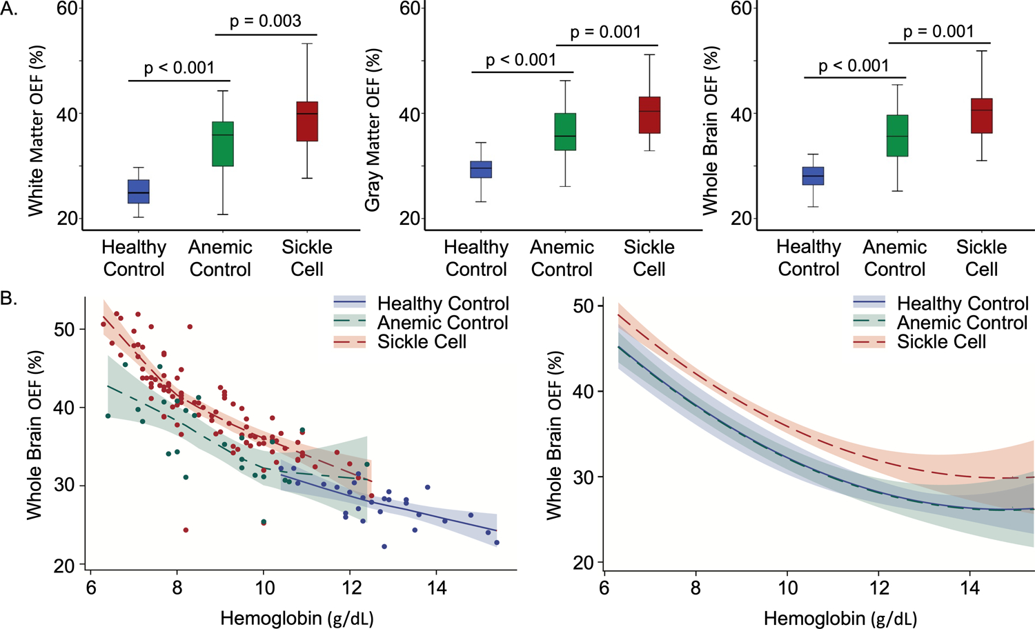
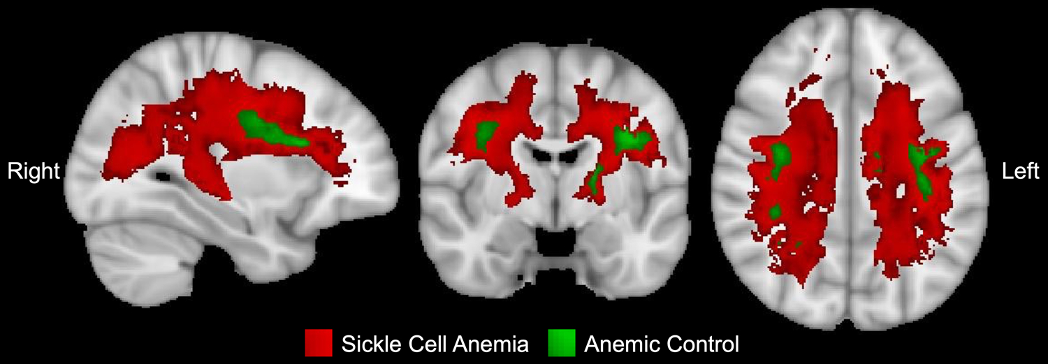
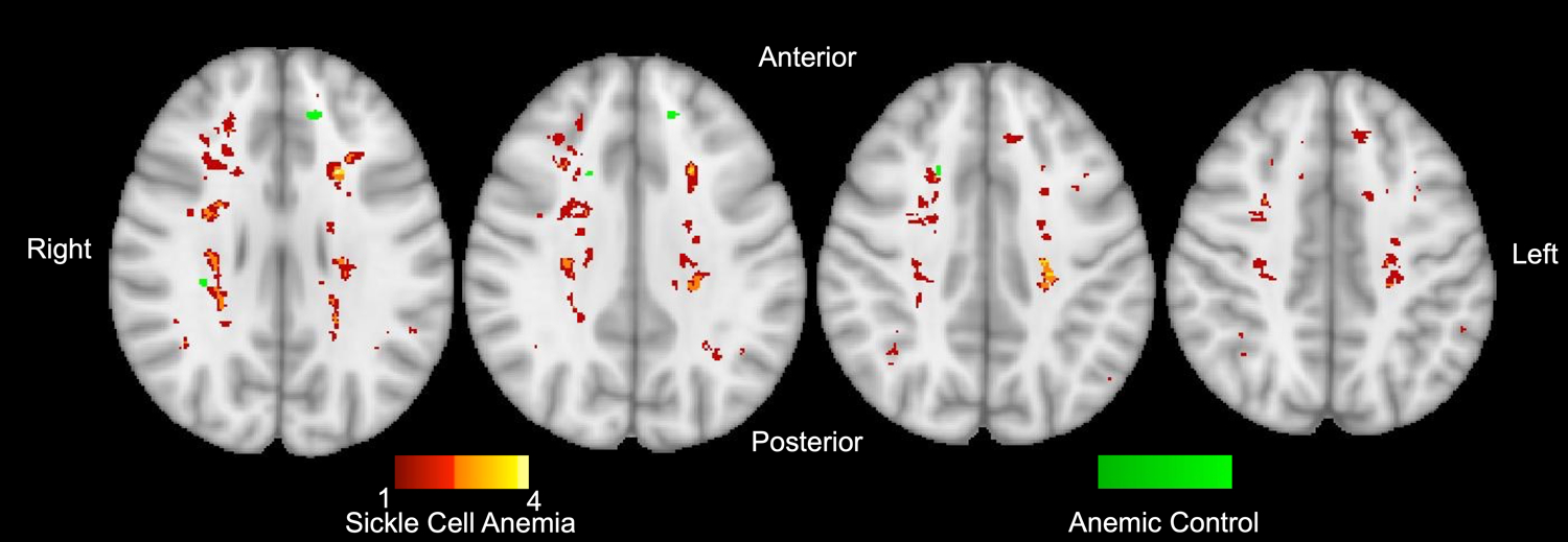
Patients with sickle cell anemia (SCA) experience cerebral metabolic stress with an increase in oxygen extraction fraction (OEF) to compensate for reduced oxygen carrying capacity due to anemia. It remains unclear if anemia alone drives this metabolic stress. Using MRI, we collected voxel-wise OEF measurements to test our hypothesis that OEF would be elevated in anemic controls without SCA (AC) compared to healthy controls (HC), but OEF would be even higher in SCA compared to AC. Brain MRIs (N = 159) were obtained in 120 participants (34 HC, 27 AC, 59 SCA). While hemoglobin was lower in AC versus HC (p < 0.001), hemoglobin was not different between AC and SCA cohorts (p = 0.459). Whole brain OEF was higher in AC compared to HC (p < 0.001), but lower compared to SCA (p = 0.001). Whole brain OEF remained significantly higher in SCA compared to HC (p = 0.001) while there was no longer a difference between AC versus HC (p = 0.935) in a multivariate model controlling for age and hemoglobin. OEF peaked within the border zone regions of the brain in both SCA and AC cohorts, but the volume of white matter with regionally elevated OEF in AC was smaller (1.8%) than SCA (58.0%). While infarcts colocalized within regions of elevated OEF, more SCA participants had infarcts than AC (p < 0.001). We conclude that children with SCA experience elevated OEF compared to AC and HC after controlling for the impact of anemia, suggesting that there are other pathophysiologic factors besides anemia contributing to cerebral metabolic stress in children with SCA.
© 2022 Wiley Periodicals LLC.
Conflict of interest statement
Potential Conflicts of Interest: M.E.F declares equity ownership in Proclara Biociences, a biopharmaceutical company developing therapies for Alzheimer’s Disease. M.E.F., A.L.F., and M.L.H received one-time compensation for scientific advisory board participation with Bluebird Bio, who is developing gene therapy trials for sickle cell disease. M.L.H performs ongoing consulting for Bluebird Bio. M.E.F. works as a consultant for Global Blood Therapeutics, a company manufacturing Oxbryta for sickle cell disease. M.L.H receives research support from Global Blood Therapeutics and from FORMA Therapeutics, a pharmaceutical company developing potential sickle cell disease treatments. M.L.H.’s spouse is employed by Pfizer, Inc. M.M.B. is employed by OpenCell Technologies, LLC., a device manufacturer for gene-editing.
Figures
References
-
- Ware RE, de Montalembert M, Tshilolo L, Abboud MR. Sickle cell disease. Lancet. 2017;390(10091):311–323. - PubMed
-
- Bernaudin F, Verlhac S, Arnaud C, et al. Chronic and acute anemia and extracranial internal carotid stenosis are risk factors for silent cerebral infarcts in sickle cell anemia. Blood. 2015;125(10):1653–1661. - PubMed
-
- Ohene-Frempong K, Weiner SJ, Sleeper LA, et al. Cerebrovascular accidents in sickle cell disease: rates and risk factors. Blood. 1998;91(1):288–294. - PubMed
-
- Wang W, Enos L, Gallagher D, et al. Neuropsychologic performance in school-aged children with sickle cell disease: a report from the Cooperative Study of Sickle Cell Disease. J Pediatr. 2001;139(3):391–397. - PubMed
Publication types
MeSH terms
Substances
Grants and funding
LinkOut - more resources
Full Text Sources
Medical