Review
. 2022 Apr 26:40:559-587.
doi: 10.1146/annurev-immunol-101320-011829. Epub 2022 Feb 3.

T Cell Responses to the Microbiota

Affiliations
Review

T Cell Responses to the Microbiota

Ivaylo I Ivanov et al. Annu Rev Immunol. .

Abstract

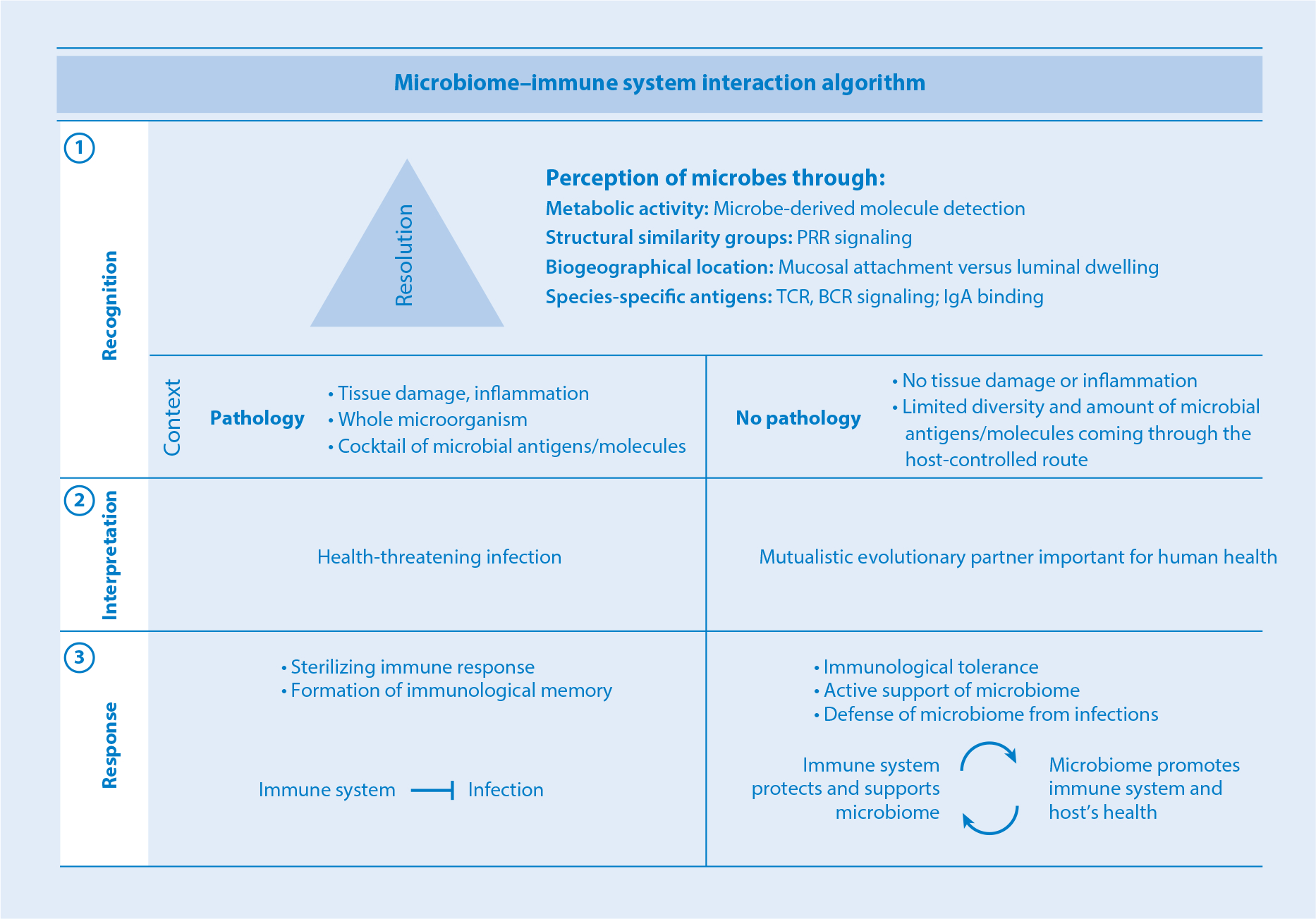
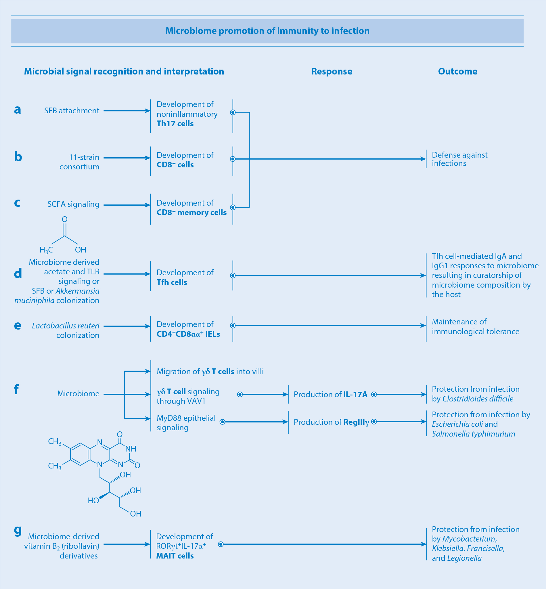
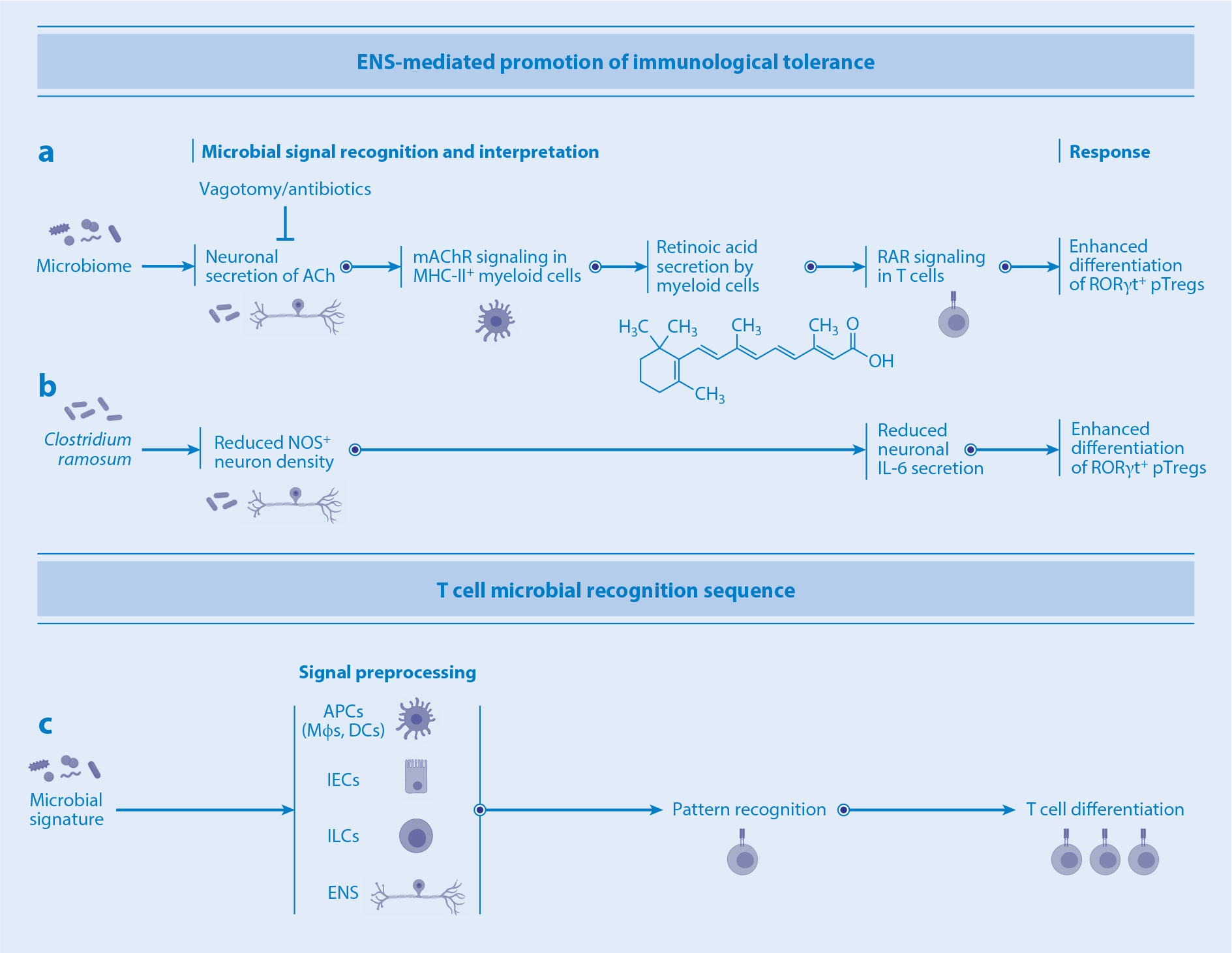
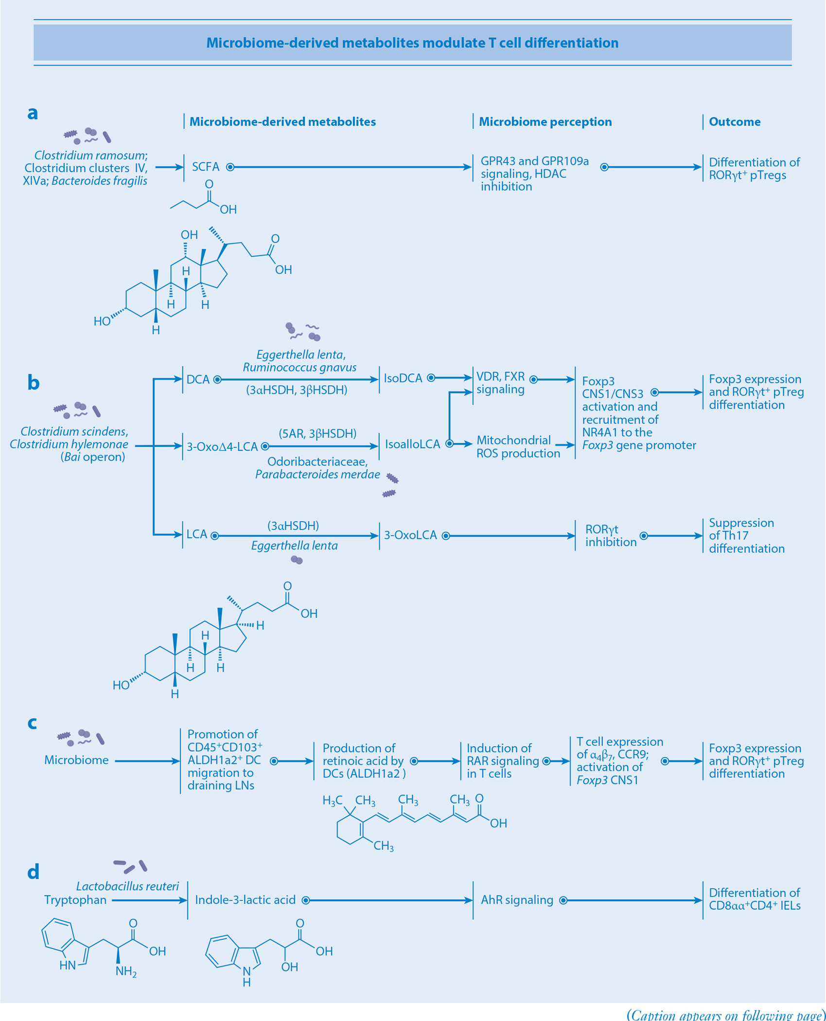
The immune system employs recognition tools to communicate with its microbial evolutionary partner. Among all the methods of microbial perception, T cells enable the widest spectrum of microbial recognition resolution, ranging from the crudest detection of whole groups of microbes to the finest detection of specific antigens. The application of this recognition capability to the crucial task of combatting infections has been the focus of classical immunology. We now appreciate that the coevolution of the immune system and the microbiota has led to development of a lush immunological decision tree downstream of microbial recognition, of which an inflammatory response is but one branch. In this review we discuss known T cell-microbe interactions in the gut and place them in the context of an algorithmic framework of recognition, context-dependent interpretation, and response circuits across multiple levels of microbial recognition resolution. The malleability of T cells in response to the microbiota presents an opportunity to edit immune response cellularity, identity, and functionality by utilizing microbiota-controlled pathways to promote human health.

Keywords: MAIT cells; Th17 cells; Tregs; dendritic cells; enteric nervous system; intestinal microbiota; segmented filamentous bacteria; γδ T cells.


Figures

Figure 1
Figure 1
Microbiota–immune system interaction algorithm. Immune microbial perception involves three steps: (①) recognition, (②) context-dependent interpretation, and (③) response. Depending on the function of the recognition circuit, different resolution may be required. At the low end of the resolution spectrum, groups of microbes are recognized via detection of microbial metabolites. The next level up involves detection of common structural patterns, for instance, via PRR signaling. Higher still are circuits involving detection of species-specific coevolutionary adaptations, such as the ability to colonize epithelium. The highest resolution characterizes antigen-specific recognition circuits. Following detection, the microbial signature is classified either as pathological or as commensal/mutualistic according to context, including the presence or absence of inflammation, tissue damage, and mode of antigen acquisition. Signatures determined to be infectious are sterilized, while those perceived as mutualistic are tolerated and receive support and protection. Abbreviations: BCR, B cell receptor; IgA, immunoglobulin A; PRR, pattern recognition receptor; TCR, T cell receptor.
Figure 2
Figure 2
How the microbiota promotes immunity to infection. (a–c) Attachment of SFB to the intestinal epithelium, colonization by an 11-strain consortium, and SCFA signaling promote differentiation of noninflammatory Th17, CD8+, and memory CD8+ T cells, respectively, ultimately bolstering the host’s and microbiota’s protection from infections. (d) Microbiome-derived acetate and TLR signaling or colonization with SFB or Akkermansia muciniphila promote differentiation of Tfh cells, which in turn supports the development of IgA and IgG1 responses. (e) Colonization with Lactobacillus reuteri promotes differentiation of regulatory CD4+CD8αα+ IELs, thus enhancing intestinal immunological tolerance. (f) Microbial signals shape the biogeographical distribution of γδ T cells within the epithelial layer. Microbial signaling through guanine nucleotide exchange factor VAV1 on γδ T cells stimulates production of IL-17A, bolstering protection against Clostridioides difficile infection. MyD88 epithelial signaling triggers secretion by γδ T cells of the antimicrobial peptide RegIIIγ, conferring protection against Escherichia coli and Salmonella typhimurium infections. (g) Microbiota-derived vitamin B2 derivatives promote development of MAIT cells, conferring protection against infection by Mycobacterium, Klebsiella, Francisella, and Legionella. Abbreviations: IEL, intraepithelial lymphocyte; Ig, immunoglobulin; IL, interleukin; MAIT cell, mucosal-associated invariant T cell; SCFA, short-chain fatty acid; SFB, segmented filamentous bacteria; Tfh cell, T follicular helper cell; Th17, T helper 17; TLR, Toll-like receptor.
Figure 3
Figure 3
Innate immune cells and enteric neuronal modulation of T cell differentiation. (a) Microbial signals stimulate enteric neurons to secrete ACh, leading to the production of retinoic acid by myeloid cells. Retinoic acid in turn promotes pTreg differentiation via RAR signaling. (b) Clostridium ramosum reduces NOS+ IL-6-secreting neurons, promoting pTreg differentiation. (c) The T cell microbial recognition sequence involves preprocessing of microbial signals by APCs and/or IECs, ILCs, or the ENS prior to the recognition and differentiation stages. Abbreviations: ACh, acetylcholine; APC, antigen-presenting cell; DC, dendritic cell; ENS, enteric nervous system; IEC, intestinal epithelial cell; IL, interleukin; ILC, innate lymphoid cell; mAChR, muscarinic acetylcholine receptor; MHC, major histocompatibility complex; Mφ, macrophage; NOS, nitric oxide synthase; pTreg, peripherally induced T regulatory cell; RAR, retinoic acid receptor.
Figure 4
Figure 4
Microbiota-derived metabolites modulate T cell differentiation. (a) SCFAs derived from Clostridium ramosum and Clostridia clusters IV and XIVa signal through GPR43 and GPR109a and inhibit HDAC in T cells, thus promoting pTreg differentiation. (b) Bai operon–carrying bacteria such as Clostridium scindens and Clostridium hylemonae produce DCA, 3-oxoΔ4-LCA, and LCA. DCA is subsequently converted to isoDCA by 3αHSDH- and 3βHSDH-carrying bacteria such as Eggerthella lenta and Ruminococcus gnavus. IsoDCA signals through the T cellular receptors VDR and FXR, triggering activation of CNS1/CNS3 regions of the Foxp3 gene, a process that ultimately leads to pTreg differentiation. 3-OxoΔ4-LCA is converted to isoalloLCA by the 5AR- and 3βHSDH-carrying bacteria Odoribacteraceae and Parabacteroides merdae. IsoalloLCA triggers mitochondrial ROS production and VDR, FXR, and NR4A1 signaling in T cells, promoting their differentiation into pTregs via CNS1/CNS3 activation. LCA is converted into 3-oxoLCA by 3αHSDH-carrying E. lenta. 3-OxoLCA then suppresses RORγt, inhibiting the Th17 route of differentiation. (c) Microbial signals promote the migration of CD45+CD103+ DCs to gLNs as well as the production of retinoic acid by DCs utilizing the ALDH1a2 enzyme. Retinoic acid receptor signaling on T cells leads to activation of Foxp3 CNS1 and expression of integrins α4β7 and CCR9, ultimately leading to pTreg differentiation. (d) Lactobacillus reuteri converts dietary tryptophan into indole-3-lactic acid, stimulating AhR signaling and resulting in CD8αα+CD4+ IEL differentiation. Abbreviations: AhR, aryl hydrocarbon receptor; CNS1/3, conserved noncoding sequence 1/3; DC, dendritic cell; DCA, deoxycholic acid; FXR, farnesoid X receptor; gLN, lymph node; HDAC, histone deacetylase; HSDH, hydroxysteroid dehydrogenase; IEL, intraepithelial lymphocyte; LCA, lithocholic acid; NR4A1, nuclear receptor subfamily 4 group A member 1; pTreg, peripherally induced T regulatory cell; RAR, retinoic acid receptor; ROS, reactive oxygen species; SCFA, short-chain fatty acid; Th17, T helper 17; 5AR, 5α-reductase.

References

    1. Ivanov II, Atarashi K, Manel N, Brodie EL, Shima T, et al. 2009. Induction of intestinal Th17 cells by segmented filamentous bacteria. Cell 139(3):485–98 - PMC - PubMed
    1. Gaboriau-Routhiau V, Rakotobe S, Lécuyer E, Mulder I, Lan A, et al. 2009. The key role of segmented filamentous bacteria in the coordinated maturation of gut helper T cell responses. Immunity 31(4):677–89 - PubMed
    1. Sczesnak A, Segata N, Qin X, Gevers D, Petrosino JF, et al. 2011. The genome of Th17 cell–inducing segmented filamentous bacteria reveals extensive auxotrophy and adaptations to the intestinal environment. Cell Host Microbe 10(3):260–72 - PMC - PubMed
    1. Atarashi K, Tanoue T, Ando M, Kamada N, Nagano Y, et al. 2015. Th17 cell induction by adhesion of microbes to intestinal epithelial cells. Cell 163(2):367–80 - PMC - PubMed
    1. Goto Y, Panea C, Nakato G, Cebula A, Lee C, et al. 2014. Segmented filamentous bacteria antigens presented by intestinal dendritic cells drive mucosal Th17 cell differentiation. Immunity 40(4):594–607 - PMC - PubMed

Publication types

LinkOut - more resources