Clinical Trial
. 2022 Apr 1;132(7):e152571.
doi: 10.1172/JCI152571.

Targeting TGF-β for treatment of osteogenesis imperfecta

Affiliations
Clinical Trial

Targeting TGF-β for treatment of osteogenesis imperfecta

I-Wen Song et al. J Clin Invest. .

Abstract

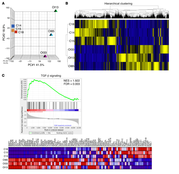
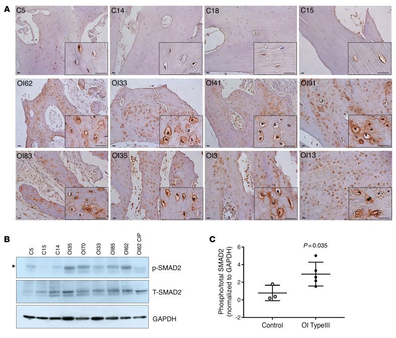
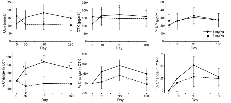
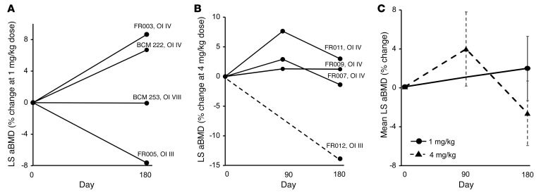
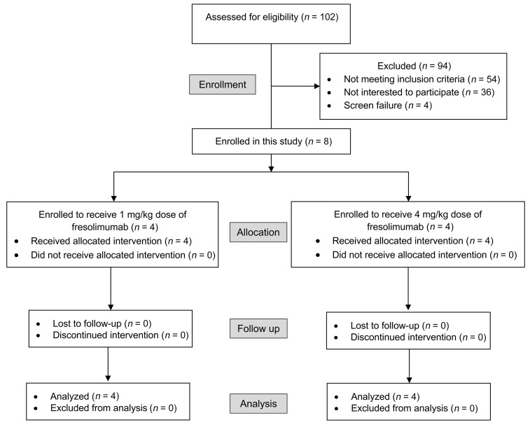
BACKGROUNDCurrently, there is no disease-specific therapy for osteogenesis imperfecta (OI). Preclinical studies demonstrate that excessive TGF-β signaling is a pathogenic mechanism in OI. Here, we evaluated TGF-β signaling in children with OI and conducted a phase I clinical trial of TGF-β inhibition in adults with OI.METHODSHistology and RNA-Seq were performed on bones obtained from children. Gene Ontology (GO) enrichment assay, gene set enrichment analysis (GSEA), and Ingenuity Pathway Analysis (IPA) were used to identify dysregulated pathways. Reverse-phase protein array, Western blot, and IHC were performed to evaluate protein expression. A phase I study of fresolimumab, a TGF-β neutralizing antibody, was conducted in 8 adults with OI. Safety and effects on bone remodeling markers and lumbar spine areal bone mineral density (LS aBMD) were assessed.RESULTSOI bone demonstrated woven structure, increased osteocytes, high turnover, and reduced maturation. SMAD phosphorylation was the most significantly upregulated GO molecular event. GSEA identified the TGF-β pathway as the top activated signaling pathway, and IPA showed that TGF-β1 was the most significant activated upstream regulator mediating the global changes identified in OI bone. Treatment with fresolimumab was well-tolerated and associated with increases in LS aBMD in participants with OI type IV, whereas participants with OI type III and VIII had unchanged or decreased LS aBMD.CONCLUSIONIncreased TGF-β signaling is a driver pathogenic mechanism in OI. Anti-TGF-β therapy could be a potential disease-specific therapy, with dose-dependent effects on bone mass and turnover.TRIAL REGISTRATIONClinicalTrials.gov NCT03064074.FUNDINGBrittle Bone Disorders Consortium (U54AR068069), Clinical Translational Core of Baylor College of Medicine Intellectual and Developmental Disabilities Research Center (P50HD103555) from National Institute of Child Health and Human Development, USDA/ARS (cooperative agreement 58-6250-6-001), and Sanofi Genzyme.

Keywords: Bone disease; Clinical Trials; Drug therapy; Therapeutics.


Figures

Figure 1
Figure 1. Transcriptomic and bioinformatics analyses demonstrate activation of TGF-β signaling in OI type III bone.
(A) Principal component analysis (PCA) plot of transcriptomic data from non-OI and OI type III bones in 3 principal component dimensions. (B) Hierarchical clustering based on Euclidian distance using RPKM of all non-OI and OI type III bone data. Blue, downregulated; yellow, upregulated. (C) Gene set enrichment plot demonstrated activation of TGF-β signaling. C18, C14, and C15 represent 3 biologically distinct non-OI bone samples. OI85, OI33, and OI31 represent 3 biologically distinct OI type III bone samples. The expression pattern of genes involved in the TGF-β gene set in the analysis database is shown. NES, normalized enrichment score; FDR, false discovery rate. Blue, downregulated; red, upregulated.
Figure 2
Figure 2. Increased phosphorylated SMAD2 in OI type III bone.
(A) IHC staining of phosphorylated SMAD2 (pSMAD2) in non-OI and OI type III bone sections. Higher-magnification images are shown in black boxes on the bottom right. Increase in pSMAD2 signal was detected in all OI samples, especially in the osteocytes. Scale bar: 20 μm. (B) Western blot of p-SMAD2 and total SMAD2 (T-SMAD2) in protein extracted from non-OI and OI type III bone. A total of 50 μg protein was loaded. One OI bone sample (OI62) was treated with calf-intestinal alkaline phosphatase (CIP) to remove phosphorylation signal to serve as a negative control for accurate pSMAD2 signal (indicated by arrowhead). See complete unedited blots in the supplemental material. (C) Quantification of Western blot in B, showing the ratio of phosphorylated (phospho) versus total SMAD2. Data are shown as the mean ± SD. GAPDH was used as loading control. C, non-OI (n = 3); OI, OI type III (n = 5).
Figure 3
Figure 3. Effect of fresolimumab on bone turnover markers and bone density.
The top row shows of serum levels of osteocalcin (Ocn), C-terminal telopeptide (CTX), and N-terminal propeptide of type 1 procollagen (P1NP) at each time point. The bottom row shows percentage changes in these markers of bone turnover as compared with baseline values. The solid lines with circles represent results for the 1 mg/kg dose cohort (n = 4), and the dotted lines with squares represent results for the 4 mg/kg dose cohort (n = 4). Data are shown as the mean ± SEM. *P < 0.05, GLM association.
Figure 4
Figure 4. Effect of fresolimumab on LS aBMD.
The percentage change in LS aBMD in (A) the 1 mg/kg dose cohort (n = 4) and (B) the 4 mg/kg dose cohort (n = 4). (C) Average aBMD changes at each time point based on dose. In B, aBMD could not be assessed in FR012 at the 90-day time point; therefore, the result is shown as a dotted line. In C, the solid lines represent results for the 1 mg/kg dose cohort, and the dotted lines represent results for the 4 mg/kg dose cohort. Data are shown as the mean ± SEM.
Figure 5
Figure 5. CONSORT flow diagram depicting screening, enrollment, and follow up of participants in the trial.

References

    1. Marini JC, et al. Osteogenesis imperfecta. Nat Rev Dis Primers. 2017;3:17052. doi: 10.1038/nrdp.2017.52. - DOI - PubMed
    1. Marom R, et al. Pharmacological and biological therapeutic strategies for osteogenesis imperfecta. Am J Med Genet C Semin Med Genet. 2016;172(4):367–383. doi: 10.1002/ajmg.c.31532. - DOI - PMC - PubMed
    1. Patel RM, et al. A cross-sectional multicenter study of osteogenesis imperfecta in North America - results from the linked clinical research centers. Clin Genet. 2015;87(2):133–140. doi: 10.1111/cge.12409. - DOI - PMC - PubMed
    1. Rossi V, et al. Osteogenesis imperfecta: advancements in genetics and treatment. Curr Opin Pediatr. 2019;31(6):708–715. doi: 10.1097/MOP.0000000000000813. - DOI - PMC - PubMed
    1. Tam A, et al. A multicenter study to evaluate pulmonary function in osteogenesis imperfecta. Clin Genet. 2018;94(6):502–511. doi: 10.1111/cge.13440. - DOI - PMC - PubMed

Publication types

Substances

Associated data