. 2022 Jun;11(11):e2102345.
doi: 10.1002/adhm.202102345. Epub 2022 Feb 18.

Organoids at the PUB: The Porcine Urinary Bladder Serves as a Pancreatic Niche for Advanced Cancer Modeling

Affiliations

Organoids at the PUB: The Porcine Urinary Bladder Serves as a Pancreatic Niche for Advanced Cancer Modeling

Michael Karl Melzer et al. Adv Healthc Mater. 2022 Jun.

Abstract

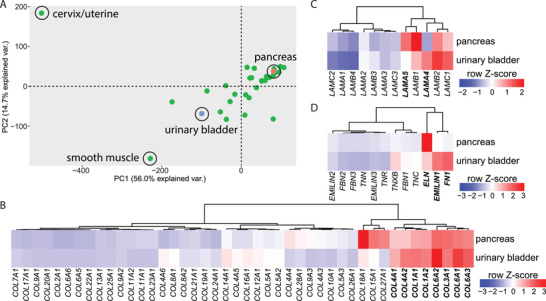
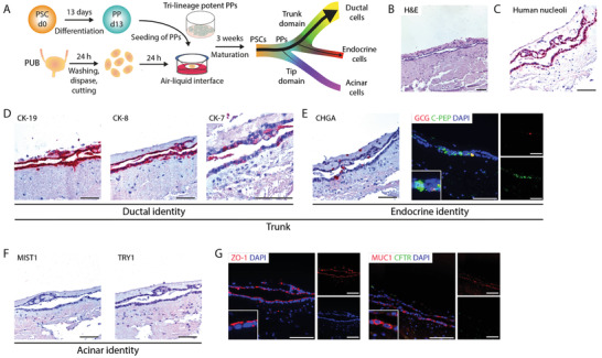
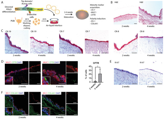
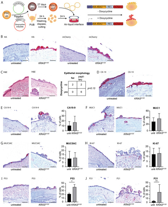
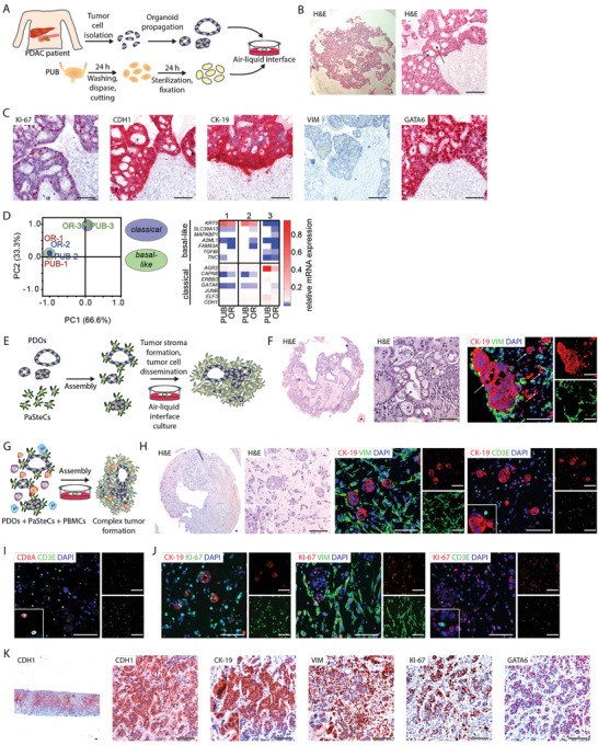
Despite intensive research and progress in personalized medicine, pancreatic ductal adenocarcinoma remains one of the deadliest cancer entities. Pancreatic duct-like organoids (PDLOs) derived from human pluripotent stem cells (PSCs) or pancreatic cancer patient-derived organoids (PDOs) provide unique tools to study early and late stage dysplasia and to foster personalized medicine. However, such advanced systems are neither rapidly nor easily accessible and require an in vivo niche to study tumor formation and interaction with the stroma. Here, the establishment of the porcine urinary bladder (PUB) is revealed as an advanced organ culture model for shaping an ex vivo pancreatic niche. This model allows pancreatic progenitor cells to enter the ductal and endocrine lineages, while PDLOs further mature into duct-like tissue. Accordingly, the PUB offers an ex vivo platform for earliest pancreatic dysplasia and cancer if PDLOs feature KRASG12D mutations. Finally, it is demonstrated that PDOs-on-PUB i) resemble primary pancreatic cancer, ii) preserve cancer subtypes, iii) enable the study of niche epithelial crosstalk by spiking in pancreatic stellate and immune cells into the grafts, and finally iv) allow drug testing. In summary, the PUB advances the existing pancreatic cancer models by adding feasibility, complexity, and customization at low cost and high flexibility.

Keywords: organ culture models; pancreatic cancer; stem cell differentiation; urinary bladder.


Conflict of interest statement

The authors declare no conflict of interest.

Figures

Figure 1
Figure 1
Transcriptomic analysis of the extracellular matrix of different organs reveals similarity between pancreas and urinary bladder. A) Principal component analysis of different organs based on the normalized RNA expression levels of extracellular matrix (ECM)‐filtered genes. Data were extracted from the Human Protein Atlas (https://www.proteinatlas.org/). Gene expression levels of different ECM genes including B) collagens, C) laminins, and D) tenascins, fibronectin, fibrillins, elastin, and emilins in urinary bladder and pancreas. Gene expression values depict row‐wise normalized Z‐scores and clustering was based on Ward.D2. PC, principal component; var., variance.
Figure 2
Figure 2
Pancreatic progenitors differentiate towards trunk lineage on the porcine urinary bladder. A) Schematic illustration of the culture of pancreatic progenitor (PP) cells on de‐epithelialized porcine urinary bladder. B) Hematoxylin and eosin (H&E) stained histological sections of PPs cultured on porcine urinary bladder (n = 2). Immunohistochemistry stainings for C) human nucleoli and D) duct‐specific cytokeratins CK‐19, CK‐8, and CK‐7 on PPs cultured on porcine urinary bladder (n = 2). E) Immunohistochemistry stainings for endocrine‐specific chromogranin A (CHGA) and immunofluorescence stainings for C‐peptide (C‐PEP, green) and glucagon (GCG, red) on PPs cultured on porcine urinary bladder (n = 2). Cells were counterstained with DAPI (blue). F) Immunohistochemistry stainings of acinar‐specific markers MIST1 and TRY1 on PPs cultured on porcine urinary bladder (n = 2). G) Immunofluorescence stainings for ZO‐1 (red) and for MUC1 (red) and CFTR (green) (n = 2). Cells were counterstained with DAPI (blue). Scale bars represent 100 µm. PSC, pluripotent stem cells; PUB, porcine urinary bladder.
Figure 3
Figure 3
Pancreatic duct‐like organoids mature to adult ductal cells on the porcine urinary bladder. A) Schematic representation illustrating the lineage‐specific generation of pancreatic duct‐like organoids (PDLOs) originated from pluripotent stem cells and their engraftment on the porcine urinary bladder. B) Hematoxylin and eosin (H&E) stained histological sections of PDLOs cultured on porcine urinary bladder for two and four weeks (n = 4). C) Immunohistochemistry for duct‐specific cytokeratins CK‐19, CK‐7, and CK‐8 (n = 4) and D) immunofluorescence stainings for ductal maturation marker MUC1 (red) and CFTR (green) on PDLOs cultured on porcine urinary bladder for two and four weeks, with quantification of CFTR‐positive cells (n = 4). Data are shown as mean ± SD. Cells were counterstained with DAPI (blue). *, p < 0.05. E) Immunohistochemistry staining for KI‐67 on PDLOs cultured on porcine urinary bladder for two and four weeks (n = 4). F) Immunofluorescence stainings for cell polarity marker ZO‐1 (red) and COL4A1 (green) in PDLOs cultured on porcine urinary bladder for two and four weeks (n = 4). Scale bars represent 100 µm. PPs, pancreatic progenitors; PSCs, pluripotent stem cells; PUB, porcine urinary bladder.
Figure 4
Figure 4
KRASG12D induces papillary neoplastic growth, dysplastic marker expression, and oncogene‐induced senescence. A) Schematic representation illustrating the propagation of pancreatic duct‐like organoids (PDLOs) harboring a Tet‐On expressing oncogenic HA‐tagged KRASG12D and mCherry on porcine urinary bladder. B) Immunohistochemistry stainings for HA‐tag and mCherry on vehicle and doxycycline‐treated Tet‐On expressing KRASG12D PDLOs cultured for four weeks on porcine urinary bladder (n = 4 for untreated (untr), n = 6 for doxycycline‐treated). C) Hematoxylin and eosin (H&E) stained histological sections of vehicle and doxycycline‐treated Tet‐On expressing KRASG12D PDLOs cultured for four weeks on porcine urinary bladder and contingency table comparing presence of papillary structures and doxycycline treatment (n = 4 for untreated, n = 6 for doxycycline‐treated). Immunohistochemistry stainings for D) CK‐19, E) CA19‐9, F) MUC1, G) MUC5AC, H) KI‐67, I) P53, and J) P21, and quantifications of respective positive cells in vehicle and doxycycline‐treated Tet‐On expressing KRASG12D PDLOs cultured for four weeks on porcine urinary bladder (n = 4 for untreated (untr), n = 6 for doxycycline‐treated). Data are shown as mean ± SD. *, p < 0.05, **, p < 0.01. Scale bars represent 100 µm. PSCs, pluripotent stem cells; PUB, porcine urinary bladder.
Figure 5
Figure 5
Pancreatic cancer patient‐derived organoids form stroma‐containing tumors on porcine urinary bladder. A) Schematic representation depicting the procedure for isolation, propagation, and engraftment of patient‐derived organoid (PDO) on porcine urinary bladder (PUB). B) Hematoxylin and eosin (H&E) stained histological sections of PDOs propagated on PUB for five days (n = 6). Arrows indicate tumor glands. C) Immunohistochemistry stainings for CDH1, CK‐19, VIM, KI‐67, and GATA6 of PDOs propagated on PUB for five days (n = 6). D) qRT‐PCR analysis (principal component analysis and heatmap of normalized RNA expression levels) of a relevant 14‐gene classical/basal‐like panel in PDOs propagated either on PUB or as 3D in vitro cultures (OR). E) Schematic representation illustrating the generation of assembloids consisting of PDOs and pancreatic stellate cells (PaSteCs) on PUB. F) H&E stained histological sections and immunofluorescence stainings for CK‐19 (red) and VIM (green) of PDO‐PaSteC assembloids cultured on PUB (n = 7). Cells were counterstained with DAPI (blue). Arrows indicate tumor glands/cancer cells. G) Schematic illustration depicting the generation of PDO, PaSteC, and peripheral blood mononuclear cells (PBMCs)‐containing assembloids on PUB. H) H&E stained histological sections and immunofluorescence stainings for CK‐19 (red) and VIM (green; left panel) or CD3E (green, right panel) of PDO‐PaSteC assembloids propagated on PUB for five days (n = 4). Immunofluorescence stainings for I) CD3E (green) and CD8A (red), and for J) KI‐67 (green on left panel; red on middle and right panels) and CK‐19 (red, left panel), VIM (green, middle panel), or CD3E (green, right panel) of PDO‐PaSteC‐PBMC assembloids propagated on PUB for five days (n = 4). K) Immunohistochemistry stainings for CDH1, CK‐19, VIM, KI‐67, and GATA6 of a pancreatic ductal adenocarcinoma patient liver metastasis biopsy. Scale bars represent 100 µm. PDAC, pancreatic ductal adenocarcinoma.

References

    1. Rahib L., Smith B. D., Aizenberg R., Rosenzweig A. B., Fleshman J. M., Matrisian L. M., Cancer Res. 2014, 74, 2913. - PubMed
    1. Beutel A. K., Schütte L., Scheible J., Roger E., Müller M., Perkhofer L., Kestler A. M. T. U., Kraus J. M., Kestler H. A., Barth T. F. E., Lemke J., Kornmann M., Ettrich T. J., Gout J., Seufferlein T., Kleger A., Cancers 2021, 13, 2539. - PMC - PubMed
    1. Frappart P. O., Walter K., Gout J., Beutel A. K., Morawe M., Arnold F., Breunig M., Barth T. F. E., Marienfeld R., Schulte L., Ettrich T., Hackert T., Svinarenko M., Rösler R., Wiese S., Wiese H., Perkhofer L., Müller M., Lechel A., Sainz B., Hermann P. C., Seufferlein T., Kleger A., United Eur. Gastroenterol. J. 2020, 8, 594. - PMC - PubMed
    1. Driehuis E., Van Hoeck A., Moore K., Kolders S., Francies H. E., Gulersonmez M. C., Stigter E. C. A., Burgering B., Geurts V., Gracanin A., Bounova G., Morsink F. H., Vries R., Boj S., Van Es J., Offerhaus G. J. A., Kranenburg O., Garnett M. J., Wessels L., Cuppen E., Brosens L. A. A., Clevers H., Proc. Natl. Acad. Sci. USA 2019, 116, 26580. - PMC - PubMed
    1. Siegel R. L., Miller K. D., Fuchs H. E., Jemal A., Ca‐Cancer J. Clin. 2021, 71, 7. - PubMed

Publication types