Review
. 2022 Feb 3;19(1):9.
doi: 10.1186/s12987-021-00282-z.

The glymphatic hypothesis: the theory and the evidence

Affiliations
Review

The glymphatic hypothesis: the theory and the evidence

Stephen B Hladky et al. Fluids Barriers CNS. .

Abstract

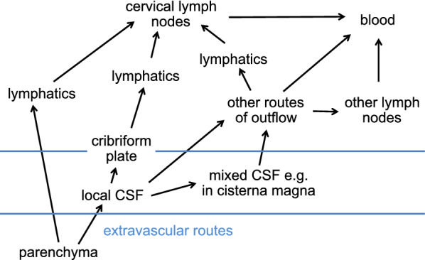
The glymphatic hypothesis proposes a mechanism for extravascular transport into and out of the brain of hydrophilic solutes unable to cross the blood-brain barrier. It suggests that there is a circulation of fluid carrying solutes inwards via periarterial routes, through the interstitium and outwards via perivenous routes. This review critically analyses the evidence surrounding the mechanisms involved in each of these stages. There is good evidence that both influx and efflux of solutes occur along periarterial routes but no evidence that the principal route of outflow is perivenous. Furthermore, periarterial inflow of fluid is unlikely to be adequate to provide the outflow that would be needed to account for solute efflux. A tenet of the hypothesis is that flow sweeps solutes through the parenchyma. However, the velocity of any possible circulatory flow within the interstitium is too small compared to diffusion to provide effective solute movement. By comparison the earlier classical hypothesis describing extravascular transport proposed fluid entry into the parenchyma across the blood-brain barrier, solute movements within the parenchyma by diffusion, and solute efflux partly by diffusion near brain surfaces and partly carried by flow along "preferred routes" including perivascular spaces, white matter tracts and subependymal spaces. It did not suggest fluid entry via periarterial routes. Evidence is still incomplete concerning the routes and fate of solutes leaving the brain. A large proportion of the solutes eliminated from the parenchyma go to lymph nodes before reaching blood but the proportions delivered directly to lymph or indirectly via CSF which then enters lymph are as yet unclear. In addition, still not understood is why and how the absence of AQP4 which is normally highly expressed on glial endfeet lining periarterial and perivenous routes reduces rates of solute elimination from the parenchyma and of solute delivery to it from remote sites of injection. Neither the glymphatic hypothesis nor the earlier classical hypothesis adequately explain how solutes and fluid move into, through and out of the brain parenchyma. Features of a more complete description are discussed. All aspects of extravascular transport require further study.

Keywords: Aquaporin 4; Blood–brain barrier; Bulk flow; Cerebrospinal; Diffusion; Extravascular transport; Fluid circulation; Glymphatic; Hydrophilic solute; Interstitial; Periarterial; Perivascular; Perivenous; Subependymal space.


Conflict of interest statement

The authors declare that they have no competing interests.

Figures

Fig. 1
Fig. 1
Principal features of the proposed glymphatic circulation. Periarterial inflow of cerebrospinal fluid, shown on the left, enters the interstitial fluid in the parenchyma by crossing a layer of glial endfeet assisted by the presence of aquaporin 4 (AQP4) water channels in the endfoot membrane facing the perivascular space. The fluid then flows through the interstitial spaces propelling solutes towards the perivenous conduits, shown on the right, leading to outflow from the brain. Flow in the perivascular space of a blood vessel is in the same direction as the blood flow, but the orientations of arterioles and venules vary and are not strictly antiparallel. Similar portrayals of the glymphatic hypothesis with varying artistic embellishments have been published repeatedly [, , , , , , –241]. Above and in many other published figures flow is portrayed as sweeping solutes towards the venules where they become more concentrated. It is argued in Sect. 5.4 that such a sweeping effect is unlikely to occur
Fig. 2
Fig. 2
Diagram illustrating stages of fluid circulation considered in the discussion of the glymphatic hypothesis. Circled numbers refer to the sections in this review where the stages are considered in detail
Fig. 3
Fig. 3
Meningeal layers associated with a cortical penetrating artery and an emerging vein. This is based on evidence obtained using electron microscopy. Note that the artery has a pial sheath as it courses along the surface of the cortex and this sheath follows the artery without break as it penetrates the cortex. By contrast the sheath around the emergent vein is not present along the course within the cortex. A, arachnoid membrane; SAS subarachnoid space (which on the dorsal surfaces of the cortex may be collapsed other than where it covers a blood-vessel); PF pial perforations. For more recent discussion of the presence (arterial) or absence (venous) of a pial sheath within the parenchyma see [127]. Reproduced with permission from Zhang et al., J. Anat. 1990 [100]
Fig. 4
Fig. 4
Trajectories of microspheres overlaid onto an image of surface blood vessels in the subarachnoid space. The trajectories are clustered along and parallel to the surfaces of arteries as if they are restricted to a periarterial space. Scale bar: 40 μm. (Reproduced from Mestre et al., Nature Comm. 2018 [89] Creative Commons Attribution 4.) From the relative straightness of the trajectories and the profile of velocities, maximum half-way across the width of the periarterial space, it has been concluded that periarterial spaces within the subarachnoid space are occupied by free fluid rather than by a porous matrix or gel [54]
Fig. 5
Fig. 5
Possible routes of solute transport along arteries both in the subarachnoid space and in the parenchyma. Extramural influx may occur (dashed green line) via a “periarterial space"between a sheath composed of pia and artery wall [11] or (solid green line) via the subpial space [129, 130]. Whatever their route, the solutes must cross the pia (green double-headed arrows) at some stage leading into the parenchyma. Efflux of solutes may occur by reversal of the extramural route (green dashed line) [12, 62, 96, 138] in which case, they would reach intramural sites within the smooth muscle layer by diffusion through the wall (double-headed red arrows in the main figure, dashed red lines in the insert). Alternatively [8, 129, 130, 139, 242], efflux may occur via an intramural route (solid red lines in the main figure and insert), which requires movement of solutes over long distances via the basement membranes of the smooth muscle layer of the arterial wall (light grey in the insert). For this route to be dominant there must be some feature of the arterial walls that prevents escape of solutes from the smooth muscle layer to the extramural periarterial space. The thick black lines represent the glia limitans at the surface of the brain parenchyma and surrounding the arteries. Structures shown in the figure are modified from those shown in Fig. 6 of [130]
Fig. 6
Fig. 6
Human MRI images showing changes in gadobutrol concentration following an intrathecal injection. At = 0, 1 µmol of gadobutrol was injected into the subarachnoid space of the spinal cord. It reached the cisterna magna in about 3 h and spread over the surface of the brain in the next 7-8 h. Note that the gadobutrol concentrations on the dorsal surface of the brain persist for longer than in the basal cisterns as expected if elimination of gadobutrol occurs primarily from the cisterns, e.g. across the cribriform plate, but not from the dorsal subarachnoid spaces. The partial analysis of these data in [117] shows that in various regions of the brain the concentrations in grey matter are still increasing up to 12 h and subsequently decrease over days in parallel with that in CSF adjacent to the region. However, even at their maximum they are less (2 to 5 fold depending on region) than the concentrations that were achieved in the adjacent subarachnoid spaces. At the very latest times, the concentrations in parenchyma appear to exceed the then current concentrations in adjacent subarachnoid spaces. The subject was awake from the time of administration until after the scan at 10 h and had a normal nights sleep before each of the last three scans. Figure taken with permission from Watts et al., Am. J. Neuroradiol., 2019 [117]
Fig. 7
Fig. 7
Schematic diagram of a cross section of the theoretical array of parenchymal blood vessels in the model used by Ray et al. [193]. In this model the vessel array is approximated by a regular repeating pattern of arterioles (red circles) and venules (blue circles) running perpendicular to the cortical surface. Streamlines (thin black lines) connect arterioles the sources of flow, and venules,- the sinks. Conveniently for the calculation of flow from the flow velocity the midplanes between the planes of arterioles and venules separate the sources from the sinks and the direction of flow is perpendicular to the midplanes (see footnote 26). Note that the cross-sectional area available for perivascular flow along the arterioles and venules is much smaller than the area available for flow through the interstitium. Thus, with the same flow inwards along periarterial spaces, through the interstitium and outwards along perivenous spaces, the flow velocity would be much higher in the perivascular spaces than in the interstitium
Fig. 8
Fig. 8
Flow diagram of routes taken by large hydrophilic solutes emerging from the parenchyma by extravascular routes. The eventual destination is blood but there are multiple routes that a solute can follow. The initial extravascular pathways be they periarterial, perivenous, subependymal or white matter tracts may lead to CSF. In addition, particularly the intramural perivascular pathways may lead to lymphatics. Solutes that reach CSF at the parenchymal surface in anterior and/or ventral portions of the brain may be taken via the olfactory nerve to the cribriform plate and thus to lymphatics. Alternatively the solutes may be mixed into CSF that at some point traverses the cisterna magna, perhaps on route to spinal sites of elimination. A significant portion of the CSF may also be directed to lymph. It cannot be assumed that solutes reaching the cervical lymph nodes have not first mixed with CSF
Fig. 9
Fig. 9
Comparisons of the classical hypothesis, the glymphatic hypothesis and a scheme based on current evidence. They each summarise processes that may be important in extravascular supply and removal of solutes. a In the classical hypothesis [59, 63, 138] ISF is produced by secretion across the blood–brain barrier and flows out of the parenchyma along “preferred routes” including periarterial spaces, white matter tracts, subependymal spaces and possibly perivenous spaces. Solute movement within interstitial spaces is by diffusion. The velocity of the flow within the interstitium is too small to produce observable movements of solutes. b In the glymphatic hypothesis [11, 36] CSF enters the parenchyma via periarterial routes, flows into the interstitial spaces where it mixes with ISF and sweeps solutes to perivenous spaces. ISF flows out of the parenchyma along perivenous spaces. In simple extensions of the hypothesis, outflow may also occur via white matter tracts and subependymal spaces. c Scheme based on current evidence of possible processes involved in supply and removal of solutes in the brain parenchyma. Solutes may move in both directions via periarterial spaces and possibly also via perivenous spaces. There may or may not be net inflow along periarterial spaces and outflow along perivenous spaces. There are also other routes for outflow of fluid and efflux of solutes including white matter tracts and subependymal spaces (compare [34]). Fluid flow may be important in efflux of solutes via extravascular pathways but the flow velocity in interstitial spaces is too small to produce observable movements of solutes. Contrary to what is inferred in many figures portraying the glymphatic circulation, e.g. Fig. 1, there is no sweeping of solutes towards perivenous spaces. In all three schemes solutes emerging from the parenchyma by extravascular routes may be delivered directly to lymph or to CSF. From CSF they can leave the brain via lymph or possibly blood flow

References

    1. Hladky SB, Barrand MA. Fluid and ion transfer across the blood–brain and blood–cerebrospinal fluid barriers; a comparative account of mechanisms and roles. Fluids Barriers CNS. 2016;13:19. - PMC - PubMed
    1. Hladky SB, Barrand MA. Elimination of substances from the brain parenchyma: efflux via perivascular pathways and via the blood–brain barrier. Fluids Barriers CNS. 2018;15:30. - PMC - PubMed
    1. Hladky SB, Barrand MA. Mechanisms of fluid movement into, through and out of the brain: evaluation of the evidence. Fluids Barriers CNS. 2014;11:26. - PMC - PubMed
    1. Tarasoff-Conway JM, Carare RO, Osorio RS, Glodzik L, Butler T, Fieremans E, Axel L, Rusinek H, Nicholson C, Zlokovic BV, et al. Clearance systems in the brain-implications for Alzheimer disease. Nat Rev Neurol. 2015;11:457–70. - PMC - PubMed
    1. Abbott NJ, Pizzo ME, Preston JE, Janigro D, Thorne RG. The role of brain barriers in fluid movement in the CNS: is there a ‘glymphatic’ system? Acta Neuropathol. 2018;135:387–407. - PubMed

LinkOut - more resources