. 2022 Feb 8;119(6):e2113489119.
doi: 10.1073/pnas.2113489119.

Different α-synuclein prion strains cause dementia with Lewy bodies and multiple system atrophy

Affiliations

Different α-synuclein prion strains cause dementia with Lewy bodies and multiple system atrophy

Jacob I Ayers et al. Proc Natl Acad Sci U S A. .

Abstract

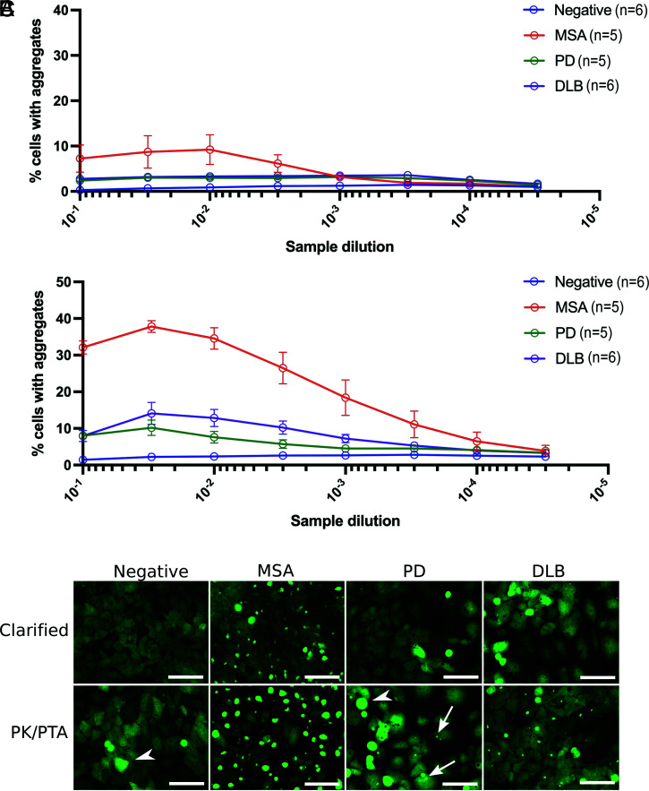
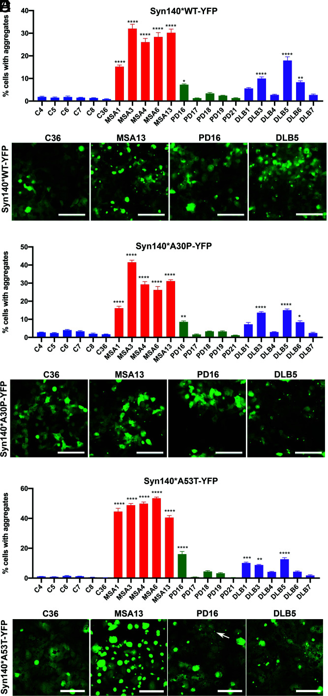
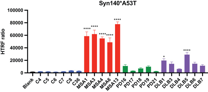
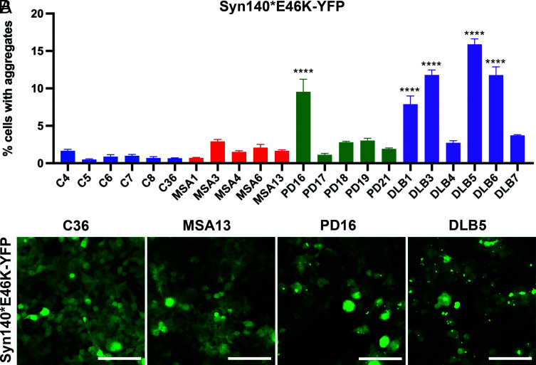
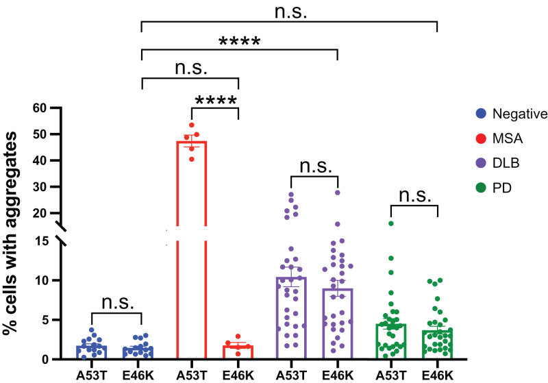
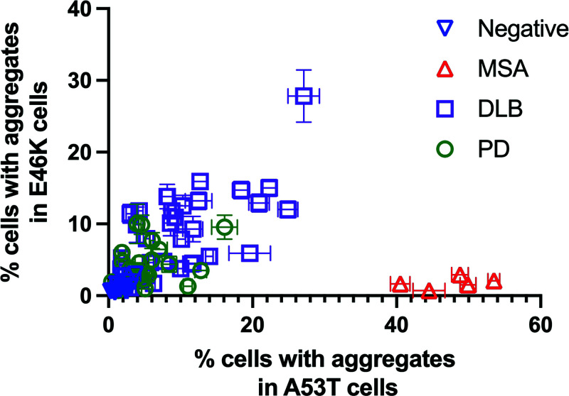
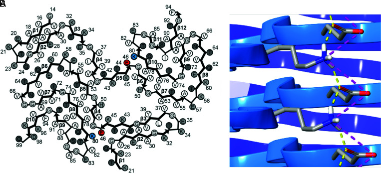
The α-synuclein protein can adopt several different conformations that cause neurodegeneration. Different α-synuclein conformers cause at least three distinct α-synucleinopathies: multiple system atrophy (MSA), dementia with Lewy bodies (DLB), and Parkinson's disease (PD). In earlier studies, we transmitted MSA to transgenic (Tg) mice and cultured HEK cells both expressing mutant α-synuclein (A53T) but not to cells expressing α-synuclein (E46K). Now, we report that DLB is caused by a strain of α-synuclein prions that is distinct from MSA. Using cultured HEK cells expressing mutant α-synuclein (E46K), we found that DLB prions could be transmitted to these HEK cells. Our results argue that a third strain of α-synuclein prions likely causes PD, but further studies are needed to identify cells and/or Tg mice that express a mutant α-synuclein protein that is permissive for PD prion replication. Our findings suggest that other α-synuclein mutants should give further insights into α-synuclein prion replication, strain formation, and disease pathogenesis, all of which are likely required to discover effective drugs for the treatment of PD as well as the other α-synucleinopathies.

Keywords: dementia with Lewy bodies; neurodegeneration; prions; strains; synucleinopathies.


Conflict of interest statement

Competing interest statement: S.B.P. is a member of the Scientific Advisory Boards of ViewPoint Therapeutics and New Ventures Inc. and a member of the Supervisory Board of Priavoid, none of which have contributed financial or any other support to these studies.

Figures

Fig. 1.
Fig. 1.
Effect of PK/PTA treatment on infectivity of human synucleinopathy samples in cultured HEK293T cells expressing syn140*A53T-YFP. (A) Cultured cells were infected with half-log dilutions of CBH from cognitively normal control brains (negative) and brains from MSA, PD, and DLB patients. Fluorescent puncta were measured 4 d following infection and presented as % cells with aggregates. The data points are the averages of signals from each disease cohort. (B) These same homogenates were treated with PK/PTA-precipitated samples and incubated in syn140*A53T-YFP cells to test for infectivity and measured 4 d following infection. The data revealed a significant increase in infectivity among the PK/PTA-precipitated fractions derived from MSA, PD, and DLB samples. The data points are the averages of signals from each disease cohort. (C) Representative images of syn140*A53T-YFP cells infected with the indicated preparations of negative control, MSA, PD, and DLB samples. The arrows point to small puncta measured in cells infected with PD inocula. Arrowheads indicate dead or dividing cells not counted as aggregates. YFP is shown in green (Scale bars: 100 μm).
Fig. 2.
Fig. 2.
Effect of various synuclein substrates on cell infectivity of PK/PTA-precipitated preparations. (A) Syn140*WT-YFP cells reveal transmissibility of MSA, PD, and DLB α-synuclein prions. (B) Representative images of syn140*WT-YFP cells infected with PK/PTA-precipitated preparations of human samples. (C) Syn140*A30P-YFP cells were infected with PK/PTA-precipitated preparations from control, MSA, PD, and DLB cases. (D) Representative images displaying syn140*A30P-YFP puncta in infected cells. (E) Syn140*A53T-YFP cells reveal transmissibility of MSA, PD, and DLB α-synuclein prions. (F) Representative images of syn140*A53T-YFP cells infected with PK/PTA-precipitated preparations of human samples. Each sample was run in replicates of six wells. Data shown as mean ± SEM. Significance of each sample calculated against the mean of negative control samples. *P < 0.05, **P < 0.01, ***P < 0.001, ****P < 0.0001 (Scale bars: 100 μm).
Fig. 3.
Fig. 3.
Cell infectivity of PK/PTA-precipitated preparations measured by HTRF in an untagged α-synuclein cell line. Infectivity of PK/PTA preps from samples were measured in α-syn140*A53T cells using an HTRF α-synuclein aggregation assay. Cells were incubated for 4 d with samples, lysed, and measured for α-synuclein aggregation. Data shown as mean ± SEM. Significance of each sample calculated against the mean of negative control samples. *P < 0.05, ****P < 0.0001.
Fig. 4.
Fig. 4.
The E46K α-synuclein mutation allows replication of DLB prions. (A) Syn140*E46K-YFP cells reveal transmissibility of PD and DLB α-synuclein prions but show resistance to transmission of MSA α-synuclein prions. (B) Representative images of syn140*E46K-YFP cells infected with PK/PTA-precipitated preparations of human samples. Data shown as mean ± SEM. Significance of each sample calculated against the mean of negative control samples. ****P < 0.0001 (Scale bars: 100 μm).
Fig. 5.
Fig. 5.
The E46K α-synuclein mutation differentiates MSA from DLB prions. An extended set of human patient samples was run through the syn140*A53T-YFP and syn140*E46K-YFP cell lines and plotted to visualize the effect of the α-synuclein substrate on infectivity. Data shown as mean ± SEM. Significance of each sample calculated against the mean of negative control samples. ****P < 0.0001. n.s., not significant.
Fig. 6.
Fig. 6.
Permissiveness of the E46K versus A53T cell lines for each patient sample. Each patient sample is depicted in the graph by its ability to infect cells expressing the E46K mutation (y axis) in comparison to infectivity in cells expressing the A53T mutation (x axis).
Fig. 7.
Fig. 7.
Interactions between E46 and K80 in MSA cryo-EM structures. (A) MSA type 1 α-synuclein filament (Protein Data Bank structure 6XYO) revealing close proximity of E46 (highlighted in red) and K80 (highlighted in blue) in both protofilaments. Adapted by permission from Springer Nature: ref. . (B) Proximity of E46 and K80 allow for the formation of hydrogen bonds (yellow lines) and salt bridges (pink lines).

References

    1. Parkinson J., An Essay on the Shaking Palsy (Sherwood, Neely, and Jones, London, 1817).
    1. Lewy F. H., “Paralysis agitans” in Pathologische Anatomie. Handbuch der Neurologie, Lewandowsky M., Ed. (Springer Verlag, Berlin, 1912), pp. 920–933.
    1. Tretiakoff C., Contribution à l’étude de l’anatomie pathologique du locus niger de Soemmering avec quelques déductions relatives à la pathogenie des troubles du tonus musculaire et de la maladie de Parkinson (University of Paris, Paris, 1919).
    1. Polymeropoulos M. H., et al. , Mutation in the α-synuclein gene identified in families with Parkinson’s disease. Science 276, 2045–2047 (1997). - PubMed
    1. Spillantini M. G., et al. , α-synuclein in Lewy bodies. Nature 388, 839–840 (1997). - PubMed

Publication types