Review
. 2022 Jan 18:15:779125.
doi: 10.3389/fnins.2021.779125. eCollection 2021.

Neuron to Oligodendrocyte Precursor Cell Synapses: Protagonists in Oligodendrocyte Development and Myelination, and Targets for Therapeutics

Affiliations
Review

Neuron to Oligodendrocyte Precursor Cell Synapses: Protagonists in Oligodendrocyte Development and Myelination, and Targets for Therapeutics

Daniela M S Moura et al. Front Neurosci. .

Abstract

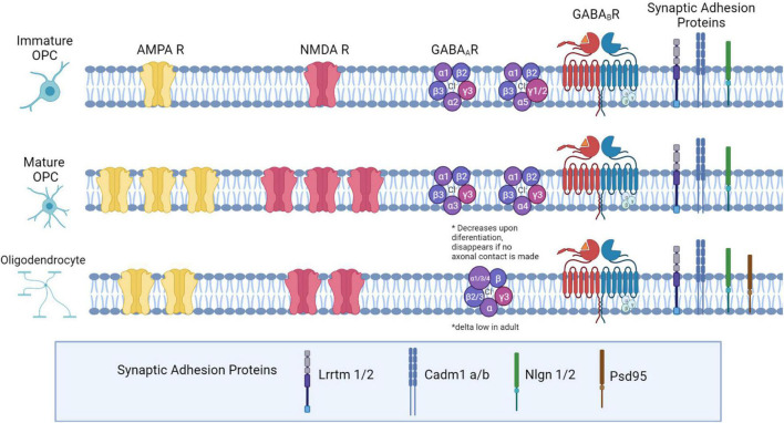
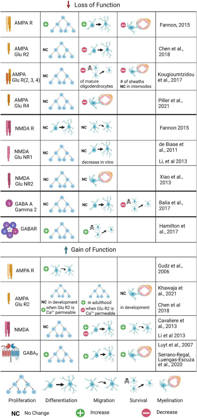
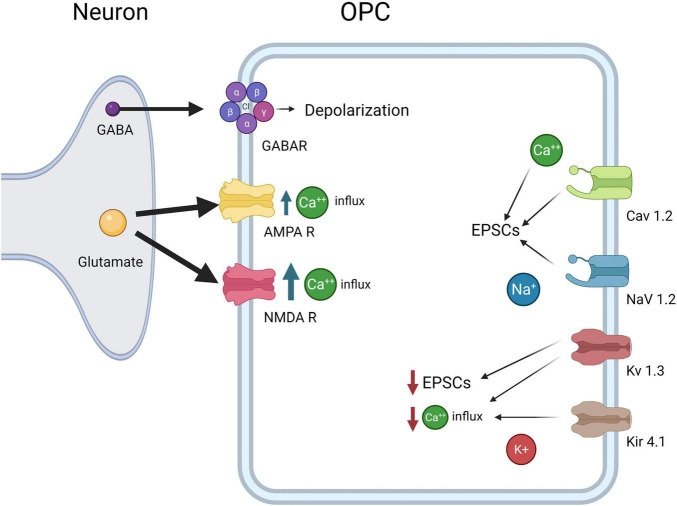
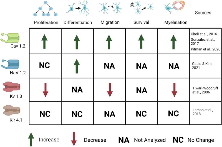
The development of neuronal circuitry required for cognition, complex motor behaviors, and sensory integration requires myelination. The role of glial cells such as astrocytes and microglia in shaping synapses and circuits have been covered in other reviews in this journal and elsewhere. This review summarizes the role of another glial cell type, oligodendrocytes, in shaping synapse formation, neuronal circuit development, and myelination in both normal development and in demyelinating disease. Oligodendrocytes ensheath and insulate neuronal axons with myelin, and this facilitates fast conduction of electrical nerve impulses via saltatory conduction. Oligodendrocytes also proliferate during postnatal development, and defects in their maturation have been linked to abnormal myelination. Myelination also regulates the timing of activity in neural circuits and is important for maintaining the health of axons and providing nutritional support. Recent studies have shown that dysfunction in oligodendrocyte development and in myelination can contribute to defects in neuronal synapse formation and circuit development. We discuss glutamatergic and GABAergic receptors and voltage gated ion channel expression and function in oligodendrocyte development and myelination. We explain the role of excitatory and inhibitory neurotransmission on oligodendrocyte proliferation, migration, differentiation, and myelination. We then focus on how our understanding of the synaptic connectivity between neurons and OPCs can inform future therapeutics in demyelinating disease, and discuss gaps in the literature that would inform new therapies for remyelination.

Keywords: gaba receptor; glutamate receptor; ion channel; multiple sclerosis; myelin; oligodendrocyte precursor cell (OPC); synapse.


Conflict of interest statement

The authors declare that the research was conducted in the absence of any commercial or financial relationships that could be construed as a potential conflict of interest.

Figures

FIGURE 1
FIGURE 1
Neurotransmitter receptor expression and synaptic adhesion protein expression across the Oligodendrocyte lineage. In immature OPCs (PO-P9), AMPA and NMDA receptors are present at low densities, with expression peaking later in development (P10-P35). As OPCs mature into OLs, they express fewer AMPA and NMDARs. GABAA receptor subunit expression changes: in early development, α1,α2, and α5 subunits are highly expressed. Later in development, α2 and α5 decrease expression, and α3 and α4 both increase in expression. β2 and β3 expression is high, and does not change during maturation, while β1 declines from low to very low expression in OPCs later in development. The δ subunit is not present in development but is expressed in a small percentage of OPCs in adulthood. All γ subunits are expressed in OPCs: γ3 is highly expressed and maintained throughout development, but γ1 and γ2 are highly expressed in OPCs early in development and decrease in adulthood. Both GABAB subunits are expressed consistently throughout development (GABAB1 and GABAB2). Both OPCs and mature OLs express the adhesion proteins Lrrtm1, Lrrtm2, Neuroligin 1, Neuroligin 2, Cadm1a, and Cadm1b, based on RNASeq expression analysis in OPCs, and based on LOF function analyses in OLs. Additionally, OLs express PSD-95.
FIGURE 2
FIGURE 2
The role of glutamatergic and GABAergic receptors in OPC development and myelination: Summary of Gain and Loss of Function Experiments. Changes in OPC proliferation, differentiation, migration, or survival are included, as well as changes in myelination. Loss of function experiments have been conducted for several AMPA and NMDA receptor subunits, as well as the GABAA γ2 subunit; gain of function experiments are limited to AMPA, AMPA GluR2, NMDA, and GABAB experiments. Where the process was tested but no differences found, no change is noted; if the process was untested it is not included for that receptor. Where conflicting results were found, both are listed.
FIGURE 3
FIGURE 3
OPC electrophysiological Properties. OPCs express CaV1.2, which results in influx in Ca2+ and increases EPSCs, NaV1.2, which results in influx in Na+ and increases in EPSCs, Kv1.3 which decreases EPSCs and Ca2+ influx, and Kir4.1, which decreases Ca2+ influx. Both glutamate receptors AMPAR and NMDA lead to increased Ca2+ concentrations inside OPCs, although NMDARs allow more Ca2+ influx than AMPARs. Finally, GABAA causes depolarization of the OPCs as the intracellular Cl- concentration is high in OPCs.
FIGURE 4
FIGURE 4
Ion channels expressed in OPCs are important for OPC development and myelination. Activation of CaV1.2 causes an increase in OPC proliferation, differentiation, migration, survival, and myelination. Activation of Nav1.2 causes an increase in OPC differentiation but no change in proliferation. Activation of Kv1.3 causes a decrease in OPC proliferation, migration, and myelination. Kir4.1 activation causes no change in OPC proliferation, differentiation, survival, or myelination, although behavioral and electrophysiological defects (not depicted here) are present.

References

    1. Allen N. J., Eroglu C. (2017). Cell biology of astrocyte-synapse interactions. Neuron 96 697–708. 10.1016/j.neuron.2017.09.056 - DOI - PMC - PubMed
    1. Almeida R. G., Lyons D. A. (2017). On myelinated axon plasticity and neuronal circuit formation and function. J Neurosci 37 10023–10034. 10.1523/JNEUROSCI.3185-16.2017 - DOI - PMC - PubMed
    1. Almeida R. G., Williamson J. M., Madden M. E., Early J. J., Voas M. G., Talbot W. S., et al. (2021). Myelination induces axonal hotspots of synaptic vesicle fusion that promote sheath growth. Curr. Biol. 31 3743–3754. 10.1016/j.cub.2021.06.036 - DOI - PMC - PubMed
    1. Arellano R. O., Sánchez-Gómez M. V., Alberdi E., Canedo-Antelo M., Chara J. C., Palomino A., et al. (2016). Axon-to-glia interaction regulates GABAA receptor expression in oligodendrocytes. Mol. Pharmacol. 89 63–74. 10.1124/mol.115.100594 - DOI - PubMed
    1. Balia M., Benamer N., Angulo M. C. (2017). A specific GABAergic synapse onto oligodendrocyte precursors does not regulate cortical oligodendrogenesis. Glia 65 1821–1832. 10.1002/glia.23197 - DOI - PubMed

LinkOut - more resources