Case Reports
. 2022 Jan 18:12:818219.
doi: 10.3389/fmicb.2021.818219. eCollection 2021.

Case Report: "Methicillin-Resistant Staphylococcus aureus Endocarditis Overlying Calcified Mitral Annular Abscess Misdiagnosed as Klebsiella pneumoniae Endocarditis"

Affiliations
Case Reports

Case Report: "Methicillin-Resistant Staphylococcus aureus Endocarditis Overlying Calcified Mitral Annular Abscess Misdiagnosed as Klebsiella pneumoniae Endocarditis"

Hiroyuki Yamamoto et al. Front Microbiol. .

Abstract

Infective endocarditis (IE) involving mitral annular calcification (MAC) is a rare disease, but is potentially lethal due to frequent serious periannular complications, and therefore requires early diagnosis and prompt treatment. However, either reaching the correct diagnosis or the detection of periannular complications, even with conventional transesophageal echocardiography (TEE), remains challenging because calcium deposition obscures clear visualization of the area around the MAC. We describe a unique case of methicillin-resistant Staphylococcus aureus (MRSA) IE involving a calcified mitral annular abscess, which was initially misdiagnosed as Klebsiella pneumoniae IE. Accurate diagnosis of MAC-related IE as well as detection of the annular abscess were made possible by 4D TEE, leading to successful cardiac surgery, which confirmed MRSA IE pathologically, and the associated annular abscess. This case highlights the usefulness of 4D TEE for the accurate diagnosis and proper surgical planning. In addition, this case raises the limitations of the modified Duke criteria in cases of definite IE with dual bacteremia.

Keywords: Duke criteria; MAC; MRSA; TEE; dual bacteremia; infective endocarditis; mitral annular abscess.


Conflict of interest statement

The authors declare that the research was conducted in the absence of any commercial or financial relationships that could be construed as a potential conflict of interest.

Figures

Figure 1
Figure 1
TTE and brain MRI. (A–C) Longitudinal TTE: (A,B) apical 4-chamber and (C) parasternal long-axis views. (A) TTE shows tiny MAC along the posterior mitral annulus 1 year ago (arrowhead). (B,C) TTE after admission shows MAC progression (arrowhead) and a large round mass superimposed on the MAC (arrow). Mass with wide attachment (10 ×11 mm in size) is characterized by heterogeneity and oscillation. (C) Notably, color Doppler TTE showed no significant mitral regurgitation. (D–G) Brain diffusion-weighted MRI detects multiple acute cerebral infarctions in both hemispheres (arrowheads). LA, left atrium; LV, left ventricle; MAC, mitral annular calcification; MRI, magnetic resonance imaging; TTE, transthoracic echocardiography.
Figure 2
Figure 2
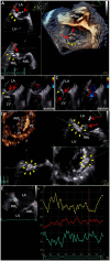
TEE of the mitral valve. (A) 3D TEE or (B,C) 2D color Doppler TEE shows a long mobile mass of vegetations which consist of body part (arrow) and tail part (dotted arrow), containing calcium-dense stippling, attached to the calcified posterior mitral annulus (arrowheads). Note the intact valve leaflets. (D) 4D TEE with Flexi-slice mode demonstrates MAC with a central echolucent area (arrowheads) around the P2 segment of the mitral valve. (E) Q-analysis presents a contrast analysis that traces the grayscale intensity in a defined region of interest as a function of time: green, yellow, and red depicts the center of MAC, the periphery of MAC, and normal cardiac muscle tissue as a control, respectively (left). Time-intensity curves are plotted for the respective region of interest (right): X axis, Time (s); and Y axis, Intensity scale (logarithmic) (dB). AML, anterior mitral leaflet; Ao, aorta; LA, left atrium; LV, left ventricle; MAC, mitral annular calcification; PML, posterior mitral leaflet; TEE, transesophageal echocardiography.
Figure 3
Figure 3
Intraoperative and pathological findings. (A) Intraoperative view of the mitral valve. A large mass of vegetations superimposed on mitral annular calcification (arrow) is observed around the P2 segment in the posterior mitral annulus (white arrowheads). (B,C) Representative macro-photographs of the extracted mitral valve leaflets (B, PML; C, AML). (D,E) Histological findings of the resected PML shows dystrophic calcification (yellow arrowheads), and surrounding neutrophilic inflammatory cell infiltration and bacterial agglomeration (red arrowheads) [(D) hematoxylin and eosin staining, Bar 200 μm; (E), gram staining, Bar 20 μm]. AML, anterior mitral leaflet; LA, left atrium; PML, posterior mitral leaflet.
Figure 4
Figure 4
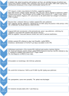
Timeline of case presentation. BCs, blood cultures; CTRX, ceftriaxone; IE, infective endocarditis; MAC, mitral annular calcification; MINO, minocycline; MRI, magnetic resonance imaging; MRSA, methicillin-resistant Staphylococcus aureus; MV, mitral valve; MVR, mitral valve replacement; ST, sulfamethoxazole/trimethoprim; TEE, transesophageal echocardiography; TTE, transthoracic echocardiography; UTI, urinary tract infection; VCM, vancomycin.

References

    1. Anderson M. J., Janoff E. N. (1998). Klebsiella endocarditis: report of two cases and review. Clin. Infect. Dis. 26, 468–474. 10.1086/516330 - DOI - PubMed
    1. Benjamin E. J., Plehn J. F., D'Agostino R. B., Belanger A. J., Comai K., Fuller D. L., et al. . (1992). Mitral annular calcification and the risk of stroke in an elderly cohort. N. Engl. J. Med. 327, 374–379. 10.1056/NEJM199208063270602 - DOI - PubMed
    1. Durack D. T., Lukes A. S., Bright D. K. (1994). New criteria for diagnosis of infective endocarditis: utilization of specific echocardiographic findings. Duke Endocarditis Service. Am. J. Med. 96, 200–209. 10.1016/0002-9343(94)90143-0 - DOI - PubMed
    1. Eicher J. C., De Nadai L., Soto F. X., Falcon-Eicher S., Dobsák P., Zanetta G., et al. . (2004). Bacterial endocarditis complicating mitral annular calcification: a clinical and echocardiographic study. J. Heart Valve Dis. 13, 217–227. - PubMed
    1. Fernicola D. J., Roberts W. C. (1993). Clinicopathologic features of active infective endocarditis isolated to the native mitral valve. Am. J. Cardiol. 71, 1186–1197. 10.1016/0002-9149(93)90644-r - DOI - PubMed

Publication types

LinkOut - more resources