. 2021 Jan;10(1):349-360.
doi: 10.21037/tcr-20-698.

SAV1, regulated by HERC4, inhibits the proliferation, migration, and invasion of hepatocellular carcinoma

Affiliations

SAV1, regulated by HERC4, inhibits the proliferation, migration, and invasion of hepatocellular carcinoma

Fang Huang et al. Transl Cancer Res. 2021 Jan.

Abstract

Background: Hepatic carcinoma is one of the most malignant cancers worldwide. Salvador 1 (SAV1) plays a key role in a variety of human carcinogenesis. This study investigated the role of SAV1 and HERC4 in hepatocellular carcinoma (HCC).

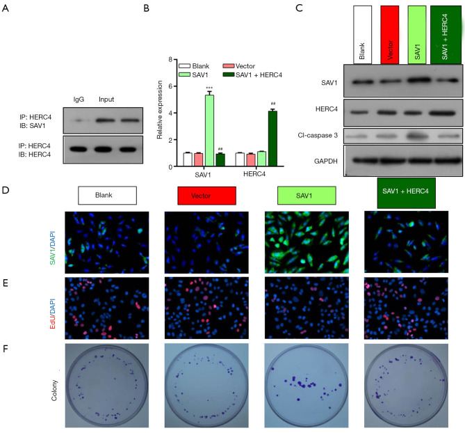
Methods: SAV1 and HERC4 expressions in HCC tissues were examined using RT-qPCR assay. The regulatory effect of HERC4 on SAV1 was verified by co-immunoprecipitation (Co-IP), RT-qPCR, Western blot, and immunofluorescent assays in HEP3B and Huh 7 cell lines. In addition, functional experimental verification was performed through Edu staining, colony formation, and Transwell assay. Finally, Xenograft tumor model was finally used in nude mice.

Results: Clinical features showed significant difference with SAV1 and HERC4 expression. HERC4 was found to be upregulated, while SAV1 was downregulated in HCC. Patients with high HERC4 or low SAV1 had a worse prognosis. Results showed that HERC4 could notably decreased the expression level of SAV1 in HCC cells. Our results showed that overexpression HERC4 could reverse the inhibitory effects of SAV1 on HCC cell proliferation, migration, and invasion. SAV1 overexpression repressed tumor growth and enhance caspase 3 expression.

Conclusion: SAV1 can be directly downregulated by HERC4, indicating that the HERC4/SAV1 axis might have great promise for targeted therapies of HCC.

Keywords: HERC4, Salvador 1 (SAV1); Hepatocellular carcinoma (HCC); invasion; migration.


Conflict of interest statement

Conflicts of Interests: All authors have completed the ICMJE uniform disclosure form (available at http://dx.doi.org/10.21037/tcr-20-698). The authors have no conflicts of interest to declare.

Figures

Figure 1
Figure 1
HERC4 was highly expressed, while SAV1 was lowly expressed in HCC tissues. The levels of HERC4 (A) and SAV1 (B) were identified by RT-qPCR assay in 15 pairs of HCC and para-carcinoma tissues, P<0.01. (C) Western blot analysis of HERC4 and SAV1 in HCC and para-carcinoma tissues. (D) Kaplan-Meier plots exhibiting the overall survivals in HCC patients with high or low HERC4 and SAV1 expressions, respectively. N, non-tumor tissues; T, tumor tissues; SAV1, Salvador 1; HERC4, HECT And RLD Domain Containing E3 Ubiquitin Protein Ligase 4.
Figure 2
Figure 2
SAV1 significantly prevented HCC cell proliferation through interaction with HERC4. (A) Direct binding of SAV1 and HERC4 was investigated by CO-IP Assay using anti-HERC4 antibodies. Input denotes positive control and IgG denotes negative control. The confirmations of HERC4 and SAV1 levels were by RT-qPCR (B) and Western blot assays (C) in HEP 3B cells after transfection with SAV1 or/and HERC4 plasmids, ***, P<0.001 vs. vector group; ##, P<0.01 vs. SAV1 group. (D) Expression and distribution of SAV1 was evaluated by IF assay, magnification, 100×, scale bar =100 µm. (E) The impacts of SAV1 and HERC4 overexpression on HEP 3B cell proliferation were examined by Edu staining, 200× (F) The impacts of SAV1 and HERC4 overexpression on HEP 3B cell proliferation were examined by colony formation. DAPI, 4',6-diamidino-2-phenylindole; SAV1, Salvador 1; HERC4, HECT And RLD Domain Containing E3 Ubiquitin Protein Ligase 4; GAPDH, glyceraldehyde-3-phosphate dehydrogenase; EdU, 5-Ethynyl-2'-deoxyuridine. IP, immunoprecipitation.
Figure 3
Figure 3
SAV1 significantly prevented HCC cell proliferation through interaction with HERC4 in Huh 7 cell line. (A) Direct binding of SAV1 and HERC4 was investigated by CO-IP Assay using anti-HERC4 antibody. Input denotes positive control and IgG denotes negative control. The confirmations of HERC4 and SAV1 levels were completed by RT-qPCR (B) and Western blot assays (C) in HEP 3B cells after transfection with SAV1 or/and HERC4 plasmids, ***, P<0.001 vs. vector group; ##, P<0.01 vs. SAV1 group. (D) The expression and distribution of SAV1 was evaluated by IF assay, magnification, 100×, scale bar =100 µm. (E) The impacts of SAV1 and HERC4 overexpression on HEP 3B cell proliferation were examined by Edu staining, 200× (F) The impacts of SAV1 and HERC4 overexpression on HEP 3B cell proliferation were examined by colony formation. DAPI, 4',6-diamidino-2-phenylindole; SAV1, Salvador 1; HERC4, HECT And RLD Domain Containing E3 Ubiquitin Protein Ligase 4; GAPDH, glyceraldehyde-3-phosphate dehydrogenase; EdU, 5-Ethynyl-2'-deoxyuridine. IP, immunoprecipitation.
Figure 4
Figure 4
Overexpression of SAV1 suppressed the migration and invasion of HCC cells by being regulated by HERC4 in HEP3B and Huh 7 cell line. Transwell assay was conducted in HEP 3B cells to assess the influences of SAV1 and HERC4 transfection on cell migration (A) and invasion (B). The migrated (C) and invaded cells (D) were counted according to the resulting graphs of Transwell assay, Cells were photographed at a magnification of 100× after stained with crystal violet. *, P<0.05; **, P<0.01 vs. vector group; #, P<0.05 vs. SAV1 group. SAV1, Salvador 1; HERC4, HECT And RLD Domain Containing E3 Ubiquitin Protein Ligase 4. Please add the magnification and staining method for cell imagines.
Figure 5
Figure 5
Overexpression of SAV1 inhibited tumor growth in nude mice. (A,B,C) SAV1 overexpression suppressed tumor growth. (D) SAV1 overexpression enhanced expression of Caspase 3 in tumor. (E) SAV1 overexpression attenuated Ki67 expression in tumor. A DAB staining was performed to the slide and then photographed at magnification 100×. *, P<0.05; **, P<0.01 vs. NC group. NC, Negative control; d, days; SAV1, Salvador 1; HERC4, HECT And RLD Domain Containing E3 Ubiquitin Protein Ligase 4. Please add the magnification and staining method for cell imagines.

Similar articles

Cited by

References

    1. Sui W, Gan Q, Liu F, Chen H, Liu J, Dai Y. The differentially expressed circular ribonucleic acids of primary hepatic carcinoma following liver transplantation as new diagnostic biomarkers for primary hepatic carcinoma. Tumour Biol 2018;40:1010428318766928. 10.1177/1010428318766928 - DOI - PubMed
    1. Budny A, Kozlowski P, Kaminska M, et al. Epidemiology and risk factors of hepatocellular carcinoma. Pol Merkur Lekarski 2017;43:133-9. - PubMed
    1. Xiong XX, Qiu XY, Hu DX, et al. Advances in Hypoxia-Mediated Mechanisms in Hepatocellular Carcinoma. Mol Pharmacol 2017;92:246-55. 10.1124/mol.116.107706 - DOI - PubMed
    1. Xia J, Song P, Sun Z, et al. Advances of diagnostic and mechanistic studies of gamma-glutamyl transpeptidase in hepatocellular carcinoma. Drug Discov Ther 2016;10:181-7. 10.5582/ddt.2016.01052 - DOI - PubMed
    1. Akoad ME, Pomfret EA. Surgical resection and liver transplantation for hepatocellular carcinoma. Clin Liver Dis 2015;19:381-99. 10.1016/j.cld.2015.01.007 - DOI - PubMed