

Ultrasonographic evaluation of extracapsular vascular invasion for subcapsular nodules of the thyroid
- PMID: 35117254
- PMCID: PMC8798561
- DOI: 10.21037/tcr-20-888
Ultrasonographic evaluation of extracapsular vascular invasion for subcapsular nodules of the thyroid
Abstract
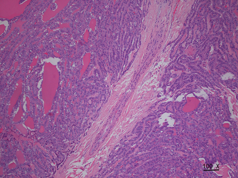
Background: Ultrasound provides a high-frequency spatial resolution. In this study, we used the combined pathological features of extrathyroid extension (ETE) measured by ultrasound to evaluate the vascular manifestations of subcapsular differentiated thyroid cancer. Our study aims to explore the value of high-frequency ultrasonography in the evaluation of extracapsular vascular invasion for the evaluation of both benign and malignant nodules and the prediction of ETE.
Methods: A total of 167 thyroid nodules were enrolled in this study. High-frequency ultrasonography was used to observe the relationship between the blood flow of the nodules and the capsules. The blood flow was divided into two types according to the relationship: non-extracapsular invasive blood flow and extracapsular invasive blood flow. Non-extracapsular blood flow was defined as any flow seen inside or around the nodule that did not extend beyond the thyroid gland. Extracapsular invasive blood flow was defined as any blood flow seen inside or around the nodule that flowed across the capsule and extended beyond the thyroid gland. A comparison of the different types of blood flow to judge the nature of thyroid nodules for predicting ETE was performed.
Results: Out of 167 nodules, 81 cases of nodules were the non-extracapsular invasive blood flow type, while the remaining 86 cases of nodules were classified as the extracapsular invasive blood flow type. Nodules with distinct types of blood flow were significantly different in malignancy rates between the nodules (P<0.001). The incidence rate of ETE was also significantly different between the malignant nodules with distinct types of blood flows.
Conclusions: Extracapsular vascular invasion is a good indicator for the evaluation of benign and malignant nodules. Using it as an indicator provides physicians with a potential tool for the prediction of ETE.
Keywords: Extrathyroid extension (ETE); blood flow; metastasis; thyroid carcinoma; ultrasonography.
2020 Translational Cancer Research. All rights reserved.
Conflict of interest statement
Conflicts of Interest: All authors have completed the ICMJE uniform disclosure form (available at http://dx.doi.org/10.21037/tcr-20-888). The authors have no conflicts of interest to declare.
Figures


References
-
- Tran B, Roshan D, Abraham E, et al. An Analysis of The American Joint Committee on Cancer 8th Edition T Staging System for Papillary Thyroid Carcinoma. J Clin Endocrinol Metab 2018;103:2199-206. - PubMed
LinkOut - more resources
Full Text Sources