

Epithelioid trophoblastic tumor found on hysteroscopy
- PMID: 35117550
- PMCID: PMC8797283
- DOI: 10.21037/tcr.2019.12.24
Epithelioid trophoblastic tumor found on hysteroscopy
Abstract
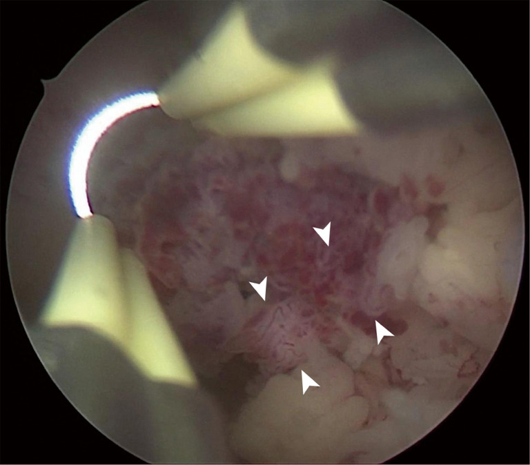
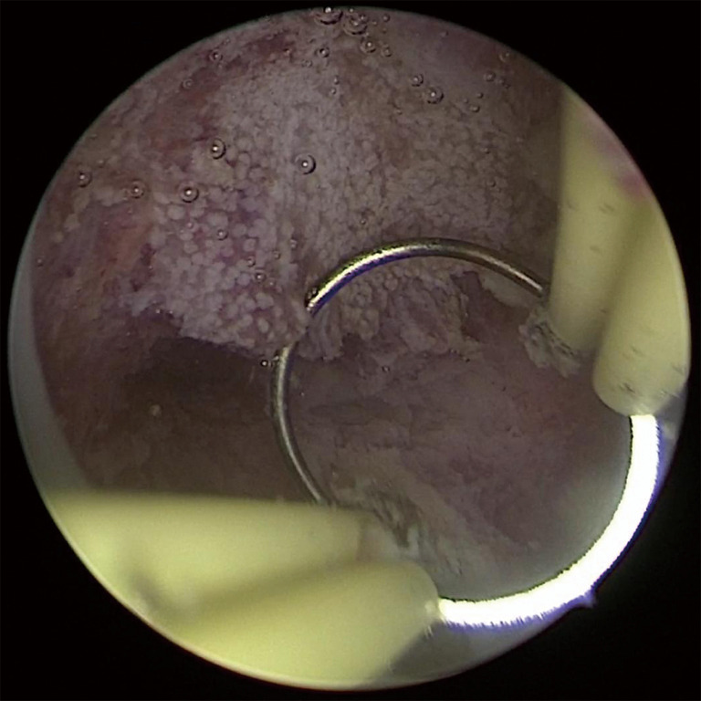
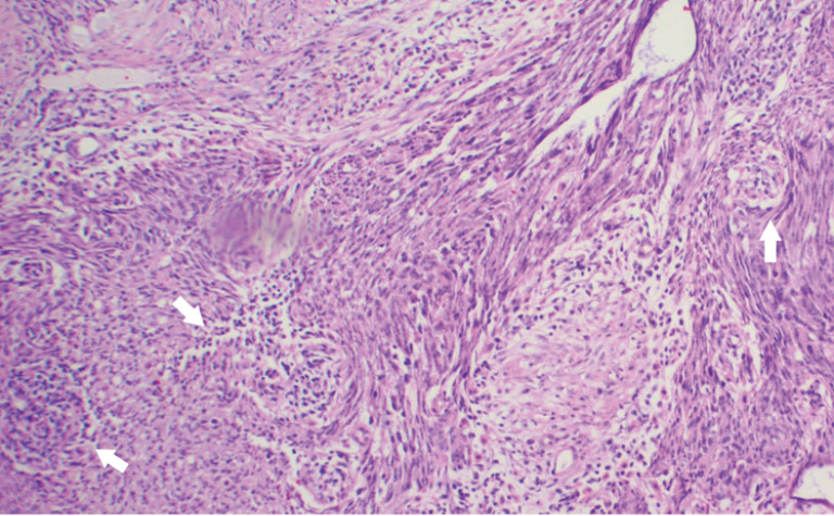
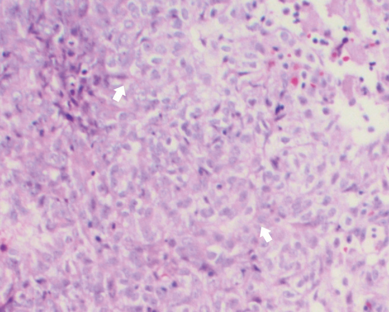
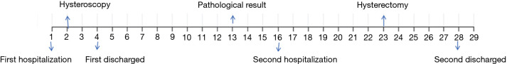
A 36-year-old woman presented with a history of prolonged menstrual period and increased menstrual volume of 4 months. Ultrasonography showed inhomogeneous echo measuring 2.5×1.9×2.2 cm3 in uterine cavity, and it can be seen that the blood flow signal enters the uterine posterior wall. Trophoblastic disease was not ruled out. But the serum β-human chorionic gonadotropin (hCG) was <0.3 mIU/mL. In order to confirm the diagnosis, the patient was planned to undergo hysteroscopy. Hysteroscopy is an ideal solution for early diagnosis. However, the drawback of hysteroscopy is that only local lesions can be removed. If the infiltration degree is deep, a second hysterectomy is required. Our authors present the first case of epithelioid trophoblastic tumor (ETT) under hysteroscopy. After neoplasm partial resection, histopathological examination revealed ETT. The patient underwent hysterectomy to prevent recurrence.
Keywords: Epithelioid trophoblastic tumor (ETT); case report; hysteroscopy.
2020 Translational Cancer Research. All rights reserved.
Conflict of interest statement
Conflicts of Interest: All authors have completed the ICMJE uniform disclosure form (available at http://dx.doi.org/10.21037/tcr.2019.12.24). The authors have no conflicts of interest to declare.
Figures

Similar articles
-
The fertility-sparing treatment and outcome of epithelioid trophoblastic tumor isolated to lung: a case report and review literature.Front Oncol. 2024 Mar 14;14:1337213. doi: 10.3389/fonc.2024.1337213. eCollection 2024. Front Oncol. 2024. PMID: 38549926 Free PMC article.
-
Uterine epithelioid trophoblastic tumor with the main manifestation of increased human chorionic gonadotropin: A case report.World J Clin Cases. 2024 Jun 6;12(16):2876-2880. doi: 10.12998/wjcc.v12.i16.2876. World J Clin Cases. 2024. PMID: 38899287 Free PMC article.
-
Epithelioid trophoblastic tumor that requires fertility preservation: A case report and review of literature.Taiwan J Obstet Gynecol. 2020 Sep;59(5):736-739. doi: 10.1016/j.tjog.2020.07.019. Taiwan J Obstet Gynecol. 2020. PMID: 32917327
-
Epithelioid trophoblastic tumor: a case report and literature review.Rom J Morphol Embryol. 2016;57(4):1365-1370. Rom J Morphol Embryol. 2016. PMID: 28174805 Review.
-
Epithelioid trophoblastic tumor coexisting with choriocarcinoma around an abdominal wall cesarean scar: a case report and review of the literature.J Med Case Rep. 2020 Oct 5;14(1):178. doi: 10.1186/s13256-020-02485-8. J Med Case Rep. 2020. PMID: 33012293 Free PMC article. Review.
References
Publication types
LinkOut - more resources
Full Text Sources