. 2022 Jan 18:9:797684.
doi: 10.3389/fped.2021.797684. eCollection 2021.

Advantages of Widefield Optical Coherence Tomography in the Diagnosis of Retinopathy of Prematurity

Affiliations

Advantages of Widefield Optical Coherence Tomography in the Diagnosis of Retinopathy of Prematurity

Thanh-Tin P Nguyen et al. Front Pediatr. .

Abstract

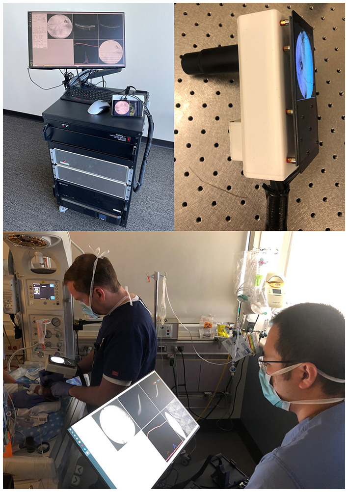
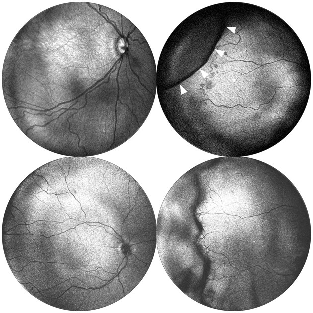
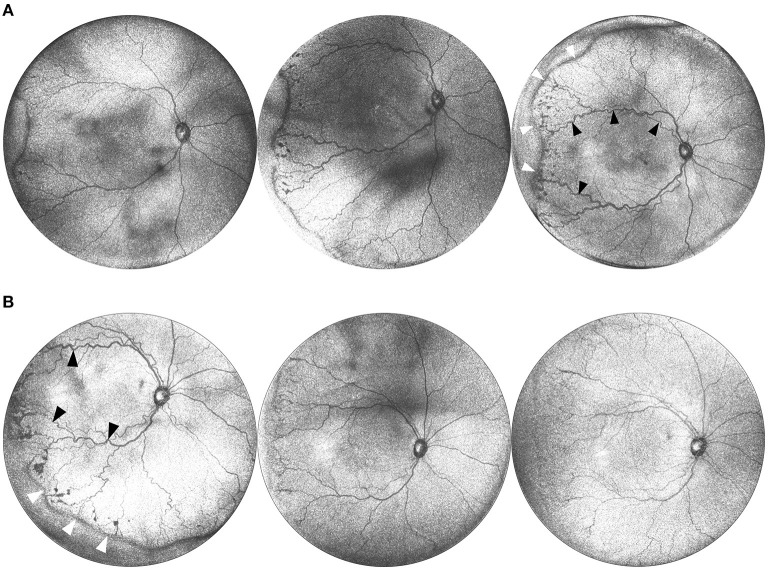
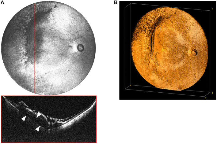
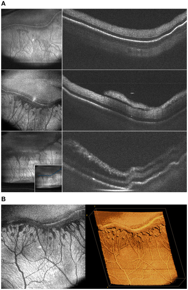
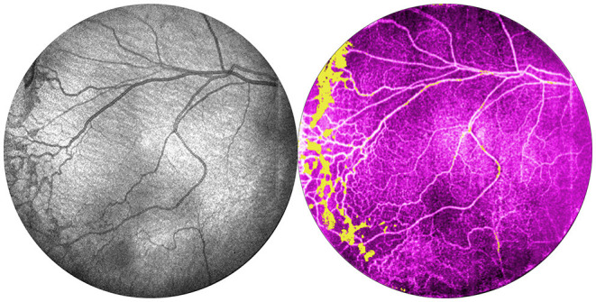
Recent advances in portable optical coherence tomography (OCT) and OCT angiography (OCTA) have resulted in wider fields of view (FOV) and shorter capture times, further expanding the potential clinical role of OCT technology in the diagnosis and management of retinopathy of prematurity (ROP). Using a prototype, handheld OCT device, retinal imaging was obtained in non-sedated infants in the neonatal intensive care unit (NICU) as well as sedated infants in the operating room of Oregon Health & Science University (OHSU) Hospital. In this observational study, we provide an overview of potential advantages of OCT-based disease assessment in ROP. We observed that next-generation OCT imaging (a) may be sufficient for objective diagnosis and zone/stage/plus disease categorization, (b) allows for minimally-invasive longitudinal monitoring of disease progression and post-treatment course, (c) provides three-dimensional mapping of the vitreoretinal interface, and (d) with OCTA, enables dye-free visualization of normal and pathologic vascular development.

Keywords: handheld optical coherence tomography; optical coherence tomography; optical coherence tomography with angiography; pediatric retina; retinopathy of prematurity.


Conflict of interest statement

Oregon Health & Science University (OHSU), DH, and YaJ have significant financial interests in Optovue, a company that may have a commercial interest in the results of this research and technology. These potential conflicts of interest have been reviewed and managed by OHSU. DH and YaJ have received royalties for patent files through OHSU, as well as loaned equipment for research from Optovue. The remaining authors declare that the research was conducted in the absence of any commercial or financial relationships that could be construed as a potential conflict of interest.

Figures

Figure 1
Figure 1
Portable handheld OCT device. Upper-left image shows the prototype device with OCT module and computer. Upper-right image shows a close-up of the probe with a 5.5-inch display capable of real-time en face visualization. Bottom image shows the imaging process of an infant in the neonatal intensive care unit.
Figure 2
Figure 2
Posterior and peripheral en face images obtained via portable 55° and 105° FOV OCT. Images in the top row were obtained using a 55° FOV system from a baby born at 24 weeks gestation (770 grams) and imaged at 33 weeks postmenstrual age. The images in the bottom row were obtained using a 105° FOV system from a baby born at 30 weeks gestation (1,390 g), and imaged at 40 weeks postmenstrual age. Posterior images are shown in the left column and demonstrate the expanded FOV. Peripheral images are shown in the right column and were obtained with the aid of scleral depression using the 55° system. White arrows indicate the indentation of the scleral depressor.
Figure 3
Figure 3
(A) Serial widefield OCT images taken on three successive weeks. These images are from a baby born at 25 weeks gestation (449 g) and imaged at 36, 37, and 38 weeks postmenstrual age. Serial imaging shows the posterior and peripheral retina in one image, and tracks the progression of arterial and venous tortuosity and fibrovascular proliferation. Black arrows highlight areas of increased dilation and tortuosity. White arrows indicate the peripheral ridge which has increased over the time period, along with popcorn neovascularization posterior to the ridge. (B) Serial images pre and post treatment. This is the same infant from (A) at postmenstrual age 38, 39, and 40 weeks. The leftmost image shows a posterior en face view taken immediately prior to treatment with 0.625 mg intravitreal bevacizumab. Middle image shows regression of disease 1 week after treatment, and rightmost image shows further regression 2 weeks after treatment. Black arrows indicate areas of vascular tortuosity pre-treatment that improve post-treatment. White arrows indicate areas of pathologic neovascularization that appears less dense post-treatment.
Figure 4
Figure 4
(A) Widefield en face OCT image of a stage 4A tractional retinal detachment secondary to ROP. These images are from a baby born at 25 weeks gestation (571 g) and imaged at 50 weeks postmenstrual age. Red horizontal line indicates location of the corresponding cross-sectional B-scan. White arrows designate area of retinal detachment with subretinal fluid. (B) Volumetric rendering of retinal detachment shown in 4A. This visualization enables topographic representation of the three-dimensional relationships in severe retinopathy of prematurity, including highlighting the elevation of the blood vessels and fibrovascular proliferation causing the retinal detachment.
Figure 5
Figure 5
(A) Changes in the morphology of fibrovascular proliferation demonstrated by cross-sectional B-scans taken along the length of the ridgeline every 2 weeks. These images were obtained from a baby born 25 weeks gestation and seen at 36, 39, and 41 weeks postmenstrual age. Left column shows en face projection of peripheral pathology. Right column shows progression of neovascularization and retinal traction on B-scan. The inset in the bottom-left image shows sample manual tracing of the ridgeline B-scan used to generate these cross-sections. (B) Volumetric rendering of ridgeline in stage 3 ROP. This is the same patient as (A) seen at 39 weeks. The image on the left shows an en face view of peripheral fibrovascular proliferation in stage 3 ROP. The image on the right shows three-dimensional rendering of the same volume.
Figure 6
Figure 6
Comparison of OCT and OCTA images of retinal vasculature. This is the same patient as Figure 1 (24 weeks gestation, 770 g) seen at 35 weeks gestation. From left: OCT en face image, and corresponding OCTA en face image with flow signal in the vitreous shown in yellow.

Similar articles

Cited by

References

    1. Muni RH, Kohly RP, Charonis AC, Lee TC. Retinoschisis detected with handheld spectral-domain optical coherence tomography in neonates with advanced retinopathy of prematurity. Arch Ophthalmol. (2010) 128:57–62. 10.1001/archophthalmol.2009.361 - DOI - PubMed
    1. Joshi MM, Trese MT, Capone A. Optical coherence tomography findings in stage 4A retinopathy of prematurity: a theory for visual variability. Ophthalmology. (2006) 113:657–60. 10.1016/j.ophtha.2006.01.007 - DOI - PubMed
    1. Vinekar A, Avadhani K, Sivakumar M, Mahendradas P, Kurian M, Braganza S, et al. . Understanding clinically undetected macular changes in early retinopathy of prematurity on spectral domain optical coherence tomography. Investig Ophthalmol Visual Sci. (2011) 52:5183–8. 10.1167/iovs.10-7155 - DOI - PubMed
    1. Erol MK, Ozdemir O, Turgut Coban D, Bilgin AB, Dogan B, Sogutlu Sari E, et al. . Macular findings obtained by spectral domain optical coherence tomography in retinopathy of prematurity. J. Ophthalmol. (2014) 2014:468653. 10.1155/2014/468653 - DOI - PMC - PubMed
    1. Maldonado RS, Yuan E, Tran-Viet D, Rothman AL, Tong AY, Wallace DK, et al. . Three-dimensional assessment of vascular and perivascular characteristics in subjects with retinopathy of prematurity. Ophthalmology. (2014) 121:1289–96. 10.1016/j.ophtha.2013.12.004 - DOI - PMC - PubMed

LinkOut - more resources