. 2022 Jan 18:9:748151.
doi: 10.3389/fbioe.2021.748151. eCollection 2021.

Cryogenic 3D Printing of ß-TCP/PLGA Composite Scaffolds Incorporated With BpV (Pic) for Treating Early Avascular Necrosis of Femoral Head

Affiliations

Cryogenic 3D Printing of ß-TCP/PLGA Composite Scaffolds Incorporated With BpV (Pic) for Treating Early Avascular Necrosis of Femoral Head

Feng Li et al. Front Bioeng Biotechnol. .

Abstract

Avascular necrosis of femoral head (ANFH) is a disease that is characterized by structural changes and collapse of the femoral head. The exact causes of ANFH are not yet clear, but small advances in etiopathogenesis, diagnosis and treatment are achieved. In this study, ß-tricalcium phosphate/poly lactic-co-glycolic acid composite scaffolds incorporated with bisperoxovanadium [bpV (pic)] (bPTCP) was fabricated through cryogenic 3D printing and were utilized to treat rat models with early ANFH, which were constructed by alcohol gavage for 6 months. The physical properties of bPTCP scaffolds and in vitro bpV (pic) release from the scaffolds were assessed. It was found that the sustained release of bpV (pic) promoted osteogenic differentiation and inhibited adipose differentiation of bone marrow-derived mesenchymal stem cells. Micro-computed tomography scanning and histological analysis confirmed that the progression of ANFH in rats was notably alleviated in bPTCP scaffolds. Moreover, it was noted that the bPTCP scaffolds inhibited phosphatase and tensin homolog and activated the mechanistic target of rapamycin signaling. The autophagy induced by bPTCP scaffolds could partially prevent apoptosis, promote osteogenesis and angiogenesis, and hence eventually prevent the progression of ANFH, suggesting that the bPTCP scaffold are promising candidate to treat ANFH.

Keywords: avascular necrosis of femoral head; bisperoxovanadium; phosphatase and tensin homolo; rats; ß-tricalcium phosphate.


Conflict of interest statement

The authors declare that the research was conducted in the absence of any commercial or financial relationships that could be construed as a potential conflict of interest.

Figures

FIGURE 1
FIGURE 1
Schematic illustration of fabricating bpV (pic)/TCP/PLGA composite scaffolds via cryogenic 3D printing. The scaffolds were seeded with rBMSCs for in vitro biological evaluate and implanted into the femoral heads of rats with ANFH to treat ANFH in vivo.
FIGURE 2
FIGURE 2
Morphology and mechanical properties of TCP/PLGA and bpV/TCP/PLGA scaffolds. (A) The morphology of the 3D-printed PTCP and bPTCP scaffolds and SEM micrographs of different scaffolds at different magnification; (B) FTIR spectra of the 3D-printed PTCP and bPTCP scaffolds. (C) Compressive strengths and elastic modulus of both scaffolds. (D) In vitro degradation behavior of both scaffolds. (E) In vitro release behavior of bPTCP scaffolds in a 8-week test period. (F) Results of inverted fluorescence microscope observation of cultured cells for 3 days, the round dots in the figure were living cells (yellow arrow).
FIGURE 3
FIGURE 3
bPTCP scaffolds promotes osteogenic differentiation of rats BMSCs. (A) mRNA expression of osteogenic genes in BMSCs stimulated with osteogenic medium supplemented control or two different concentrations of extracts from the bPTCP scaffolds [bpV (pic)-1, bpV (pic)-2] for 7 days. B. ALP staining of the BMSCs stimulated with osteogenic medium supplemented control or two different concentrations of extracts from the bPTCP scaffold for 7 days. C. Western blot results of OSX and OCN in BMSCs stimulated with osteogenic medium supplemented control or extracts from the bPTCP scaffold for 7 days *p < 0.05.
FIGURE 4
FIGURE 4
bPTCP scaffolds inhibited adipogenic differentiation of rats BMSCs. (A) mRNA expression of adipogenesis genes in BMSCs stimulated with adipogenic medium supplemented control or two different concentrations of extracts from the bPTCP scaffold for 7 days. (B) Oil red O staining of BMSCs stimulated with adipogenic medium supplemented control or two different concentrations of extracts from the bPTCP scaffold for 7 days. (C) Western blot results of PPARγ and CEBPα in BMSCs stimulate with adipogenic medium supplemented control or extracts from the bPTCP scaffold for 7 days *p < 0.05.
FIGURE 5
FIGURE 5
bPTCP scaffolds induced autophagy and inhibited apoptosis via activating AKT/mTOR signaling in vitro. (A) Western blot results of p-PTEN, p-AKT, p-S6 in BMSCs stimulate with control or extracts from the bPTCP scaffold for 24 h. (B) Western blot results of autophagy and apoptosis marker (LC3B and cleaved-caspase3) in BMSCs stimulate with control or extracts from the bPTCP scaffold for 24 h.
FIGURE 6
FIGURE 6
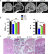
bPTCP scaffolds alleviated the progression of ANFH in rats. (A) Micro-CT scanning images of femoral heads from ANFH rats with different treatments. (B) Quantification of the value of BMD and BV/TV in both groups; (C) H and E staining of the femoral head in ANFH rats from different groups. n = 10 for each group. Scale bar = 50 μm, *p < 0.05. * *p < 0.01, * **p < 0.001.
FIGURE 7
FIGURE 7
bPTCP scaffolds prevented ANFH by inducing autophagy and inhibiting apoptosis in vivo. Representative images of IF staining of p-PTEN (A), p-S6 (B), CD31 (C) LC3B (D) and TUNEL (E) in the femoral head of ANFH rats from different groups. ns, not significant, *p < 0.05. * *p < 0.01, * **p < 0.001.

References

    1. Aoki M., Fujishita T. (2017). Oncogenic Roles of the PI3K/AKT/mTOR Axis. Curr. Top. Microbiol. Immunol. 407, 153–189. 10.1007/82_2017_6 - DOI - PubMed
    1. Cui L., Zhuang Q., Lin J., Jin J., Zhang K., Cao L., et al. (2015). Multicentric Epidemiologic Study on Six Thousand Three Hundred and Ninety Five Cases of Femoral Head Osteonecrosis in China. Int. Orthopaed. 40 (2), 267–276. 10.1007/s00264-015-3061-7 - DOI - PubMed
    1. Derubeis A. R., Cancedda R. (2004). Bone Marrow Stromal Cells (BMSCs) in Bone Engineering: Limitations and Recent Advances. Ann. Biomed. Eng. 32 (1), 160–165. 10.1023/b:abme.0000007800.89194.95 - DOI - PubMed
    1. Guo X., Jiang H., Zong X., Du L., Zhao J., Zhang D., et al. (2020). The Implication of the Notch Signaling Pathway in Biphasic Calcium Phosphate Ceramic‐induced Ectopic Bone Formation: A Preliminary experiment. J. Biomed. Mater. Res. 108 (5), 1035–1044. 10.1002/jbm.a.36878 - DOI - PubMed
    1. Li Z., Liao W., Zhao Q., Liu M., Xia W., Yang Y., et al. (2013). Angiogenesis and Bone Regeneration by Allogeneic Mesenchymal Stem Cell Intravenous Transplantation in Rabbit Model of Avascular Necrotic Femoral Head. J. Surg. Res. 183 (1), 193–203. 10.1016/j.jss.2012.11.031 - DOI - PubMed